

Method for preparing 3D cartilage organoid block
- Summary
- Abstract
- Description
- Claims
- Application Information
AI Technical Summary
Benefits of technology
Problems solved by technology
Method used
Image

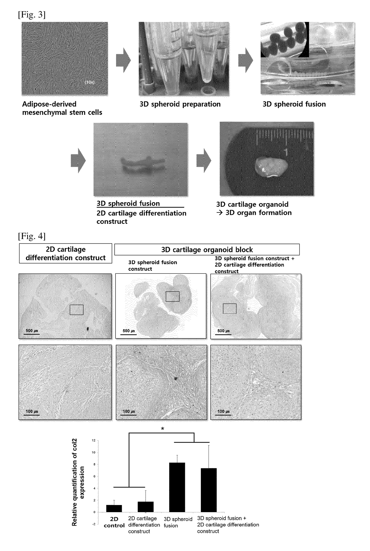
Examples
example 1
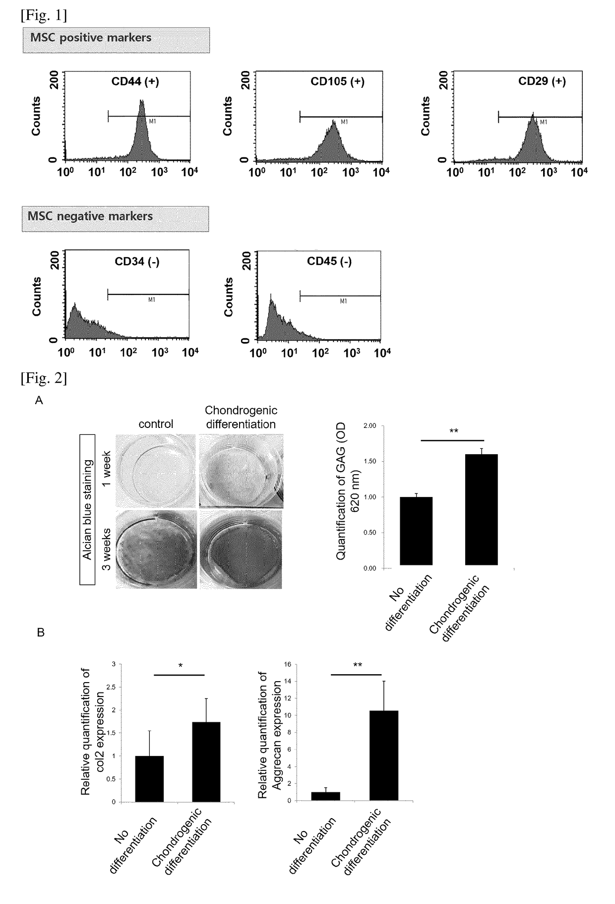
of Human Mesenchymal Stem Cells, Incubation and Assessment of Stemness
[0032]Adipose tissues were cut to small pieces with a mess, and then the obtained pieces were washed three times with phosphate buffered saline (PBS)(Sigma, St. Louis, Mo.). Then, the small pieces of adipose tissues were put into a 50 ml conical tube. PBS was added to the tube and stirring was carried out, and then centrifuged. The soup was discarded, and Dulbecco's modified Eagle medium (DMEM) was added up to a volume of 50 ml, and thereafter the mixture was allowed to react for 90 minutes at 37° C. Unsolved adipose tissues suspended in the upper layer was removed after centrifugation for 10 minutes at 2,000 rpm. Then, washing with DMEM, centrifugation and removal were repeated. The isolated adipose-derived stem cells were incubated and proliferated with serum-free stem cell culture medium (chemically defined media) at 37° C. in a 5% CO2 incubator. The DMEM including 10% FBS can be used for the proliferation. The...
example 2
sment of Differentiation of Mesenchymal Stem Cells into Cartilage
[0033]The obtained stem cells were seeded at 1×10⁴ cells / cm2 and incubated at 37° C. in a 5% CO2 incubator, while the cells were treated with cartilage differentiation medium every 2 days, in order to differentiate the mesenchymal stem cells into cartilage cells. The cartilage differentiation medium comprised 50 ug / ml ascorbate 2-phosphate, 100 nM dexamethasone, 1% ITS, and 10 ng / ml TGF-beta1. The GAG matrix formation level was fixed by treating the cells in 2D cartilage plate with 10% formaldehyde for 30 minutes, and then the cells were treated with 3% acetic acid solution for 3 minutes, followed by staining with 500 μl Alcian blue (pH 2.5) solution for 30 minutes. The stained sample was washed several times with distilled water and detected using a microscope. In order to obtain quantitative GAG value, the Alcian blue stained plate was left with 3% acetic acid for 10 minutes, and 100 μl of soup was collected to deter...
example 3
Stimulation Using Real Time PCR
[0034]In order to analyze gene modification between undifferentiated stem cells and cartilage differentiation cells, the expression of cartilage differentiation gene was assessed. For this purpose, Col II and Aggrecan were used as gene markers relating to cartilage cells, and GAPDH was employed as a housekeeping gene. The Real-time PCR was performed as follows. That is, the cells obtained from each group were washed with PBS, and then they were collected with Trypsin-EDTA and RNAs were extracted with TRIzol (Life Technologies, Inc. Grand Island, N.Y.) method. The extracted RNAs 1 μg were used to prepare cDNA, and the change of gene expression was investigated. The primer sets and respective differentiation markers are as shown in Table 1 below.
TABLE 1markerSequences*OriginsCol IISEQ ID NO. 1sense5′-TTC AGC TAT GGA GAT GAC AAT C-3′NM_001844SEQ ID NO. 2antisense5′-AGA GTC CTA GAG TGA CTG AG-3′AggrecanSEQ ID NO. 3sense5′-GAA TCT AGC AGT GAG ACG TC-3′NM_01...
PUM
 Abstract
Description
Claims
Application Information