Direct affinity measurement of human igg1 binding multimeric antigens

a multi-mer, affinity-based technology, applied in the field of direct affinity measurement of human igg1 binding multi-mer antigens, can solve the problems of difficult to analyze the diverse chromatographic and electrophoretic bands, separate antibody species, and long time-consuming and laborious problems of applying and optimizing rp-hplc methods

Inactive Publication Date: 2019-01-17
F HOFFMANN LA ROCHE & CO AG
View PDF0 Cites 1 Cited by
  • Summary
  • Abstract
  • Description
  • Claims
  • Application Information

AI Technical Summary

Benefits of technology

[0037]selecting one or more antibodies based on the binding affinity determined in the previous step.

Problems solved by technology

Although mAbs can be successfully analyzed by means of various separation and testing techniques, it has for a long time been difficult to apply and optimize RP-HPLC methods (RP-HPLC, Reversed Phase-High Performance Liquid Chromatography) to separate antibody species.
However, various modifications of the antibody are often present simultaneously during the course of a degradation process, which makes it more difficult to analyze the diverse chromatographic and electrophoretic bands.

Method used


Image

  • Direct affinity measurement of human igg1 binding multimeric antigens
  • Direct affinity measurement of human igg1 binding multimeric antigens
  • Direct affinity measurement of human igg1 binding multimeric antigens

Examples


example 1

Transient Fab Expression and Purification

[0164]The antibody light chain and heavy chain Fd-fragments were ordered as gene syntheses and cloned via unique restriction sites using standard cloning procedures into separate expression vectors for each chain enabling secretory expression in HEK cells growing in suspension. Transfection (1:1 plasmid ratios) into HEK293-F cells (Invitrogen, Cat. No. 510029) was performed according to the cell supplier's instructions using Maxiprep (Qiagen, Cat. No. 12163) preparations of the antibody vectors, Opti-MEM I medium (Invitrogen, Cat. No. 31985) 293fectin (Invitrogen, Cat. No. 31985070), and an initial cell density of 1-2×10E+06 viable cells / mL in serum-free FreeStyle 293 expression medium (Invitrogen, Cat. No. 12338018). Antibody containing cell culture supernatants were harvested after 7 days of cultivation in shake flasks by centrifugation at 14,000× g for 30 min. and filtered through a 0.22 μm sterile filter (Thermo Scientific, Cat. No. 566-0...

example 2

Enzymatic Cleavage of Bevacizumab with Papain

Without Purification:

[0165]The antibody was diluted in 20 mM Histidine, 140 mM NaCl, pH 6.0 to a final concentration of 1 mg / mL, added 2 μL 250 mM L-cysteine (Sigma-Aldrich, Schnelldorf, Germany) and 10.9 μL diluted papain (7.34 U / mL in 20 mM Histidine, 140 mM NaCl, pH 6.0), and incubated 1 h at 37° C.

With Purification:

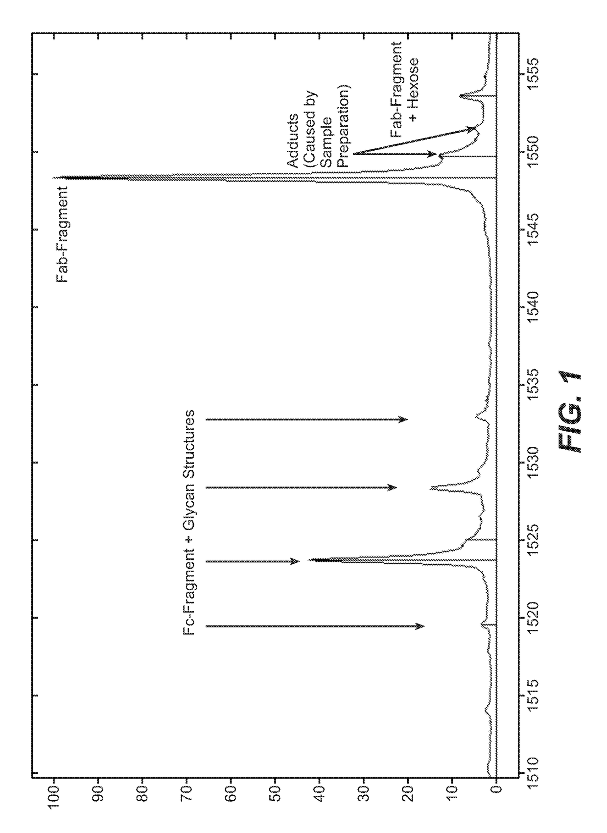
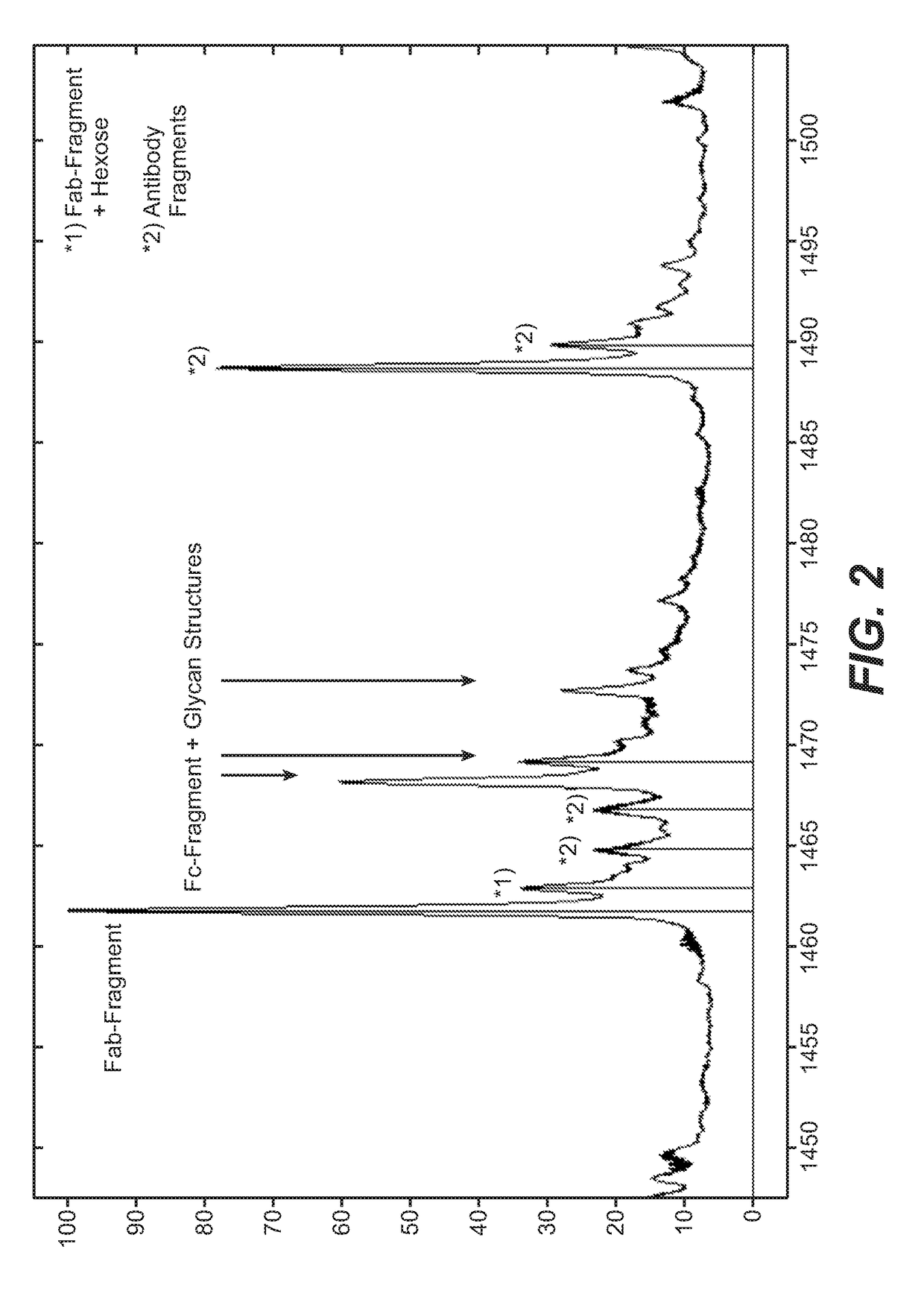
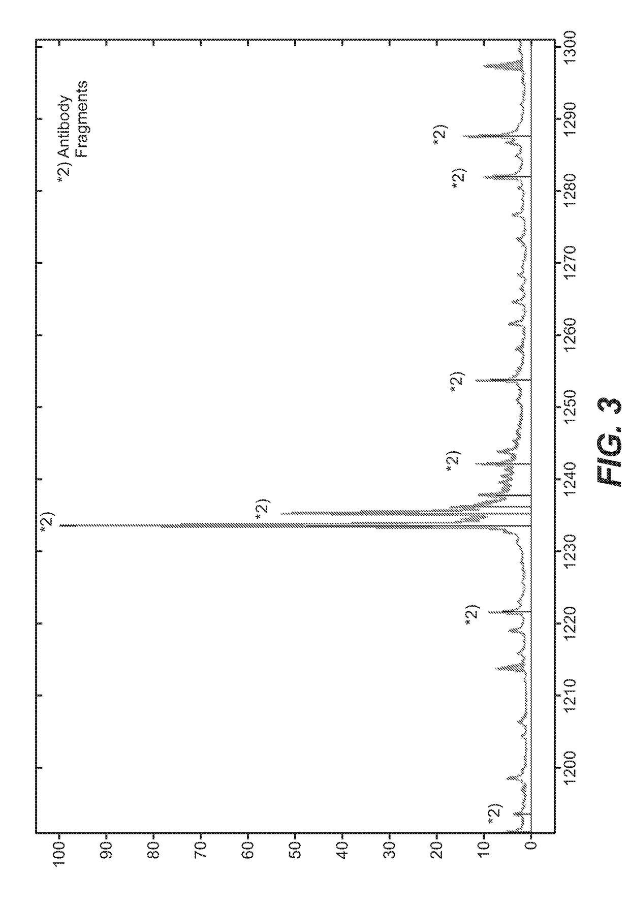
[0166]The antibody was incubated with Papain (0.8 U / mg mAb; Sigma-Aldrich / Roche) in presence of 5 mM Cystein for 170 minutes at 37° C. To isolate the Fab from non-cleaved antibodies, Fc-fragments and Papain, the mixture was applied to a CaptureSelect IgG-CH1 and MabSelectSuRe affinity chromatography (GE Healthcare) according to manufacturer protocol. Finally, a size exclusion chromatography using a Superdex 75 10 / 300 GL column (GE Healthcare) was performed using 140 mM NaCl, 20 mM histidine (pH 6.0) as running buffer. Protein concentration of the Fab was determined by measuring the optical density (OD) at 280 nm, using the ...

example 3

Enzymatic Cleavage of Bevacizumab with Lysine-Gingipain of Porphyromonas Gingivalis

[0167]GingisKHAN was reconstituted in 200 μL ddH2O resulting in 2000 U / 200 μL, and the 10× reducing agent was freshly prepared in 50 μL ddH2O (final concentration: 20 mM Cysteine) prior to each digestion. 100 μg antibody was diluted to a final concentration of 1 mg / mL in 100 mM Tris, pH 8.0 and subsequently digested with 10 μL GingisKHAN and 11 μL of freshly prepared 10× reducing agent at 37° C. for 1 hour.


PUM

PropertyMeasurementUnit
temperatureaaaaaaaaaa
pHaaaaaaaaaa
pHaaaaaaaaaa

Abstract

Herein is reported a method for determining the binding affinity of the binding sites of a bivalent full length antibody of the human IgG1 subclass to a homo-multimeric antigen comprising the steps of i) incubating a mixture comprising the antibody and a polypeptide that is derived from lysine-gingipain of porphyromonas gingivalis at a pH of from pH 7.5 to pH 8.5, in the presence of a reducing agent, at a temperature of from 30° C. to 42° C., for time of from 10 min. to 240 min. to cleave the antibody into Fabs and Fc-region, and ii) determining the binding affinity of the Fabs of the antibody for its antigen using a surface plasmon resonance method by directly applying the incubated reaction mixture obtained in the previous step in the surface plasmon resonance method and therewith determining the binding affinity of the binding sites of the bivalent full length antibody of the human IgG1 subclass.

Description

CROSS-REFERENCE TO RELATED APPLICATIONS[0001]This application is a continuation of International Patent Application No. PCT / EP2016 / 079756, having an international filing date of Dec. 5, 2016, the entire contents of which are incorporated herein by reference, and which claims benefit under 35 U.S.C. § 119 to European Patent Application No. 15198556.1, filed on Dec. 9, 2015.SEQUENCE LISTING[0002]The instant application contains a Sequence Listing which has been submitted electronically in ASCII format and is hereby incorporated by reference in its entirety. Said ASCII copy, created on Jun. 4, 2018 is named P33225-US_Sequence_Listing.txt and is 27,995 bytes in size.FIELD OF THE INVENTION[0003]Herein is reported a fast and easy method for determining affinity constants of human IgG1s binding di- or multimeric antigens using surface plasmon resonance. The method is based on a highly specific and quantitative digestion with lysine-gingipain of porphyromonas gingivalis generating a homogen...

Claims


Application Information

Patent Timeline
Patent Type & Authority Applications(United States)
IPC IPC(8): G01N33/557G01N33/537G01N21/552G01N33/566G01N33/569C07K16/12
CPCG01N33/557G01N33/5375G01N21/553G01N33/566G01N33/56911C07K2317/52C07K2317/55C07K2317/92C07K2317/31C07K2317/35C07K16/1203G01N33/56955C12Q1/37G01N33/6857G01N2333/96466C07K16/00C07K16/22
Inventor MOLHOJ, MICHAELGASSNER, CHRISTIANMOELLEKEN, JOERGENDESFELDER, MANUEL
Owner F HOFFMANN LA ROCHE & CO AG
Why Eureka
Try Eureka