
Method for measurement and model-free evaluation of injectable biomaterials properties

a biomaterial and model-free technology, applied in the field of new methods of testing injectable materials, can solve problems such as pain, local perturbation of tissue, damage to organs or somatic systems,

Active Publication Date: 2019-02-07
SEQVERA LTD OY
View PDF0 Cites 2 Cited by
  • Summary
  • Abstract
  • Description
  • Claims
  • Application Information

AI Technical Summary

Benefits of technology

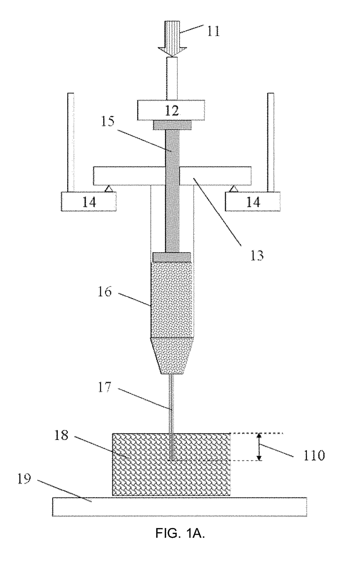
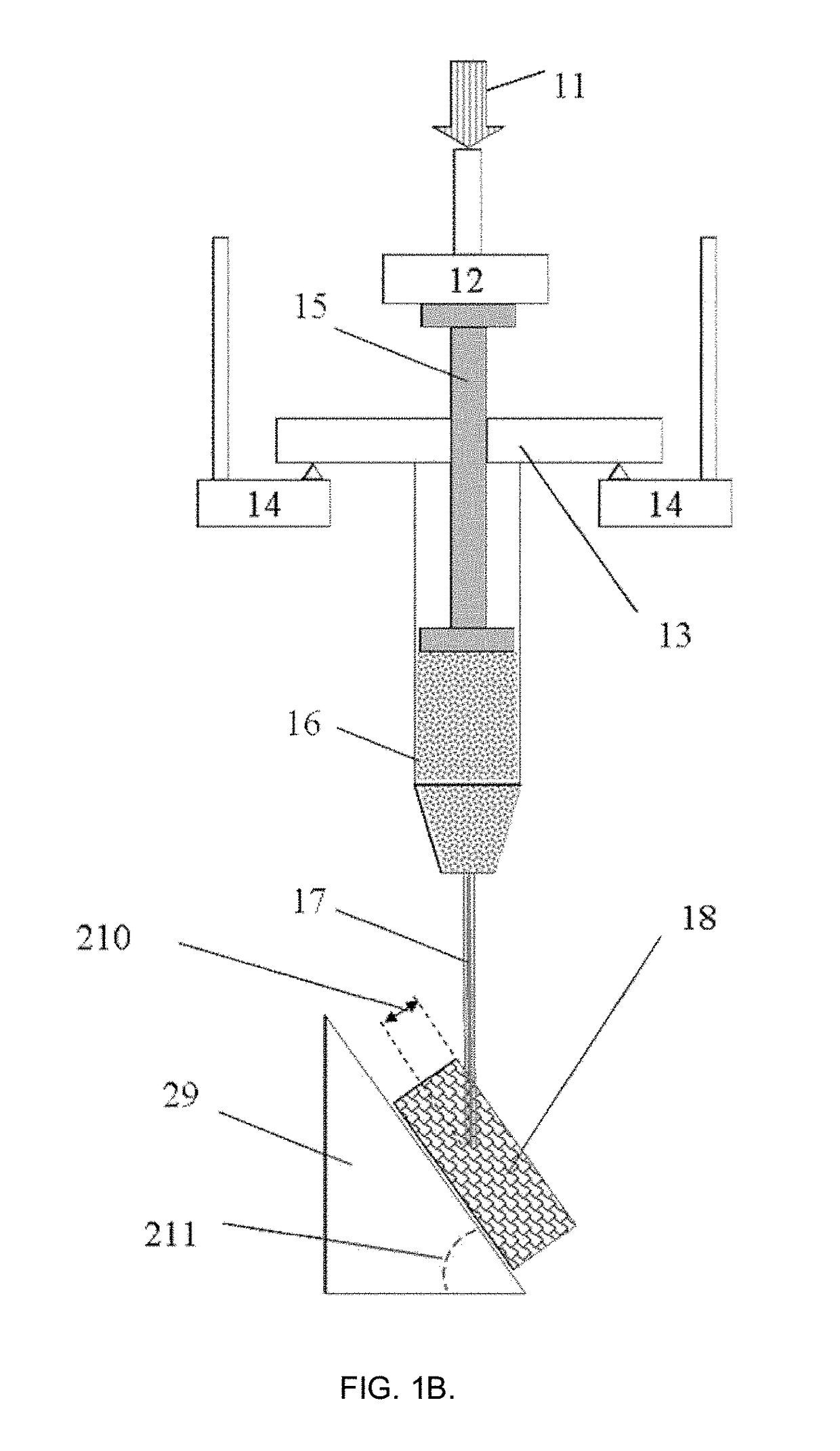
The present invention provides a method for testing the injectability of biomaterials and evaluating the performance of biomaterials-treated objects. This method involves placing the biomaterial into an injection device, positioning the device onto a sample holder, and injecting the material into the sample at a desired depth while measuring the resulting displacement, displacement rate, and strain. The method does not require any prior assumptions about the material, its structure, or behavior, and can provide information on the time-invariant properties of the material and the target object. This method can be used for various applications in medicine and cosmetics.

Problems solved by technology

Injection of any material into body tissues causes local perturbation of the tissue due to pressure peaks, influencing adjacent nerves and causing pain feeling.
Many applications like injection of a contrast into spine channel, of a corrective substance into intervertebral disk, of R-cells-laden hydrogel into pancreas, or of visco-supplementation into an articulate joint, etc., might have a catastrophic effect causing damage to the organ or somatic system due to improper injection rate.
The latter however are much less in use, complex, expensive and furthermore are designed for specific product only.
For instance, when the fluid in non-Newtonian (its viscosity is stress- or rate-dependent), automatic syringe system cannot know the local shear rate and the instant viscosity, which lead to variable supply of the injected material while the system considers it to be injected evenly.
None of these methods however is really practical—even in the case when inaccuracies arising from variation in frictional resistance within the syringe, in the syringe and needle diameter and depth of insertion, in the plunger speed, in the degree of lubrication, in the contact surface between the plunger cover and the syringe wall, in the apparent discontinuities in the plunger movement, etc., etc. are eliminated, this does not give a useful information how much fluid was actually injected at that depth in the tissue and at which rate this has happened.
However, many such tests, for example by the standard ISO 11040, were criticized by US FDA lacking essential specifications and describing only a testing method of the injection as a stand-alone product [1].
Indeed, pressure measurements using a syringe without a needle and with water as a fluid (ISO 7886, Indian standard IS 10258) do not produce data suitable for another system (especially non-Newtonian) where fluid or hydrogel is being injected into a real tissue with a specific needle size.
This method also capable only of pressure sensing and is not able to control any other parameters.
Especially in the case of biological fluids (which are known to be non-Newtonian), there is no possibility to know exact stresses and shear rates acting in the syringe space, in the needle and in the target tissue.
Despite that rheological properties of these materials were measured in advance, revealing links between shear rate and viscosity, it did not help to catch the proper variation of injectability of these fluids.
Application of these or similar control or measurement methods is not possible in practice, when the medium behaves in a non-conventional way, for instance for shape-memory hydrogels, which collapse upon applied injection pressure and then restoring their shape and volume after leaving the needle [4].
There is no single hydrodynamic flow or material theory able to clearly characterize the injectability of such systems.
Only very simplified assumptions (like constant viscosity for laminar Hagen-Poiseueille flow equation) could be employed, but it is well-known they are incapable to describe non-linear behavior of complex fluids.
None of the known existing methods discloses a procedure to measure how big volume of a material would be injected from a specific syringe with a selected needle at proper rate and time, when the needle is inserted at specified depth and angle into a relevant tissue; and which applied force pattern in time would be required for that.
After the injection stage has been completed, another problem appears—there is a lack of essential information about the properties, evolution and transient physical maturation of injected biomaterials and tissues where those biomaterials have been injected.
This is especially of concern in cosmetic operations (botox and hyaluronan injections, lipocorrections—fat transfer), as esthetic and medical result often may be far from desired due to formation of bruising, edema, erythema, discoloration, Tyndall effect, relatively short effect time requiring regular injections or surgical procedures, nodules, abnormal tissue migration, excess “hardening” or “softening”, etc.
“feels not so good”) and not quantified; further they are depending on time, patient mood and status and are not easily compared—even for a single person, there are no two fully identical skin or tissue areas which could be tested and validated.
The rationale is that oversimplified fluid models (like a shear-thinning liquid) and tissue models (like a hyper-elastic soft solid) do not provide realistic description of any part of the process nor in the whole, and do not allow correct comparison with the controls.
The data quality reported for the same material may also be confusing, as no exact information is given for conditioning changes, and usually no solid proof shown e.g. about suitability of the small strain theory or material linearity [7].
There is also no single reliable biomechanical test able to get simultaneously a spectrum of invariant functions of the tissue or sample before and after injection, including e.g. sample memory and viscostiffness spectra, characteristic times, intrinsic modulus and viscosity spectra, etc., without assumption of some simplified material model.

Method used


Image

  • Method for measurement and model-free evaluation of injectable biomaterials properties
  • Method for measurement and model-free evaluation of injectable biomaterials properties
  • Method for measurement and model-free evaluation of injectable biomaterials properties

Examples


example 1

[0092]According to this example, a 1 mL capacity Fortuna® Optima® glass syringe (Poulten & Graf GmbH, Wertheim, Germany) was filled with acrylate polymer-based hydrogel fluid used for disinfection as a model injectable biomaterial of sufficiently high viscosity (GOJO Industries Inc., Akron, Ohio, USA). The syringe was attached with a sterile 26G1 / 2 size PrecisionGlide® hypodermal needle (Becton Dickinson & Co., Franklin Lakes, N.J., USA) internal needle diameter 0.260 mm) and located at the fixed support according to FIG. 1A in the customized sample holder connected to the dynamic mechanical analyzer DMA242C (Netzsch Gerätebau GmbH, Selb, Germany). The needle was inserted on ˜1 mm depth at the right angle into a commercial skin tissue phantom (Simskin LLC, Chicago, Ill., USA), comprising of several layers mimicking epidermis, dermis, hypodermis and fat layers. Tests were carried out at 25±1° C. and normal atmospheric pressure in air.

[0093]The test began with moving the probe-sensor ...

example 2

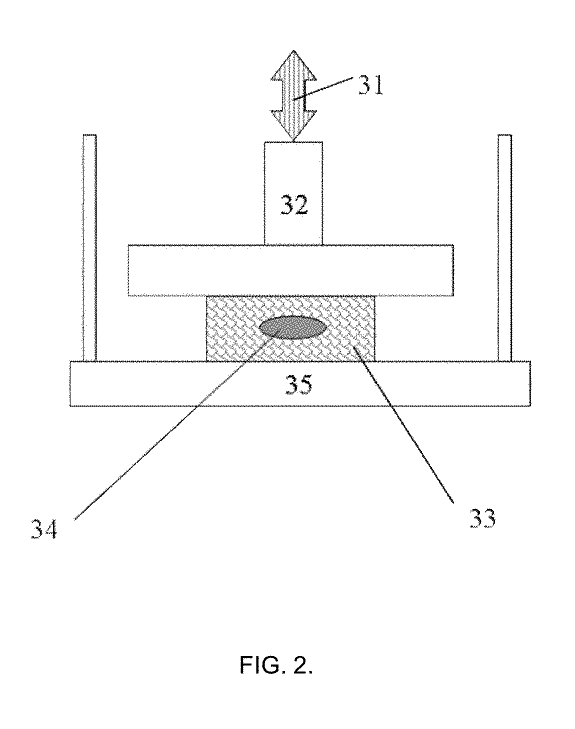
[0099]The tissue phantom with injected 50±10 μL of the test material as shown in Example 1 was further tested on its biomechanical properties in the manner depicted in FIG. 2. The tissue phantom specimen with external dimensions (l×w×h) (11±0.5)×(12±0.5)×(6.5±0.3) mm was positioned on the sample holder plate and brought into the contact with the probe sensor of 15 mm diameter using compression mode sample holder. The precise size of the tissue phantom specimen was measured with non-contact optical method (±0.5 μm) with laser micrometer (Metralight Inc., CA, USA). The measurements were done before injection and after injection to check that no apparent significant variations (such as specimen twisting or buckling) are present. The other essential details of this measurement and theoretical description are also presented in the non-provisional U.S. patent application Ser. No. 15 / 655,331 of 20 Jul. 2017.

[0100]After letting the probe to establish the contact with the specimen and taring...

example 3

[0107]In this example, the injectability of the hydrogel material was measured in a similar way to Example 1 but with the purpose to obtain values needed to get constant injection rate. It is anticipated that for maximal patient comfort the injection rate should be kept as constant as possible (minimizing injection pain). This is difficult to achieve with manual syringe or an automated injection gun where injection force (pressure) is set up initially.

[0108]The tests were made using DMA242E “Artemis” (Netzsch Gerätebau GmbH, Germany) customized by the applicant. The tests were done at 22±1° C. and RH 25% in a climate-control room under laminar flow cabinet of ISO Class 5 (USP compliant). Plastic syringes (Galderma AB, Sweden) syringes of 1 mL capacity (as used for Restylan® Skin Booster dermal filler) were thoroughly washed several times with deionized water, dried and cleaned from any residual matter and connected with sterile needles 29G×½″. Acrylic hydrophilic gel (Gojo Industrie...


PUM


Abstract

The invention discloses a method for testing of injectable biomaterials and their performance in the treated materials, to obtain functional properties of the biomaterials and of the specimen injected with that biomaterial. The purpose of the method is to measure these properties during injection of a biomaterial into a target object, then measure the properties of a target object specimen after the injection, and then combine these results using material model-free data processing with possible prediction.

Description

PRIORITY[0001]This application is nonprovisional application of U.S. Provisional Patent application No. 62 / 541,252 filed 4 Aug. 2017, the contents of which are incorporated herein by reference.FIELD OF THE INVENTION[0002]The present invention relates to a new method of testing injectable materials, especially biomaterials, allowing measurement, model-free evaluation and possible prediction of plurality of those material properties during and after injection to describe the behavior or performance of that biomaterial at proper conditions.BACKGROUND OF THE INVENTION[0003]Injectable biomaterials and fluids are of a daily use in medical practice, healthcare and cosmetology. Injection of any material into body tissues causes local perturbation of the tissue due to pressure peaks, influencing adjacent nerves and causing pain feeling. Many applications like injection of a contrast into spine channel, of a corrective substance into intervertebral disk, of R-cells-laden hydrogel into pancrea...

Claims


Application Information

Patent Timeline
IPC IPC(8): G01N11/04A01N25/04G01N33/15
CPCG01N11/04A01N25/04G01N33/15G01N11/08G01N11/00
Inventor GASIK, MICHAEL
Owner SEQVERA LTD OY
Learn More