3-d tissue culture based method to assess mitochondrial impairment

Inactive Publication Date: 2019-03-14
INSPHERO AG
View PDF0 Cites 0 Cited by
  • Summary
  • Abstract
  • Description
  • Claims
  • Application Information

AI Technical Summary

Benefits of technology

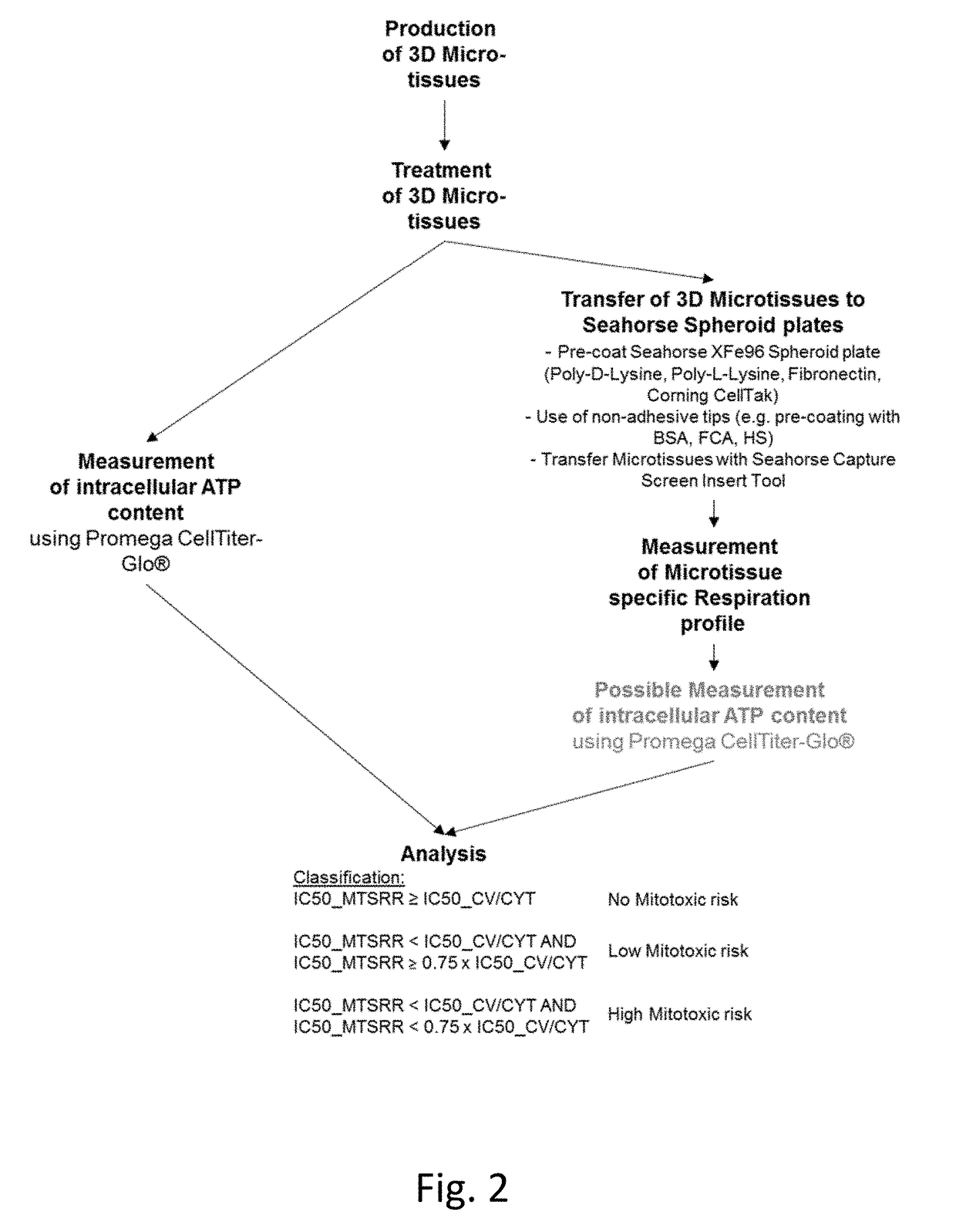
[0064]In one embodiment, the measurement of cell viability / cytotoxic effect determines the residual viability of one or more cells.
[0065]Cell viability / cytotoxic effect can for example be determined by quantitation of the ATP present in the cells. One such assay is the CellTiter-Glo Luminescent Cell Viability Assay (Promega, Madison, US), while other cell viability / cytotoxicity assays exist on the market and can likewise be used.
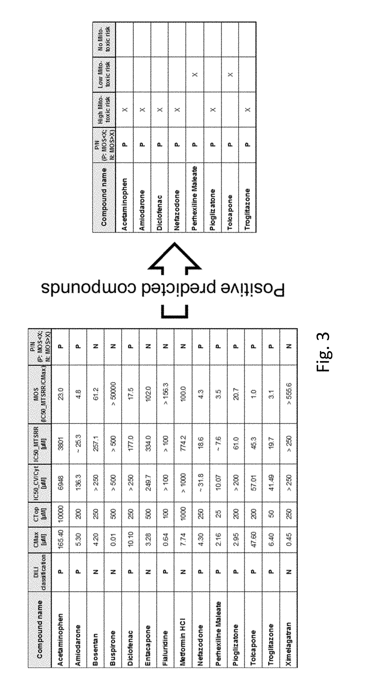
[0066]In another embodiment, the effect on microtissue-specific respiration rate and / or the effect on cell viability / cytotoxic effect is determined as inhibitory concentration, preferably as half maximal inhibitory concentration (IC50).

Problems solved by technology

Third, in a 3-dimensional cell culture or tissue, drugs to be tested have to enter the tissue by diffusion from the outside, thus affecting cells in the outer cell mass more than cells in the inner cell mass.

Method used


Image

  • 3-d tissue culture based method to assess mitochondrial impairment
  • 3-d tissue culture based method to assess mitochondrial impairment
  • 3-d tissue culture based method to assess mitochondrial impairment

Examples


example 1

Generation of 3D Microtissue

[0086]1.1. Thawing of Cryopreserved Hepatocytes[0087]Take chosen cell-vial from cryo-tank and transfer into water bath (37° C.); set timer on 2 minutes[0088]Pipette 40 ml of the Wash / Thawing medium into 50 ml tube[0089]Transfer the hepatocytes into the tube, wash with 1 ml medium[0090]Place 50 ml tube into centrifuge; start centrifugation for 5 min at 50 rcf (corresponds to 600 rpm) at RT.[0091]Remove supernatant[0092]Wash pellet with 20 ml wash buffer[0093]Place carefully 3 ml of Wash / Thawing medium into the 50 ml tube[0094]Re-suspend cell pellet with 2D cell culture medium[0095]Count hepatocytes with e.g. Trypan Blue

[0096]1.2. Hepatocyte Pre-Plating[0097]Use a collagen coated cell-culture dish for pre-seeding[0098]Seed Hepatocytes in a 6 cm dish: 100000-250000 hepatocytes / cm2; 0.05-0.5ml per cm2 [0099]Place at 37° C. in a CO2 containing incubator[0100]Optionally: Exchange medium after attachment with Serum-free 2D-culture medium[0101]Harvest hepatocytes...

example 2

Measuring Mitochondrial Toxicity with the Seahorse XF Analyzer

2.1 Treatment of 3D Microtissues

[0138]After generating 3D primary human hepatocytes microtissues (IPHH_02) of a diameter size of 370 um, the microtissues have been incubated with varying concentration of compounds for 48 h Thereby, the compounds were dissolved in 3D InSight™ Human Liver Maintenance Medium-TOX

[0139]2.2 Seahorse XF Analyzer Measurement

A. Seahorse Assay Medium Preparation

[0140]

Stock conc. Assay conc.Volume Component(mM)(mM)(mL)DMEM XF Base——50Glucose1000100.5Na-Pyruvate10010.5UltraGlutamine20020.5Adjust pH to 7.4

[0141]B. Coating of Seahorse Spheroid plates[0142]20 uL of Poly-D-Lysine (100 ug / mL diluted in cell culture grade water) were added to each well of the Seahorse Spheroid plate (Incubation for 20min @ RT)[0143]After coating, the Seahorse Spheroid plate was washed with 200 uL of sterile water and wash was removed (Washing step repeated once)[0144]Seahorse Spheroid plate has been air dried for a few min...


PUM

PropertyMeasurementUnit
volumesaaaaaaaaaa
diameter sizeaaaaaaaaaa
tissue-specific respiration rateaaaaaaaaaa

Abstract

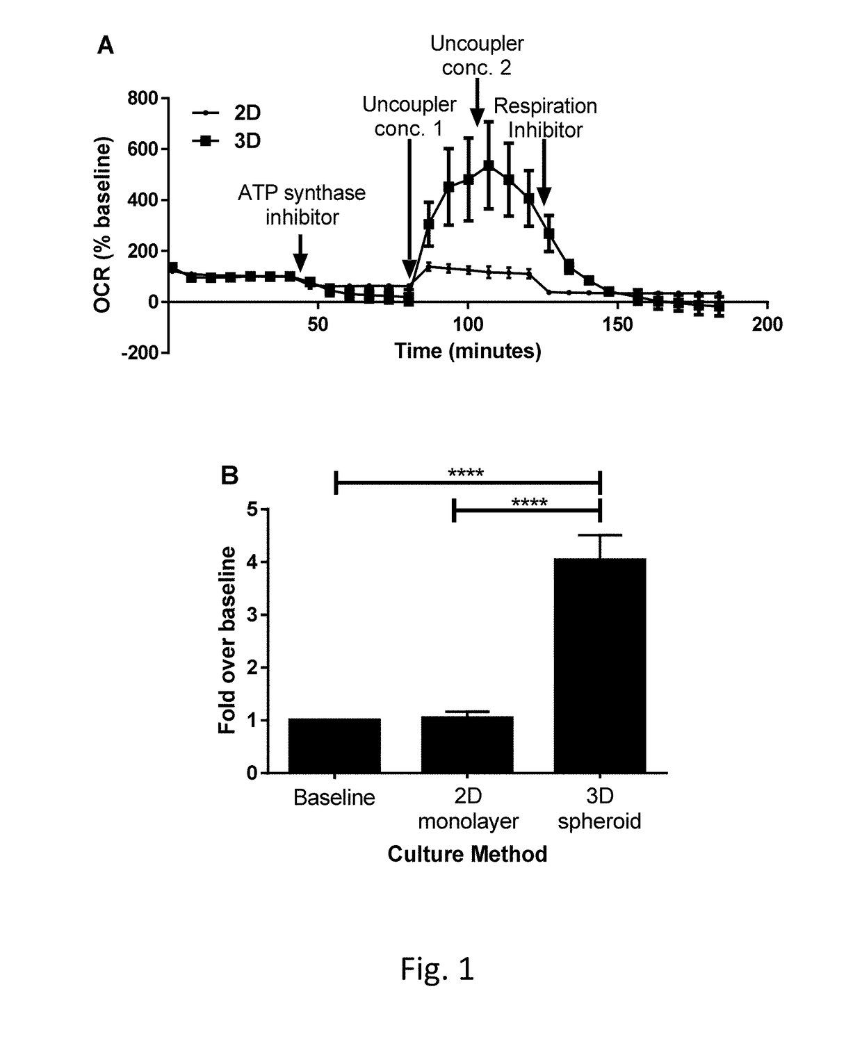
The present invention relates to a method and/or assay for the assessment of the metabolic effect of a candidate compound. The method and/or assay comprises exposing one or more 3-dimensional cell culture or tissue to one or more candidate compounds, and measuring, in at least one 3-dimensional cell culture or tissue, the effect of such exposure on the 3-dimensional cell culture or tissue-specific respiration rate (MTSRR) (FIG. 1B).

Description

FIELD OF THE INVENTION[0001]The present invention relates to a method and / or assay for the assessment of the metabolic effect of a candidate compound. The method and / or assay comprises exposing one or more 3-dimensional cell culture or tissue to a candidate compound.[0002]Mitochondria play a critical role in generating most of the cell's energy as ATP. They are also involved in other metabolic processes such as urea generation, haem synthesis and fatty acid beta-oxidation. Disruption of mitochondrial function by drugs can result in cell death by necrosis or can signal cell death by apoptosis (e.g., following cytochrome c release).[0003]Drugs that injure mitochondria usually do so by inhibiting respiratory complexes of the electron chain; inhibiting or uncoupling oxidative phosphorylation; inducing mitochondrial oxidative stress; or inhibiting DNA replication, transcription or translation.[0004]It is important to test for mitochondrial toxicity early in drug development as impairment...

Claims


Application Information

Patent Timeline
Patent Type & Authority Applications(United States)
IPC IPC(8): G01N33/50C12N5/0775C12N5/078C12N5/077C12N5/095C12N5/071
CPCG01N33/5079C12N5/0667C12N5/0634G01N33/5047C12N5/0656C12N5/0695C12N5/0671G01N33/5014C12M23/12G01N33/50G01N33/5008
Inventor KELM, JENSMESSNER, SIMON
Owner INSPHERO AG
Why Eureka
Try Eureka