Determining a probabilistic diagnosis of cancer by analysis of genomic copy number variations

a technology of copy number variation and probability diagnosis, applied in the field of probability diagnosis of cancer by analysis of genomic copy number variation, can solve the problems of low resolution of cdna array methods, inefficient and incomplete one-by-one query method, and inability to achieve a level of resolution greater than can be achieved, and achieve the effect of high probability

Inactive Publication Date: 2019-10-24
COLD SPRING HARBOR LAB INC
View PDF0 Cites 2 Cited by
  • Summary
  • Abstract
  • Description
  • Claims
  • Application Information

AI Technical Summary

Benefits of technology

[0019]Another aspect of the present invention provides a genomic segment useful as a copy number probe for assessing probable clinical outcome for an individual patient having a tumor associated with breast cancer. In certain embodiments, a genomic segment corresponds or relates to: an EGFR locus as shown, e.g., in FIG. 15 or Table 8, which indicates chromosomal positions of probes specific to the EGFR locus; a Her2 locus as shown, e.g., in FIG. 4 or Table 8, which indicates chromosomal positions of probes specific to the Her2 locus; and an INK4 (CDKN2A) locus located at chromosome 9p21.97 or, e.g., as shown in Table 8, which indicates chromosomal positions of probes specific to the CDKN2A locus.
[0020]A further aspect of the present invention provides a method for assessing probable clinical outcome for an individual patient having a disorder, condition or disease, such as a tumor, the method including: obtaining a segmented genomic profile, GP(i), of DNA extracted from one or more diseased (e.g., tumor) cells from a first individual patient, said GP(i) representing a subpopulation of chromosome rearrangements present in the extracted diseased (e.g., tumor) cell DNA derived by measuring relative copy number of one or more segments representing a portion of the genome comprising one or more of the genomic segment of the present invention.
[0021]Another aspect of the present invention provides a method for identifying one or more potential oncogenic loci associated with a particular tumor type or disease. In certain embodiments, the method includes the steps of comparing genomic profiles generated according to the methods of present invention, and identifying as oncogenic loci segments of the genome that correlate with high probability, alone or in combination, to probable clinical outcome for an individual patient having the particular tumor type or disease.
[0022]Another aspect of the present invention relates to a method for determining whether a subject tumor in an individual patient is related to a tumor that occurred earlier (earlier tumor) in the same patient. In certain embodiments, the method includes obtaining a segmented genomic profile, GP(T2), of DNA extracted from one or more cells of the subject tumor, said GP(T2) representing chromosome rearrangements present in the extracted DNA derived by measuring relative copy number of a plurality of discrete segments of the genome or one or more portions of the genome. In certain embodiments, the method further includes comparing the GP(T2) to a GP(T1), wherein the GP(T1) is a segmented genomic profile of DNA extracted from one or more cells of the same patient's earlier tumor and representing chromosomal rearrangements present in DNA extracted from the earlier tumor derived by measuring relative copy number of a plurality of discrete segments of the genome or a portion of the genome. Accordingly, a match in one or more chromosomal rearrangements present in both GP(T2) and GP(T1) is used to determine that the subject tumor is related to the earlier tumor.
[0023]In another aspect, the present invention provides a method for determining whether two or more tumors present in an individual patient at the same time are related to each other. In certain embodiments, the method includes obtaining a segmented genomic profile, GP(Ti), of DNA extracted from one or more cells of each respective tumor, each GP(Ti) representing chromosome rearrangements present in the extracted DNA derived by measuring relative copy number of a plurality of discrete segments of the genome or one or more portions of the genome. The method may further include comparing each GP(Ti) to each other GP(Ti); and accordingly, a match in one or more chromosomal rearrangements present in two or more GP(Ti) is used to determine that one tumor is related to the other tumor.
[0024]A further aspect of the present invention relates to a method for determining the origin of one or more tumors. In certain embodiments, said one or more tumors are present in a patient. In other alternative or further embodiments, said one or more tumors are present in a biological sample. The method may include the steps of obtaining a segmented genomic profile, GP(Ti), of DNA extracted from one or more cells of each respective tumor, each GP(Ti) representing chromosome rearrangements present in the extracted DNA derived by measuring relative copy number of a plurality of discrete segments of the genome or a portion of the genome; and comparing each GP(Ti) to one or more segmented genomic profiles in a database or clinical annotation table for tumors of known origin. Accordingly, a match in one or more chromosomal rearrangements present in one or more GP(Ti) is used to determine the origin of said one or more tumors.

Problems solved by technology

However, those methods do not afford a level of resolution of greater than can be achieved by standard microscopy, or about 5-10 megabases.
62:676-689; Gebhart et al., 1998, Int. J. Oncol. 12:1151-1155; Hacia et al., 1996, Nat. Genet. 14:441-447), this one-by-one query is an inefficient and incomplete method for genetically typing cells.
Third, a high resolution genomic analysis may lead to the discovery of new genes involved in the etiology of the disease or disorder of interest.
A major disadvantage of the BAC array and the cDNA array methods is low resolution.
For other diseases (such as certain degenerative diseases and neurobehavioral diseases), genomic changes or rearrangements are presumably deleterious to cell growth and / or survival.
This is especially apparent in the case of small primaries without axillary lymph node involvement, which usually have a good prognosis but are sometimes associated with eventual metastatic dissemination and inevitable lethality.
Expression profiling does not look directly at underlying genetic changes, and its dependence on RNA, a fragile molecule, creates some problems in standardization and cross validation of microarray platforms.
Moreover, variation in the physiological context of the cancer within the host, such as the proportion of normal stroma and the degree of inflammatory response, or the degree of hypoxia, as well as methods used for extraction and preservation of sample, are all potentially confounding factors (Eden et al., 2004).
These published microarray studies have largely validated the results of cytogenetic CGH, but have not had sufficient resolution to significantly improve our knowledge of the role of genetic events in the etiology of disease, nor assist in the treatment of the patient.

Method used


Image

  • Determining a probabilistic diagnosis of cancer by analysis of genomic copy number variations
  • Determining a probabilistic diagnosis of cancer by analysis of genomic copy number variations
  • Determining a probabilistic diagnosis of cancer by analysis of genomic copy number variations

Examples


example 1

ncer Study and Respective Patient Populations

[0168]One goal of this study was to determine whether there were features in the genomes of tumor cells that correlated with clinical outcome in a uniform population of women with “diploid” breast cancers. This population was chosen because a significant number of cases culminate in death despite their clinical and histo-pathological parameters that would predict a favorable outcome. The subject population of 99 diploid cancers drew from a bank at the Karolinska Institute (KI), and was comprised of long term and short term survivors that were similar for node status, grade and size. For part of the analysis, additional studies in progress have been drawn upon, one using 41 aneuploid (defined as >2n DNA content, see Materials and Methods) cancers from KI, and the other using an additional 103 cancers from the Oslo Micrometastasis Study, Oslo, Norway (OMS). The latter set was not scored for ploidy and has only an average of eight years foll...

example 2

ncer Study: Materials and Methods

[0173]Patient Samples

[0174]A total of 140 frozen tumor specimens was selected from archives at the Cancer Center of the Karolinska Institute, Stockholm Sweden. Samples in this particular dataset were selected to represent several distinct diagnostic categories in order to populate groups for comparison by FISH and ROMA. Samples were grouped according to ploidy, tumor size, grade and 7-year patient survival. From a total of 5782 cases, analysed for ploidy at the division for Cellular and Molecular Pathology at the Karolinska Hospital at the time of primary diagnosis (1987-1991), 1601 pseudo-diploids were available with complete clinical information including ploidy, grade, node status and clinical followup for 14 to 18 years. Of these, 4.0% or 64 cases were node-negative, non-survivors at 7 years and 8.0% or 127 cases were node positive non-survivors. Of these, 47 cases were locally available as frozen tissue and made up the group of node-negative and...

example 3

g Individual Cancer Genomes

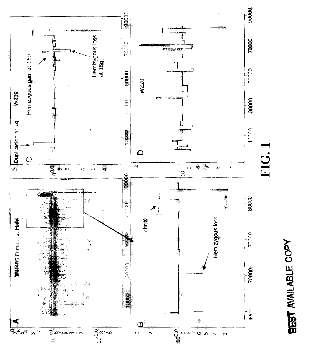
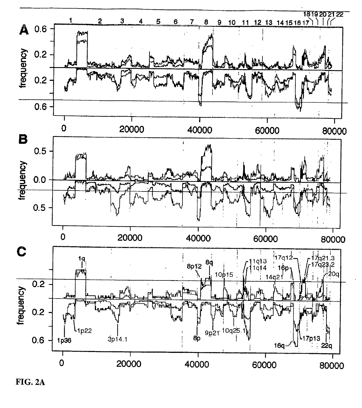
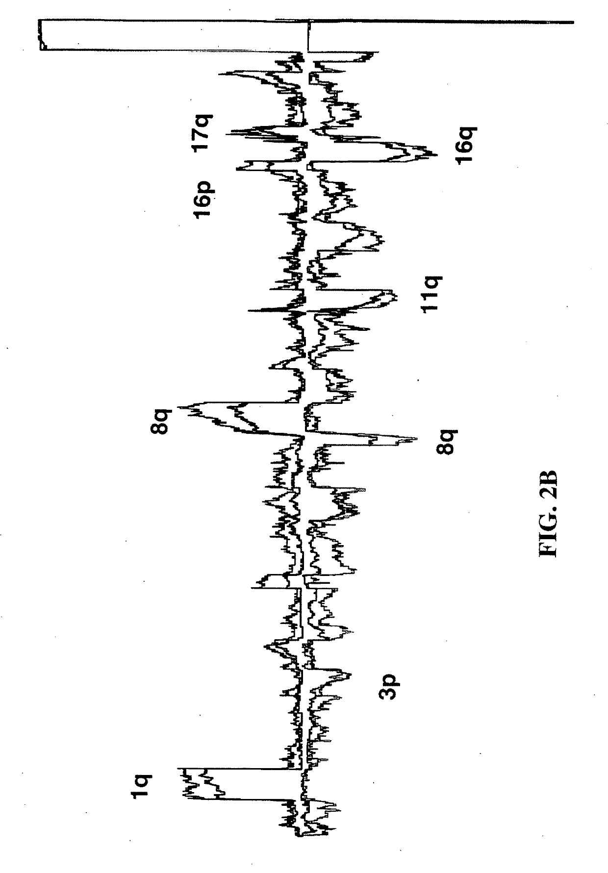
[0200]All breast cancer genomes of the present study were examined with ROMA an array based hybridization method that utilizes genomic complexity reduction based on representations. In the present case, comparative hybridization using BglII representations were performed and arrays of 85,000 oligonucleotide (50-mer) probes with a Poisson distribution throughout the genome and a mean inter-probe distance of 35 kb (Lucito et al., 2003). In all cases, tumor DNA from a patient was compared to a standard unrelated male human genome. Hybridizations were performed in duplicate with color-reversal, and data was rendered as normalized ratios of probe hybridization intensity of tumor to normal.

[0201]The normalized ratios are influenced by many factors, including the signal-to-noise characteristics that differ for each probe, sequence polymorphisms in the genomes that affect the BglII representation, DNA degradation of the sample, and other variation in reagents and ...


PUM

PropertyMeasurementUnit
timeaaaaaaaaaa
pixel sizeaaaaaaaaaa
sizeaaaaaaaaaa

Abstract

The present invention provides methods and compositions related to genomic profiling, and in particular, to assigning probabilistic measure of clinical outcome for a patient having a disease or a tumor using segmented genomic profiles such as those produced by representational oligonucleotide microarray analysis (ROMA).

Description

CROSS REFERENCE TO RELATED APPLICATIONS[0001]This application claims the benefit of and priority to U.S. Provisional Application No. 60 / 751,353, filed on Dec. 14, 2005, and No. 60 / 860,280, filed on Nov. 20, 2006, the contents of which are hereby incorporated by reference in their entirety.STATEMENT REGARDING GOVERNMENT FUNDING[0002]This invention was made with government support under 5R01-CA078544-07 awarded by the U.S. National Institutes of Health, and W81XWH04-1-0477, W81XWH-05-1-0068, and W81XWH-04-0905 awarded by the U.S. Department of Army. The U.S. government has certain rights in the invention.BACKGROUND OF THE INVENTION[0003]Global methods for genomic analysis, such as karyotyping, determination of ploidy, and more recently comparative genomic hybridization (CGH) (Feder et al., 1998, Cancer Genet. Cytogenet, 102:25-31; Gebhart et al., 1998, Int. J. Oncol. 12:1151-1155; Larramendy et al., 1997, Am. J. Pathol. 151:1153-1161; Lu et al., 1997, Genes Chromosomes Cancer 20:275-2...

Claims


Application Information

Patent Timeline
Patent Type & Authority Applications(United States)
IPC IPC(8): G06F19/18C12Q1/6886G16H50/20G16H50/30G06F19/24G06F17/18
CPCC12Q1/6886G06F17/18G16B40/00G16H50/30G16B20/00G16H50/20G16H10/40G16H20/10G16H50/70G16H70/60G16B20/10G16B25/20G16B40/30
Inventor WIGLER, MICHAEL H.HICKS, JAMESKRASNITZ, ALEXANDERZETTERBERG, ANDERS
Owner COLD SPRING HARBOR LAB INC
Why Eureka
Try Eureka