
Mouse myopia-induced model and endoplasmic reticulum stress suppressant for preventing and suppressing myopia

a technology of endoplasmic reticulum and model, which is applied in the field of mouse myopia-induced model and endoplasmic reticulum stress suppressant for preventing and suppressing myopia, can solve the problems of inability to effectively treat myopia, the molecular mechanism of occurrence and progression of myopia has not been clarified at all, and various abnormalities in the fundus

Pending Publication Date: 2020-05-14
TSUBOTA LAB
View PDF1 Cites 2 Cited by
  • Summary
  • Abstract
  • Description
  • Claims
  • Application Information

AI Technical Summary

Benefits of technology

The patent text describes a mouse model for myopia, a common eye condition in humans. The model mimics the symptoms of human myopia and has been created using gene manipulation technologies. The analysis of the model mice showed that myopia induction causes endoplasmic reticulum stress in the sclera, which in turn triggers myopia. It also highlighted that an endoplasmic reticulum stress suppressant can effectively suppress myopia. This discovery led to the development of a therapeutic agent for myopia, which was previously not effectively treatable.

Problems solved by technology

Nevertheless, the molecular mechanism of occurrence and progression of myopia has not been clarified at all, and corrections with glasses or contact lenses are conducted although the radical treatment is not available.
As a result, the retina and the choroid are elongated backward and thus load on them increases, causing various abnormalities in the fundus.
Although pathologic myopia may risk blindness, effective treatment is not currently available, and there is a need to establish treatment.
However, it is not easy to perform gene manipulation for any of these animals and thus none of them is suited for research of genetic elements for myopia.
However, the lack of myopia-induced model animals that allow for research of genetic elements contributes to the fact that treatment for suppressing myopia is not established yet.

Method used


Image

  • Mouse myopia-induced model and endoplasmic reticulum stress suppressant for preventing and suppressing myopia
  • Mouse myopia-induced model and endoplasmic reticulum stress suppressant for preventing and suppressing myopia
  • Mouse myopia-induced model and endoplasmic reticulum stress suppressant for preventing and suppressing myopia

Examples


example 1

[Example 1] Preparing of Mouse Myopia-Induced Model

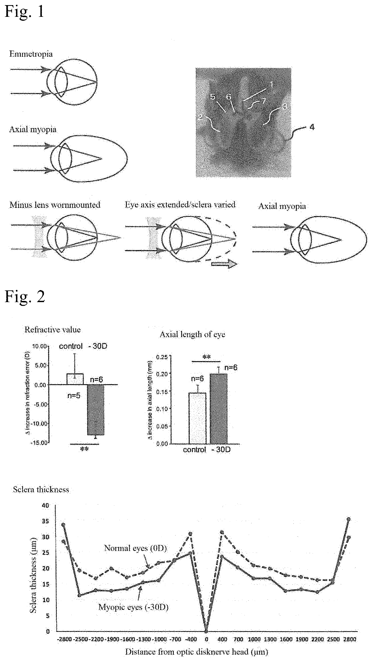
[0050]First, a description will be given below with respect to a method for preparing a mouse model of the present invention. FIG. 1 schematically shows a mechanism in which a minus lens is worn to induce the axial myopia. The emmetropia refers to a condition in which parallel rays entering the eyes focus into an image on the retina and thus one can see images clearly. On the other hand, the axial myopia refers to a condition in which parallel rays entering the eyes focus into an image in front of the retina because of long axial length of the eye, and thus one cannot see clearly. The eyes of animals, including a human, enlarge as they grow. If a juvenile mouse is worn with a minus lens, the eye axis will extend to the position at which an image is focused when the minus lens is worn, i.e., the condition in which one can see clearly with the minus lens worn. As a result, the eye axis extends, resulting in an eye condition similar to...

example 2

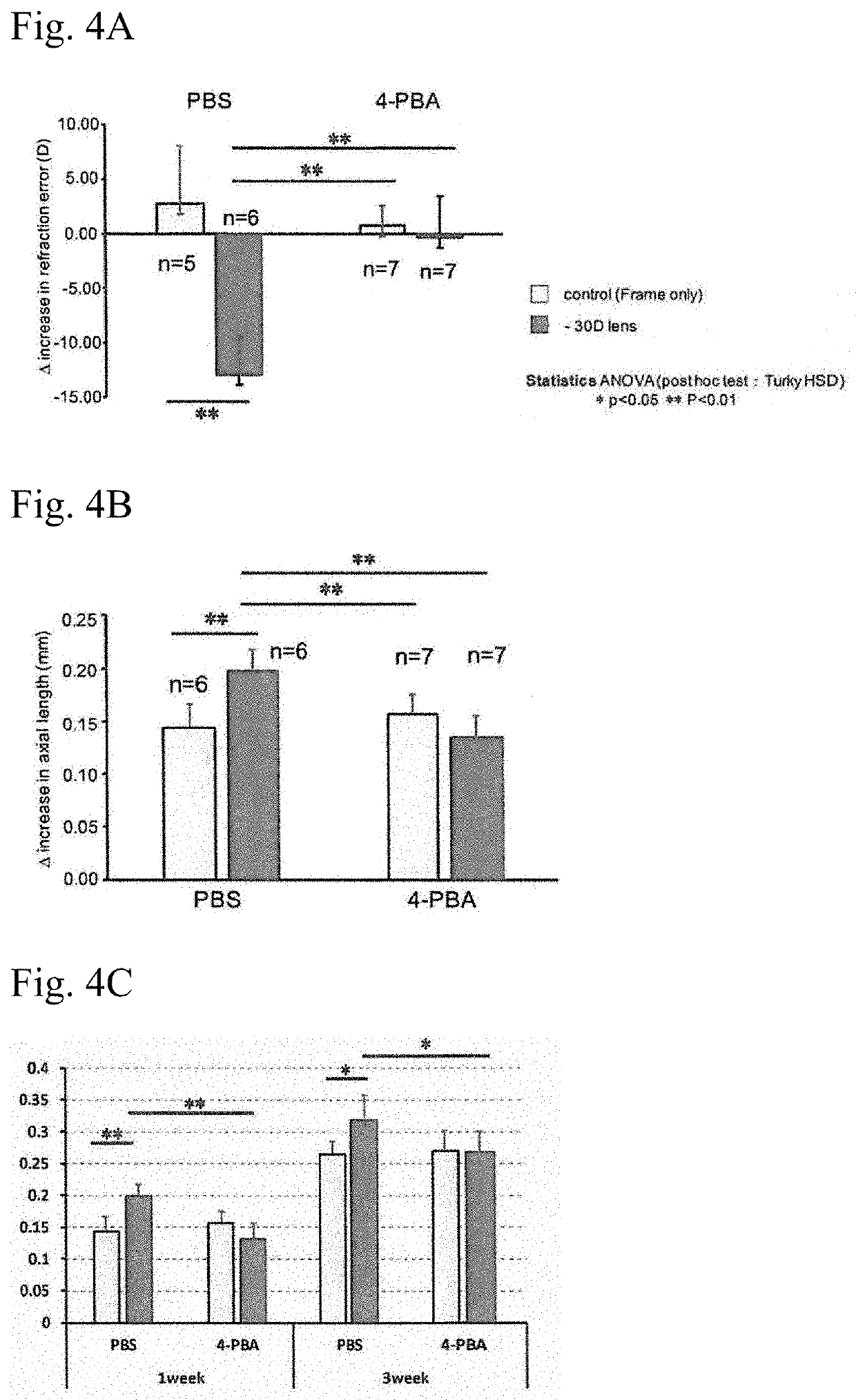
[Example 2] Screening of the Therapeutic Agent Using the Mouse Myopia-Induced Model

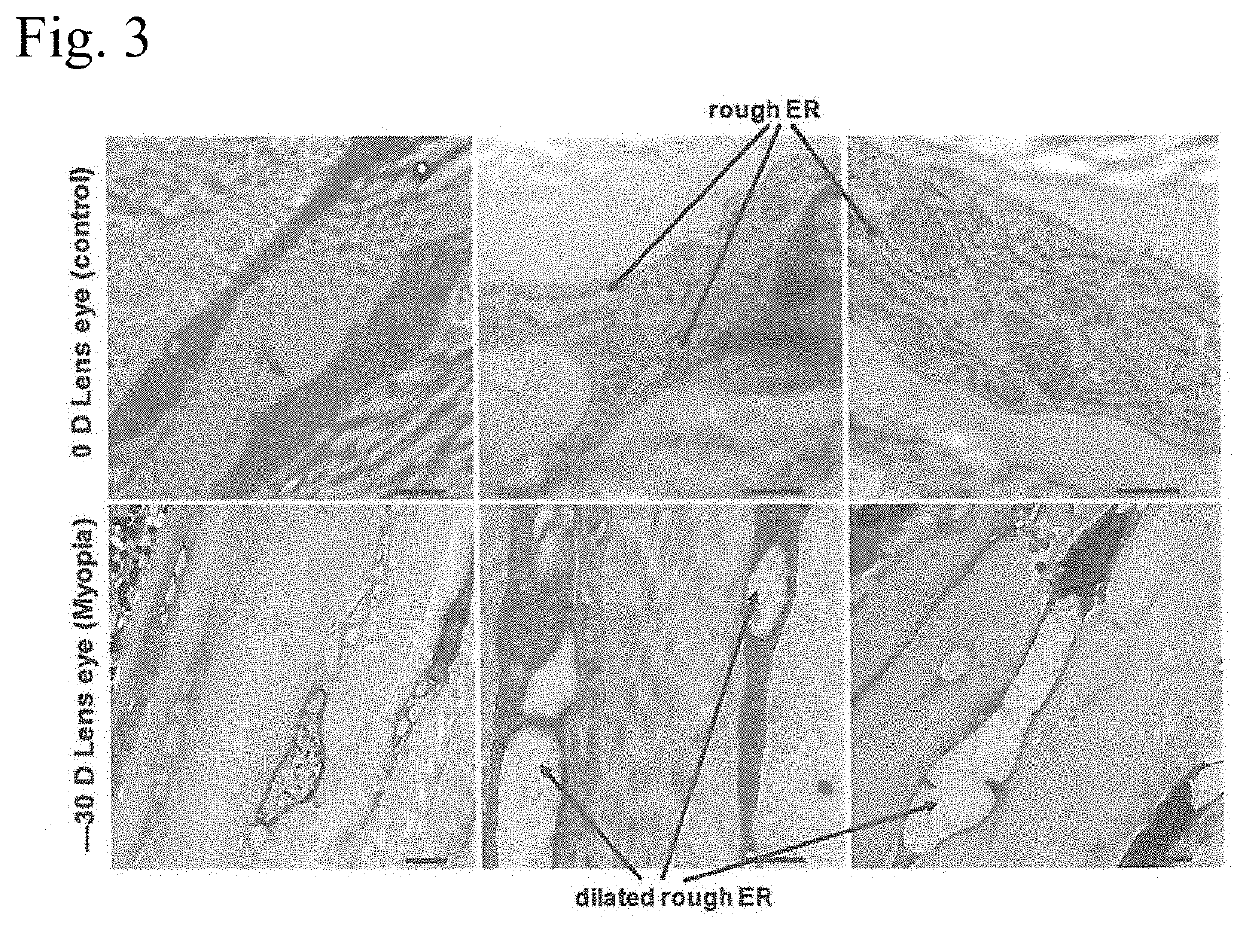
[0058]To investigate the pathological condition of the myopia-induced model in more detail, a transmission electron microscope (TEM) was used for analysis. An eyeball that was worn with a minus lens for three weeks and had axial myopia induced thereto and an eyeball that was worn with only a frame as a control were removed from a mouse and then fixed in 2.5% glutaraldehyde / physiological saline for one hour at 4° C. The cornea was removed and post-fixed in 2.5% glutaraldehyde / physiological saline overnight. It was then embedded in Epok 812 (Okenshoji Co., Ltd.) and thinly sectioned for observation under TEM (JEM-1400 plus, JEOL Ltd.). In FIG. 3, the upper shows the control and the lower shows the sclera of the sample from the mouse that was worn with the −30D lens and had myopia induced thereto. The scale is 1.0 pin, 500 nm, and 500 nm from left to right.

[0059]The upper image of the control shows that ...

example 3

[Example 3] Effects of Endoplasmic Reticulum Stress Induction on Myopia

[0073]Because the endoplasmic reticulum stress suppressant has a suppression effect on myopia induction as describe above, it is considered that the endoplasmic reticulum stress directly participates in myopia induction. Then, an analysis was conducted to check for induction of myopia by administration of an agent for inducing endoplasmic reticulum stress. The subject was a 3-week-old male C57BL6J mouse (n=12). The right eye of the mouse was administered once by eye drop with 50 μg / ml of tunicamycin (Tm) (Sigma-Aldrich Co. LLC) or 10 μM of thapsigargin (TG) (Wako Pure Chemical Industries, Ltd.), and the left eye was administered once by eye drop with PBS (Veh). Before and at one week after administration of tunicamycin and thapsigargin, the refractive value and the axial length of the eye were measured to calculate the variation (FIG. 7).

[0074]By administration of either agent of tunicamycin and thapsigargin, whi...


PUM

PropertyMeasurementUnit
ODaaaaaaaaaa
endoplasmic reticulum stressaaaaaaaaaa
angleaaaaaaaaaa

Abstract

A model that closely resembles human excessive myopia can be prepared by mounting a minus lens (2) and a protector (4) to a juvenile mouse, the minus lens having an angle and a width adjustable in response to growth of the mouse. Further, this model analysis shows that myopia induction causes endoplasmic reticulum stress in a sclera and the endoplasmic reticulum stress induces myopia. Furthermore, it is revealed that an endoplasmic reticulum stress suppressant, particularly, phenylbutyrate and tauroursodeoxycholic acid act as a myopia prevention / suppression agent.

Description

CROSS REFERENCE TO RELATED APPLICATIONS[0001]This application is a Continuation-in-Part application of International Application No. PCT / JP2018 / 008554 filed Mar. 6, 2018, claiming priority based on Japanese Patent Application No. 2017-041349, filed Mar. 6, 2017, the contents of all of which are incorporated herein by reference in their entirety.FIELD OF THE INVENTION[0002]The present invention relates to a mouse myopia-induced model to clarify the mechanism by which myopia occurs, and a method for preparing the same, an agent for suppressing myopia, and a myopia prevention / suppression medicine screening method.BACKGROUND ART[0003]It is said that East Asian people have a higher ratio of myopia than Westerners and that for Japanese, at least about ⅓ of the population, i.e., about 40 million people have myopia. Nevertheless, the molecular mechanism of occurrence and progression of myopia has not been clarified at all, and corrections with glasses or contact lenses are conducted althoug...

Claims


Application Information

Patent Timeline
Patent Type & Authority Applications(United States)
IPC IPC(8): G02C7/06A01K67/02A61K31/575A61K9/00
CPCA01K2267/03A01K2227/105A01K67/02A61K31/575G02C7/06A61K9/0048A61K31/235A61P27/10G01N33/15A61K31/192A61K31/4706A61K31/155A61K31/519A61K31/472A61K31/365A61K31/7088G01N33/5088A61K49/0008A01K67/027A01K2207/30A61K2121/00A61K2300/00
Inventor IKEDA, SHINICHIJIANG, XIAOYANTSUBOTA, KAZUOKURIHARA, TOSHIHIDE
Owner TSUBOTA LAB
Learn More