

Physiology and pathophysiology of human gut: intestine-on-chip
a microfluidic and intestine technology, applied in the field of human gut physiology and pathophysiology, can solve the problems of lack of physiologically relevant in vitro system, and achieve the effect of reducing inflammation
- Summary
- Abstract
- Description
- Claims
- Application Information
AI Technical Summary
Benefits of technology
Problems solved by technology
Method used
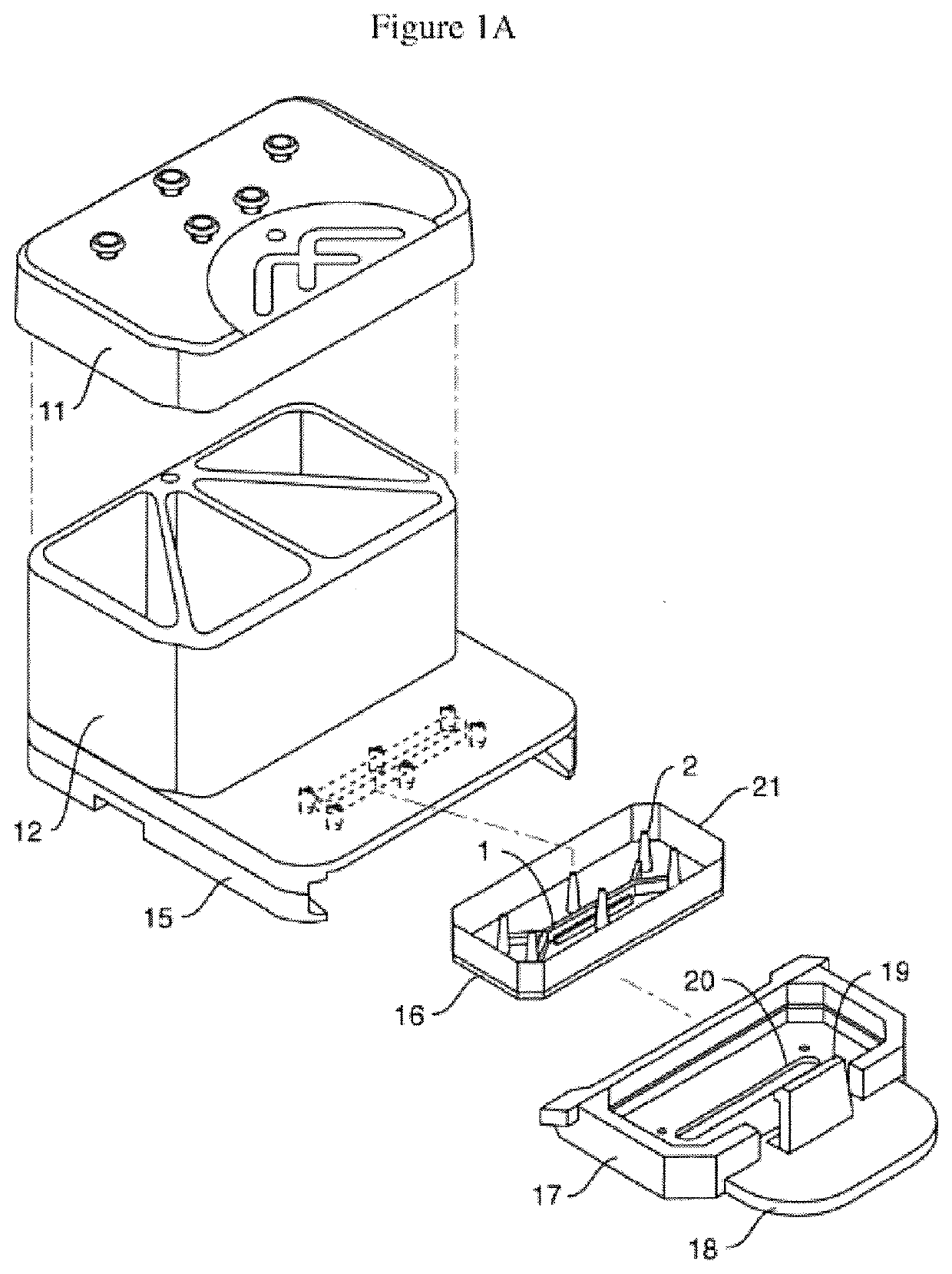
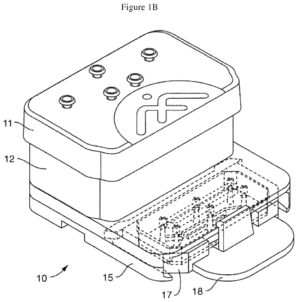
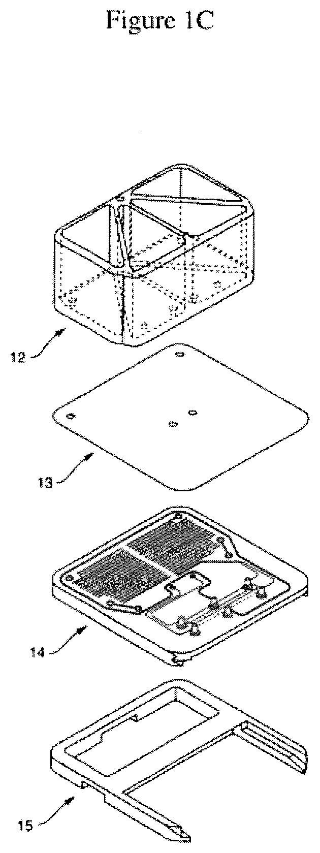
Image

Examples
example 1
Methods and Readouts (Assays)
Complex Media Composition:
[0501]Conditioned media produced by adding one or more agents: L-Wnt3a (CRL-2647), Cultrex® R-spondin1 (RSPO1) and Noggin cells. In some embodiments, conditioned media from L-cells growing in culture is used. Recombinant growth factors may be used, including but not limited to: supplements and small molecules including: EGF, N-acetyl cysteine, Gastrin, etc.
Percoll / Medium.
[0502]In some embodiments, a Percoll liquid, e.g. media formulation of 50% is used in combination with assist immune recruitment assays On Chips, including but not limited to immune-cell types: PBMCs, white blood cells, lymphocytes, macrophages, neutrophils, B cells, T cells, killer cells, etc.
Observe Cells to Assess Morphology and Viability:
[0503]Capture representative images along the length of Chip, including but not limited to inlet junctions, outlet junctions, and center of Chip. Collect samples from the back side of the Reservoirs into pre-labeled tubes or...
example 2
Seeding of Enteroids On-Chip
[0505]In one embodiment, microfluidic chips are seeded with Enteroids, obtained from biopsied tissues of different intestinal regions through collaboration with hospitals (Adult tissue), See, Table 1, and HIMEC, human small intestinal endothelial cells (commercially obtained from Cell Biologics).
1. Cell Preparation
[0506]d. For the Intestine-Chip (Enteroids), Human Small Intestinal Microvascular Endothelial Cells (HIMECs) are seeded into the bottom channel and allowed to attach prior to seeding the primary enteroids[0507]e. Prepare cell suspension and count cell number[0508]f. Seeding density is specific to the cell type[0509]iii. HIMECs: 9 million cells / ml
After counting cells, adjust cell suspension to the appropriate density for seeding.
2. Bottom Channel Seeding (HIMECs)
[0510]Use ONE Chip First—Confirm Seeding Density Before Seeding Other Chips[0511]i. Prior to seeding, wash each channel with 200 ul of cell culture medium[0512]j. Pipette 30 cell culture ...
example 3
Methods of Immune Cell Recruitment
[0554]The following Sections (i.e. steps) were used for providing immune cell recruitment assays on-chip using intestine on-chip. In some embodiments, inflammation is induced in a microfluidic intestine on-chip by inducing inflammation with cytokines.
Section 1: Inflammatory Stimulation of Intestine-Chip: Cytokine Induced Inflammation.
[0555]Seed Intestine-Chip following general protocol; At day 5, divide all of the chips into at least two subgroups: 1) Controls—which will not be treated with the inflammatory stimuli, and 2) Inflamed by treatment for 4-24 hours with an inflammatory stimuli such as TNFalpha, IL-1 beta or LPS. Then, aspirate the media in both output Reservoirs and input Reservoir of the Bottom Channel; Induce vascular inflammation in the Intestine-chip. In one embodiment, vascular inflammation is triggered by perfusing fresh EGM2-MV media, with an inflammatory stimuli added, through the Bottom Channel. Perfuse EGM2-MV media+ / −inflammato...
PUM
| Property | Measurement | Unit |
|---|---|---|
| porosity | aaaaa | aaaaa |
| porosity | aaaaa | aaaaa |
| porosity | aaaaa | aaaaa |
Abstract
Description
Claims
Application Information