Graft for repairing articular cartilage defects and method for the same
- Summary
- Abstract
- Description
- Claims
- Application Information
AI Technical Summary
Benefits of technology
Problems solved by technology
Method used
Image
Examples
example 1
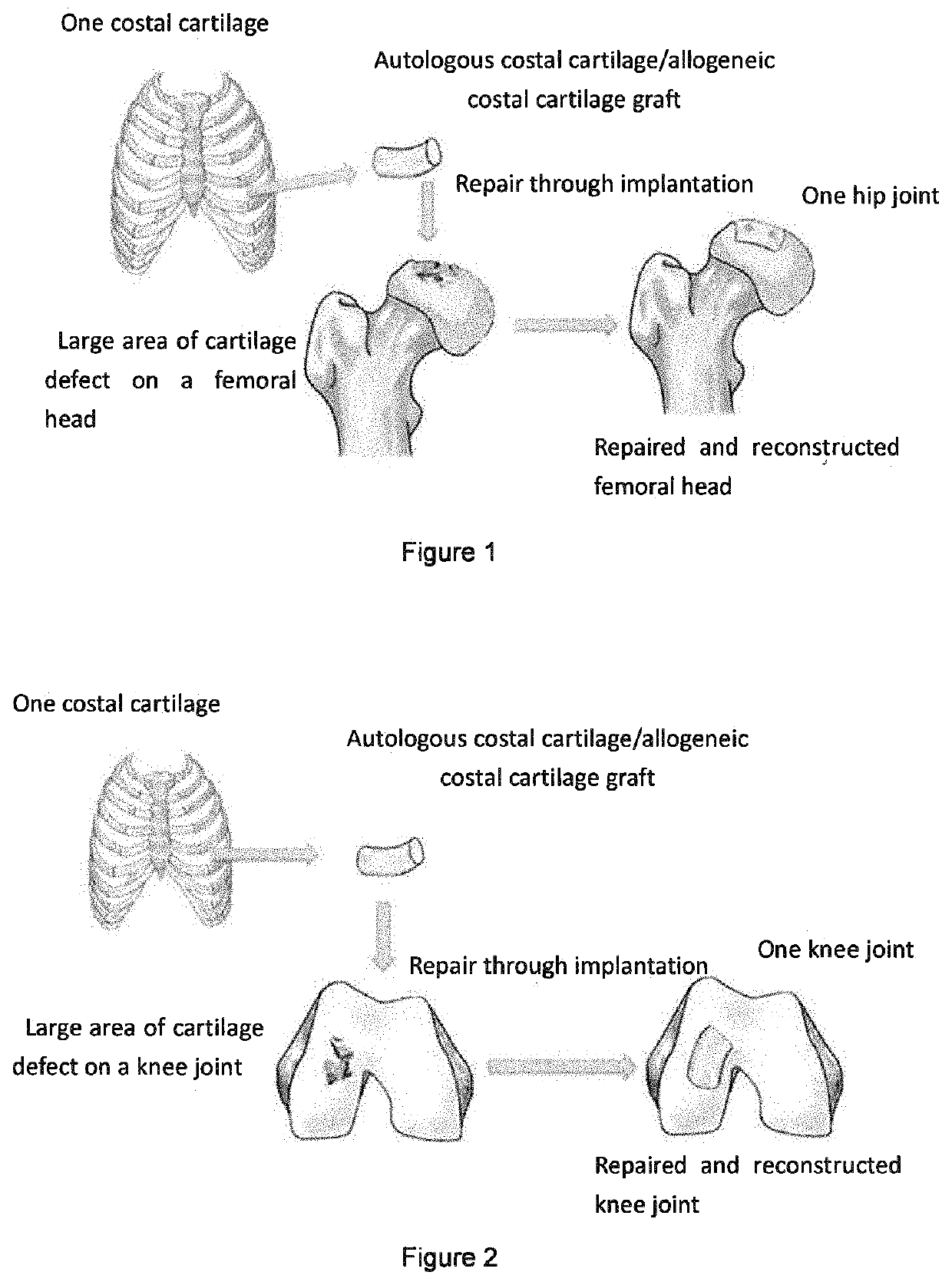
[0065]This example is a method for repairing cartilage defect on a femoral head by implanting autologous costal cartilage, and the method comprises the following steps, as shown in FIG. 1:
[0066]Under general anesthesia, the patient was placed in the supine position, incisions were made on skin, subcutaneous tissues, and fascia lata in sequence by using Smith-Peterson approach, but taking care to avoid damaging anterolateral cutaneous nerves, to fully expose the anterior joint capsule of the hip joint, and incisions were made in a “T”-shape. After fully loosening the hip joint, the femur is at adduction and extorsion, and the femoral head is exposed. The femoral head is thoroughly cleared of the degenerative necrotic bone and cartilage tissues The osteochondral detect site on the surface of the femoral head is identified. According to the severity of the bone defect, the iliac bone block on the same side in the same incision was collected to reconstruct the subchondral bone defect of...
example 2
[0067]This example is a method for repairing a cartilage defect on a knee joint by implanting autologous costal cartilage comprising the following steps and as shown in FIG. 2:
[0068]The patient was placed in a supine position under general anesthesia. After sterilization and draping, exsanguination was performed on the affected limb to reduce bleeding. A medial parapatellar incision was made longitudinally in the left knee in superficial and deep fascia, and an incision was made on the media patellar retinaculum. After the patella was elevated laterally the cartilage lesion was identified in the medial femoral condyle. The degenerative cartilage and bone tissue of the lesion were thoroughly debrided until fresh bleeding occured in the bone bed. The ipsilateral iliac bone mass was collected to reconstruct the subchondral bone defect. At the same time as the knee surgery, another group of doctors stood on the right side of patient to collect the costal cartilage. A 4 cm incision along...
example 3
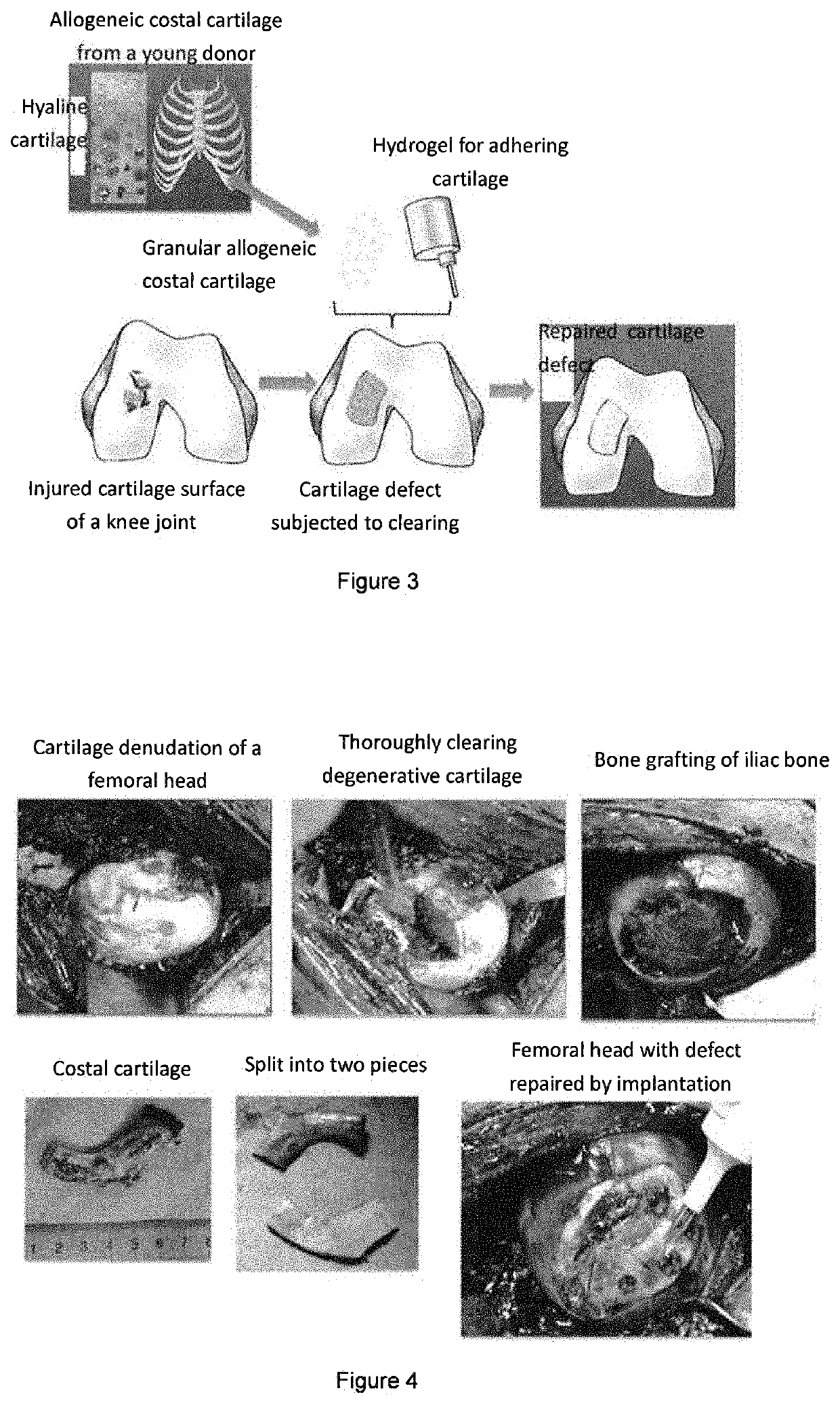
[0069]This example is a method for repairing a cartilage defect on a femoral head by implanting allogeneic costal cartilage comprising the following steps:
[0070]Collecting and storing the allogeneic cartilage: costal cartilage from a young donor was collected and placed in a preservation solution (serum-free medium, pH buffered), and the preservation solution was removed during use;
[0071]Under general anesthesia, the patient was placed in the supine position, incisions were made on skin, subcutaneous tissues, and fascia lata in sequence by using Smith-Peterson approach, but taking care to avoid damaging anterolateral femoral cutaneous nerves, to fully expose the anterior joint capsule of the hip joint, and incisions were made in a “T”-shape. After fully loosening the hip joint, the femur was at adduction and extorsion, and the femoral head was exposed. The femoral head was thoroughly cleared of degenerative necrotic bone and cartilage tissues. The osteochondral detect site on the su...
PUM
| Property | Measurement | Unit |
|---|---|---|
| Length | aaaaa | aaaaa |
| Length | aaaaa | aaaaa |
| Size | aaaaa | aaaaa |
Abstract
Description
Claims
Application Information
 - R&D
- Intellectual Property
- Life Sciences
- Materials
- Tech Scout
- Unparalleled Data Quality
- Higher Quality Content
- 60% Fewer Hallucinations
Browse by: Latest US Patents, China's latest patents, Technical Efficacy Thesaurus, Application Domain, Technology Topic, Popular Technical Reports.
© 2025 PatSnap. All rights reserved.Legal|Privacy policy|Modern Slavery Act Transparency Statement|Sitemap|About US| Contact US: help@patsnap.com


