Method for the isolation of intact extracellular vesicles

a technology of extracellular vesicles and isolation methods, which is applied in the field of isolation methods of intact extracellular vesicles, can solve the problems of inconvenient high-throughput applications, altering the results of downstream analysis, and method not suitable for harvesting evs from cell culture media, and achieves convenient microarray fluorescence, site-selective surface binding of proteins, and the effect of convenient configuration

Pending Publication Date: 2021-04-08
CONSIGLIO NAT DELLE RICERCHE
View PDF0 Cites 2 Cited by
  • Summary
  • Abstract
  • Description
  • Claims
  • Application Information

AI Technical Summary

Problems solved by technology

Ultracentrifugation in a density gradient provides preparations free from contaminating cellular debris, but this method is not suited for high-throughput applications.
Ultrafiltration is faster than ultracentrifugation but the use of centrifuge force may result in the deformation and breaking up of large vesicles potentially altering the results of downstream analysis.
However, while SEC purifies EVs, it also dilutes them.
Therefore, the method is not suitable for harvesting EVs from cell culture media, where they are often less concentrated than in plasma.
However, with these methods the presence of contaminating cellular and protein debris has been noted.
In general, a significant problem in EVs isolation is that both, blood and cell culture media, contain large numbers of nanoparticles (some non-vesicular) in the same size range as exosomes.
Therefore, separation by physical characteristics is not possible.
The problem that arises in a conventional immunoaffinity approach is that EVs are tightly bound to antibodies directed against antigen proteins on their surface.
Releasing vesicles requires strong acidic buffers and / or detergents, both conditions that damage the nanoparticles and impair certain types of downstream analysis.

Method used


Image

  • Method for the isolation of intact extracellular vesicles
  • Method for the isolation of intact extracellular vesicles
  • Method for the isolation of intact extracellular vesicles

Examples


example 1

Antibody Functionalization with ssDNA

[0072]Antibodies directed against exosome-specific tetraspanins (CD9 and CD63) were modified with a linker bearing N-oxysuccinimide ester at one end and dibenzocyclooctine (DBCO) at the other end to allow their binding to azido modified oligonucleotides as reported below.

[0073]1.1 Synthesis of ssDNA-AntiCD63: 4.4 μL of a 4 mM PBS solution of DBCO-NHS ester were added to 200 μL of sodium azide-free mouse anti-human CD63 IgG (0.70 mg / ml) and the solution was allowed to react for 30 min at room temperature. The reaction was quenched by adding 20 μL of 1M TRIS-HCl, pH=8.0, and the unreacted ester was removed, through centrifugation over an Amicon Ultra 10K filter (3 min at 12,000×g). The eluted solution was discarded and the protein was recovered in 200 μL of PBS. The oligonucleotide ssDNA-TAG-63 (8.2 μL, 100 μM) was added to 200 μL of DBCO-modified antibody (0.66 mg / ml) and the strain-promoted 1,3-dipolar dycloaddition of cycloalkynes to azide react...

example 2

DNA-Directed Immobilization (DDI) of Antibodies on Silicon Chips

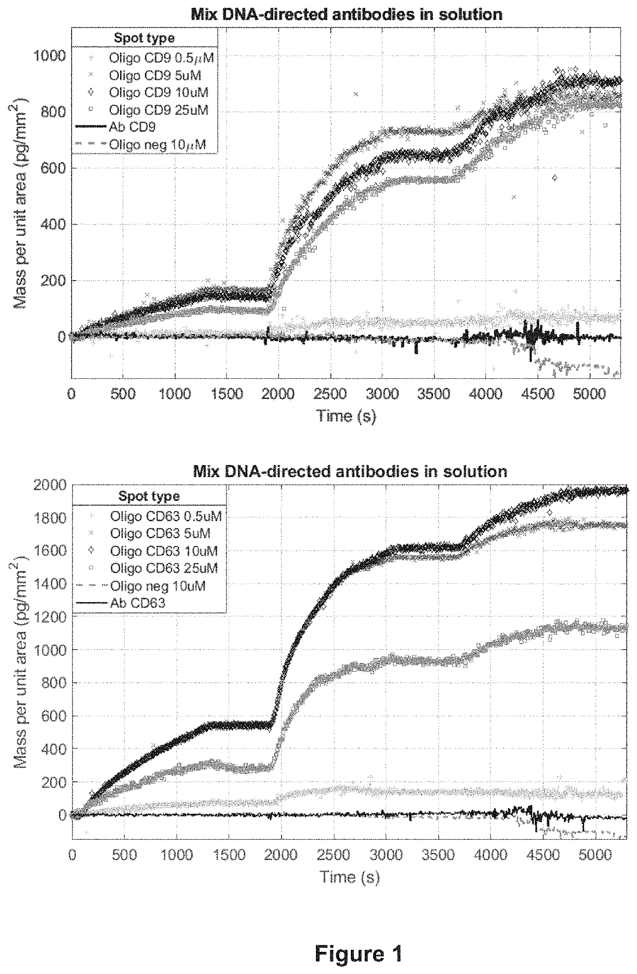
[0075]2.1 Probe spotting on the surface of microarray: both antibody and oligonucleotide solutions were prepared. Amino-modified oligonucleotides complementary to the antibody tag (ssDNA-PROBES) were dissolved at different concentrations (0.5, 5, 10 and 25 μM) in a solution of 150 mM sodium phosphate buffer containing 0.01% sucrose monolaurate at pH 8.5. The probes were then spotted onto the surface of different silicon / silicon oxide chips, depending on the detection technique used. In particular, oxide layers of 110 nm and 55 nm were use for IRIS and SP-IRIS measurements respectively. All chips were coated with copoly (DMA-NAS-MAPS), a polymer commercially available with the trade name of MCP-2 (Lucidant Polymers Inc., Sunnyvale Calif., USA) using a noncontact microarray spotter (sciFLEXARRAYER S12, Scienion, Berlin) equipped with a 80 μm nozzle. MCP-2 is a ter-copolymer of N,N-dymethilacrylamide (DMA) (97% in moles), ...

example 3

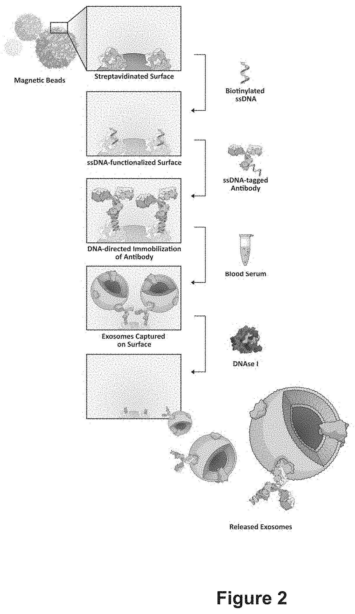
Extracellular Vesicle Purification using DNA-Directed Immobilization of AntiCD63 on Magnetic Beads (FIG. 2)

[0082]3.1 Microparticles functionalization: DNA tagged antibodies were immobilized on the surface of magnetic beads and used to immunocapture Evs. Prior to their use, streptavidin-coated magnetic beads (Dynabeads M-270 Streptavidin, Invitrogen) were washed three times with Binding and Washing buffer (B&W) (5 mM Tris-HCl, pH 7,5; 0,5 mM EDTA; 1 M NaCl) according to the manufacturer's protocol. Then 500 i—ig of beads were added to 100 μL of 1μM biotinylated ssDNA-Probe-63 solution. The suspension was stirred for 30 min at 23° C., then the solution was removed and the beads were washed twice with B&W buffer, then once with PBS.

[0083]Oligonucleotide modified beads (500 μg) were incubated with 100 μL of ssDNA-AntiCD63 antibody (160 μg / mL) for 1h at 25° C., then the solution was removed and the beads were washed twice in PBS.

[0084]3.2 Capture and release of EVs from magnetic beads: D...


PUM

PropertyMeasurementUnit
sizeaaaaaaaaaa
sizeaaaaaaaaaa
sizeaaaaaaaaaa

Abstract

The present invention relates to a method for the isolation of intact extracellular vesicles from biological tissues and fluids. Under a further aspect, the present invention relates intact extracellular vesicles, obtainable from the method herein described, and their use as a diagnostic and therapeutic tool.

Description

[0001]This application claims priority to and the benefit of Italian Patent Application No. 102019000018239, filed on 8 Oct. 2019, the content of which is incorporated herein by reference in its entirety.FIELD OF THE INVENTION[0002]The present invention relates to a method for the isolation of intact extracellular vesicles from biological tissues and fluids.[0003]Under a further aspect, the present invention relates to intact extracellular vesicles obtainable from the method herein described, and their use as a diagnostic and therapeutic tool.STATE OF THE ART[0004]Cells release in the extracellular environment vesicles that are attracting considerable interest in the scientific community due to their role in intercellular communication. Extracellular vesicles (EVs) is a generic term for all secreted vesicles. They can be further classified in subpopulations including: exosomes, (30-150 nm), microvesicles (MVs) (100-350 nm) and apoptotic bodies (500-1000 nm) according to their cellul...

Claims


Application Information

Patent Timeline
Patent Type & Authority Applications(United States)
IPC IPC(8): C12N15/10C12N9/22G01N33/50G01N33/537G01N33/543
CPCC12N15/1013C12N9/22G01N33/543G01N33/537G01N33/5076A61K35/14C12N5/0634G01N2458/10G01N2333/922G01N33/54353
Inventor CHIARI, MARCELLA
Owner CONSIGLIO NAT DELLE RICERCHE
Why Eureka
Try Eureka