Scaffold with adhesive for articular cartilage repair
a technology of articular cartilage and adhesive, which is applied in the field of articular cartilage adhesive for articular cartilage repair, can solve the problems of little or no potential for further healing, affecting mobility, and affecting the ability of active individuals and older adults to damage the articular cartilage, and achieves the effect of strengthening cell attachment and/or proliferation
- Summary
- Abstract
- Description
- Claims
- Application Information
AI Technical Summary
Benefits of technology
Problems solved by technology
Method used
Image
Examples
example 1
[0085]Swine are divided into at least two groups, with at least one control group present. Each test group has a barrier composition applied, with the control group not having a barrier composition applied. In all groups, a cartilage defect is created in the weight bearing region of the femoral medial condyle of the knee joint.
[0086]In each test group, a barrier composition is applied onto the subchondral bone. Multiple test groups can be created to test various barrier compositions. In all groups, the same matrix is applied after any barrier composition is applied. The conditions involving application of matrix and any top polymer barrier above the matrix should be identical among all test groups and the control group.
[0087]At one month, and at additional time periods, testing is undertaken to determine whether the barrier composition prevents migration of subchondral components, e.g., cells and fluids, into the cartilage lesion. Such testing can include histological analysis and a...
example 2
[0090]Scaffolds were prepared for testing in swine as follows. A honeycomb-shaped porous collagen sponge (5 mm in diameter and 1 mm in thickness, Koken, Tokyo, Japan) was soaked in 25 μl of cold 0.3% neutralized collagen solution (Vitrogen, Cohesion Tech, Palo Alto, Calif.), and then incubated at 37° C. for 1 hour. The neutralized collagen solution solidified to form an acellular scaffold composed of collagen gel within the sponge.
[0091]An engineered cell construct implanted with adhesive and sutures was prepared by harvesting a biopsy from the porcine articular cartilage, mincing the biopsy and then digesting it in 1.5 mg / ml collagenase (CLS 1, Worthington, Freehold, N.J.), dissolved in Ham's F-12 (F-12, Invitrogen) with 100 μg / ml penicillin and 100 unit / ml streptomycin (P / S, Invitrogen) on a rotator at 37° C. for 18 hours. Non-digested tissue was removed using a cell strainer (70 μm mesh, BD Biosciences, Franklin Lakes, N.J.). The isolated porcine articular chondrocytes (pACs) wer...
example 3
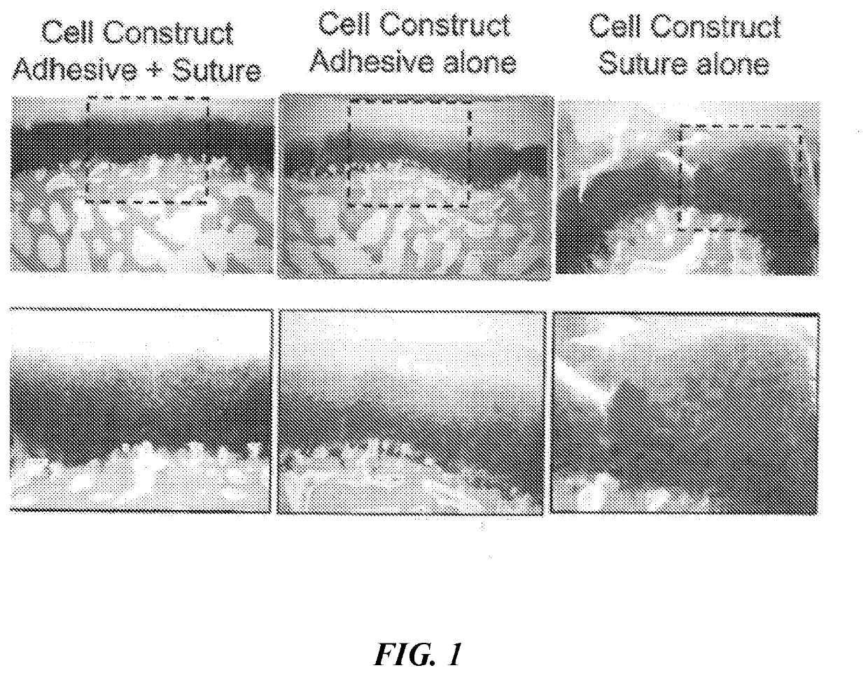
[0094]Five different engineered cell constructs were implanted into swine and their properties compared. The five constructs were a) an empty defect control (“Empty”), b) an acellular scaffold control implanted with adhesive and sutures (“Scaffold”), c) an engineered cell construct implanted with adhesive and sutures (“Cell-construct”), d) a engineered cell construct implanted with adhesive alone (“Adhesive-cell-construct”), and e) a engineered cell construct implanted with sutures alone onto subchondral bone (“Sutured-cell-construct”).
[0095]Two rounds of surgeries were conducted in the swine. The protocol for the animal study was approved by the institutional animal care and use committee of Charles River Laboratories (Worcester, Mass.). Sixteen castrated 12- to 15-month-old male swine weighing 30 to 45 kg (Micro-Yucatan, Charles River Laboratories) were acclimatized more than one week prior to the first surgery. Animal anesthesia was induced with intramuscular injections of 0.04 m...
PUM
 Abstract
Description
Claims
Application Information
 - R&D
- Intellectual Property
- Life Sciences
- Materials
- Tech Scout
- Unparalleled Data Quality
- Higher Quality Content
- 60% Fewer Hallucinations
Browse by: Latest US Patents, China's latest patents, Technical Efficacy Thesaurus, Application Domain, Technology Topic, Popular Technical Reports.
© 2025 PatSnap. All rights reserved.Legal|Privacy policy|Modern Slavery Act Transparency Statement|Sitemap|About US| Contact US: help@patsnap.com

