Method for measuring intracellular potential with a capacitance type potential measurement device

a capacitance type, intracellular potential technology, applied in the direction of instruments, biochemistry apparatus and processes, material analysis, etc., can solve the problems of inability to accurately measure the intracellular potential, the reliability of data is not as reliable as replacing the manual patch-clamp method, and the cost of auto-clamp test equipment is so high, so as to reduce the damage to cells, accurately measure easily, and fluctuate the intracellular potential

Pending Publication Date: 2021-07-29
ION CHAT RES CORP
View PDF0 Cites 1 Cited by
  • Summary
  • Abstract
  • Description
  • Claims
  • Application Information

AI Technical Summary

Benefits of technology

[0090]In the present invention, since the conductive nanoparticles introduced into the cell by a known method or the conductive nanoparticles on the cell surface mixed with the transfection reagent are attracted by the magnetic field to penetrate the cell membrane, damage to the cells can be minimized as much as possible, and the fluctuation of the intracellular potential can be observed while allowing the cells to survive in a normal state for a long period of time.
[0091]In addition, the measuring method of the present invention does not directly measure the potential change between the positive input and the negative input of the amplifier, but this method of recording the change in intracellular potentials is performed using the charge-voltage conversion using a capacitor (conductive plate) that seeds cells as a sensor. Therefore, it is not necessary to set the earth in the extracellular fluid. Since it is sufficient that the conductive plate that serves as a sensor can sense a change in charge in the cell through the conductive nanoparticles penetrating the cell membrane, and the conductive plate may be in contact with the extracellular fluid at a portion not covered with cells.
[0092]That is, it is not necessary for the measuring method of the present invention to culture in a sheet form even when applied to a cell group, and even when applied to a single cell, the action potential of a cell or its change can be accurately measured easily.
[0093]Especially, even in cells that cannot be cultured in a sheet, typically in cultured nerve cells, it is possible to record the action potential generated in the cell or its change. Therefore, in the future, in vitro electrophysiology of (cultured) nerve cells, it is expected to make a dramatic contribution to scientific research.
[0094]Furthermore, by applying it to transformed cells or cell groups that express various ion channels, drug transporters, etc., it can be used as a tool for screening drug efficacy or cytotoxicity by observing changes in intracellular potential due to the addition of various drugs.

Problems solved by technology

However, the manual patch-clamp method is very inefficient because an operator uses a microscope to operate a manipulator to perform an experiment and requires a large amount of specialized knowledge.
This has become a major hurdle in medical biology research, especially in the field of drug discovery.
On the other hand, the auto patch-clamp method uses an automated electrophysiological measuring instrument, and although its performance has improved remarkably in recent years, the reliability of data is not as reliable as replacing the manual patch-clamp method.
Furthermore, the auto patch-clamp test equipment is so expensive that its use is limited to large pharmaceutical companies.
However, in that method, the electrode loses access to the intracellular space, because the perforated cell membrane is repaired immediately.
Therefore, it is not a practical method.
Although MEA allows for easier handling of cells due to extracellular recording, this technology only allows recordings for AC-like changes (changes in membrane potential unit time, differential waveforms), so it is not applicable for recording slow changes in the intracellular membrane potential.
Therefore, this measurement method does not provide sufficient information for analysis and its application is limited.
The recording time of the intracellular potential could only be maintained for a short time, confirming it is not a practical recording method.

Method used


Image

  • Method for measuring intracellular potential with a capacitance type potential measurement device
  • Method for measuring intracellular potential with a capacitance type potential measurement device
  • Method for measuring intracellular potential with a capacitance type potential measurement device

Examples


examples

[0210]The present invention will be specifically described below with reference to Examples, but the present invention is not limited to these.

[0211]Other terms and concepts in the present invention are based on the meanings of terms commonly used in the art, and various techniques used for carrying out the present invention are notably defined as those demonstrating the source thereof. Except for this, those skilled in the art can easily and surely carry out the implementation based on publicly known documents and the like. In addition, various analyzes and the like were carried out by applying methods described in analytical instruments or reagents used, instruction manuals of kits, catalogs and the like.

[0212]The description in the technical documents, patent publications, and patent application specifications cited in this specification shall be referred to as the description of the present invention.

(Reference Example 1) Measurement of Intracellular Potential in CHO Cells Stabl...

reference example 7

(Reference Example 7) Measurement of the CHO Cell Endogenous Outward Current by Conductive Nanoparticles Penetrating the Cell Membrane of CHO Cells Cultured on Conductive Plate Electrode Using the Voltage Clamp Method

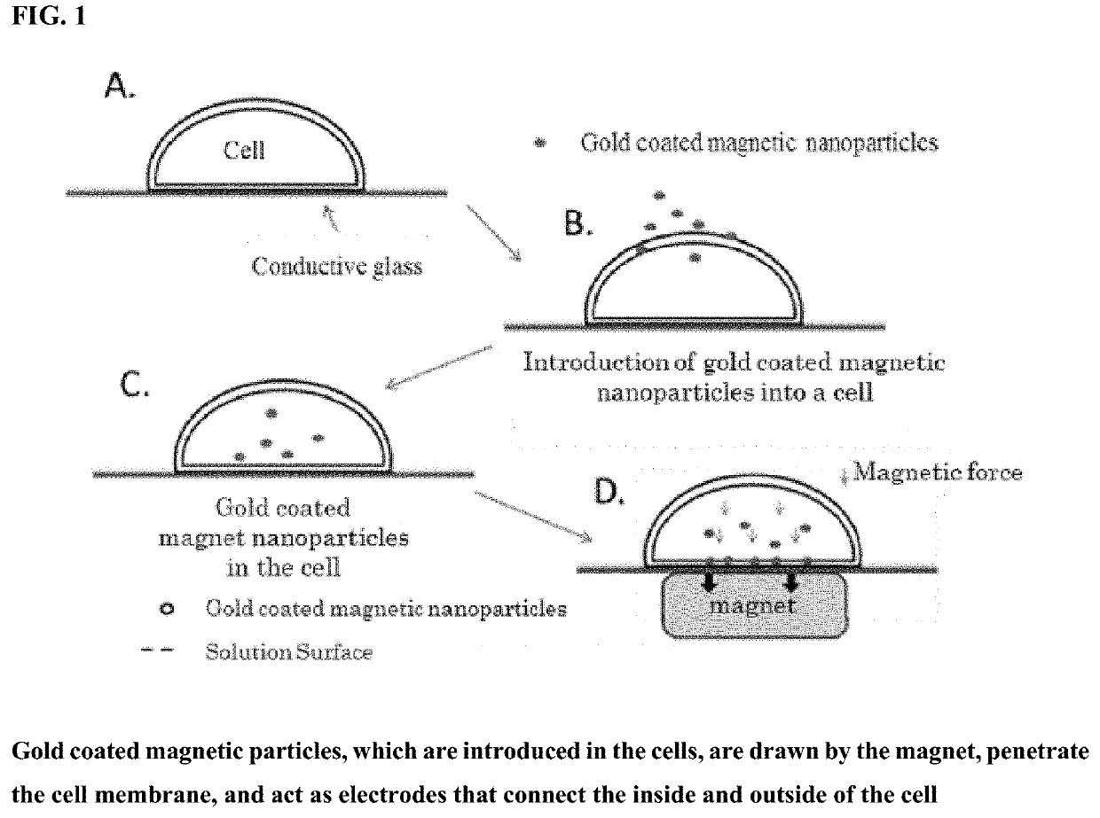
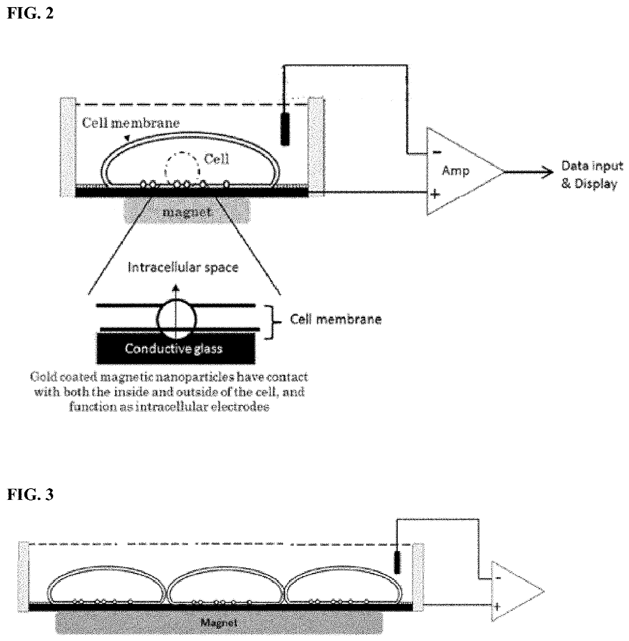
[0267]In this reference example, by using the intracellular recording electrode constructed by the conductive plate electrode and the conductive nanoparticles inside the cell, it was confirmed that the cell membrane current generated in the cell as well as the intracellular potential can be measured.

[0268]CHO cells were cultured in a culture medium containing Minimum Essential Medium Eagle (Sigma-Aldrich), 10% FBS (BioWest), 40 mM L-Glutamine (Wako Pure Chemical), and Streptolysin O (SLO) (manufactured by Wako Pure Chemical Industries, Ltd.) was reduced using DTT to prepare activated SLO at a concentration of about 5 U / μl.

[0269]CHO cells (5×10⁵) were suspended in gold-coated magnetic nanoparticle transfection solution (5 μl 5×HBPS (1 mM Ca2+, 1 mM Mg2+)) containing 20 μ...


PUM

PropertyMeasurementUnit
capacitanceaaaaaaaaaa
pore sizeaaaaaaaaaa
thicknessaaaaaaaaaa

Abstract

The purpose of the present invention is to provide a method for accurately measuring and controlling intracellular potential by a simple method that is less invasive to the cell and does not require a skilled technique. The method according to the present invention comprises: by using magnetic force from a magnetic electrode adhered to the surface of a target cell, into which conductive nanoparticles have been preliminarily introduced, or a magnet being in contact with a conductive plate electrode, attracting the conductive nanoparticles inside the cell to the side of the cell surface adhering to the electrode, and then allowing the conductive nanoparticles to pass through the cell membrane to thereby bring the end outside the cell into contact with the magnetic electrode or the conductive plate electrode; or alternatively, adhering the conductive nanoparticles adsorbed on the magnetic electrode surface to the upper side of the target cell, and allowing the conductive nanoparticles to pass through the cell membrane by attracting the conductive nanoparticles to an iron plate disposed on the lower side of the cell to thereby leave the end outside the cell being in contact with the magnetic electrode. A capacitor is fabricated by using the conductive plate in contact with the conductive nanoparticles together with a conductive plate (an aluminum foil). Alternatively, a capacitor is fabricated by disposing a magnetic body via an insulating body on the upper side of the magnetic electrode. This capacitor detects, as a sensor, a change in charge inside the target cell and converts the same into a voltage change to thereby enable the measurement of the intracellular potential. The present invention also makes it possible to monitor the effect of a test reagent, said test reagent being administered to an extracellular solution, on a cell.

Description

TECHNICAL FIELD[0001]The present invention relates to a method for measuring the intracellular membrane potential (intracellular potential) by combining the principle of a charge amplifier with a capacitance-type potential measurement device (Capacitive potential detection device).BACKGROUND ART[0002]All cells have different ionic compositions inside and outside the cell, and the intracellular potential (membrane potential) is maintained by a transporter (e.g. sodium pump) that keeps the difference in ion distribution and the difference in ionic composition. In the resting state, the membrane potential is stable (resting membrane potential). However, when the ion channels on the cell membrane surface are activated and the ion channels are opened, ions are released or flow in at once through the ion channels due to the difference in ion concentration inside and outside the cell membrane. As a result, the electric potential of the entire cell to change (depolarization or hyperpolariza...

Claims


Application Information

Patent Timeline
Patent Type & Authority Applications(United States)
IPC IPC(8): G01N33/483G01N33/50G01N33/15G01N27/22
CPCG01N33/483G01N33/5061G01N27/226G01N33/15G01N33/5058C12Q1/025G01N33/54326G01N33/48728C12M41/46
Inventor SAITO, MITSUYOSHI
Owner ION CHAT RES CORP
Why Eureka
Try Eureka