Novel uses

Pending Publication Date: 2021-11-04
INTRA CELLULAR THERAPIES INC
View PDF0 Cites 0 Cited by
  • Summary
  • Abstract
  • Description
  • Claims
  • Application Information

AI Technical Summary

Benefits of technology

[0202]BV2 cells were added to upper chamber of a 5 μm pore Transwell 96-well plate over a reservoir containing 100 μM ADP and incubated at 37° C. with 5% CO2 for 4 hours. After the incubation cells were harvested with pre-warmed cell detachment solution for 30 minutes in the same incubation conditions. 75 μl of this cell detachment solution was combined with 75 μl of culture medium in a new 96 well plate compatible with a fluorescence reader. Cell number in bottom chamber was determin

Problems solved by technology

Prolonged M1 type of macrophages is harmful for the organism and that is why tissue repair and restoration is necessary.
Inflammatory processe

Method used


Image

  • Novel uses
  • Novel uses
  • Novel uses

Examples


Example

Example 1: Periphera
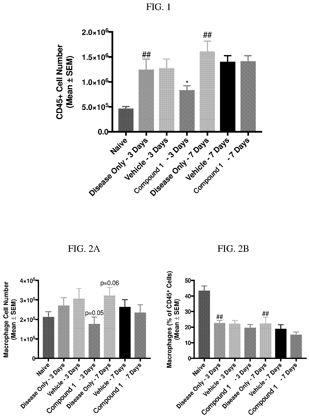
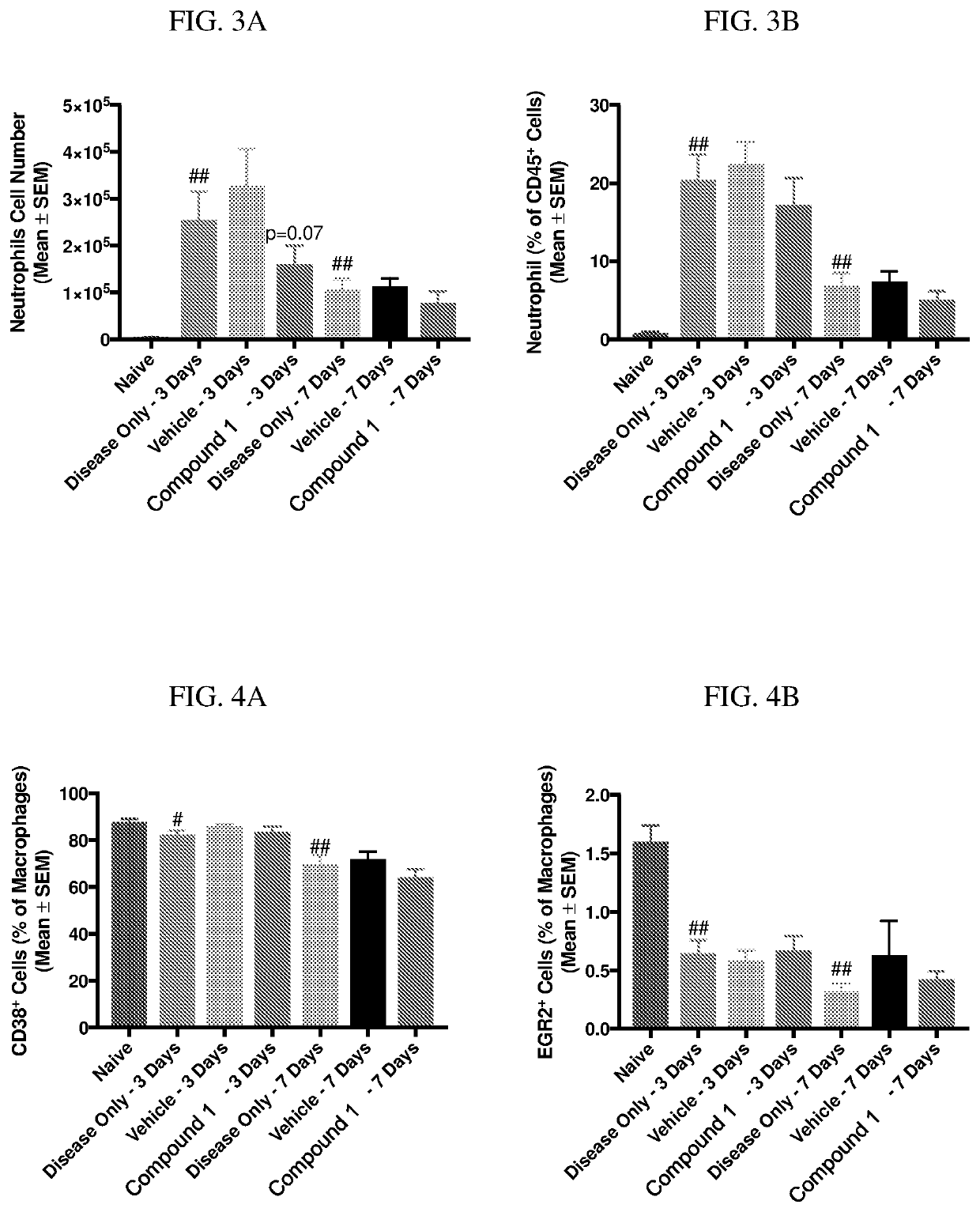
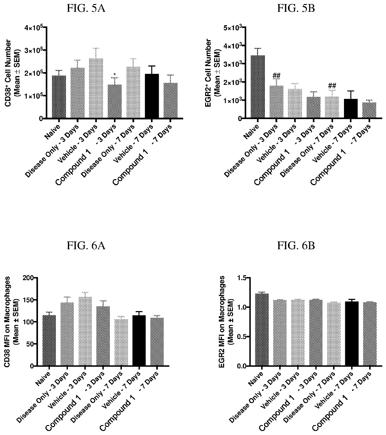
l Inflammation Assessment Using Mouse Zymosan Pleurisy Model

[0194]Zymosan is injected into the pleural cavities of mice in order to induce sterile inflammation. Infiltration of leukocytes, neutrophils, and macrophages are monitored at days 3 and 7 following injection. Detection of various cell types are identified according to the gating strategy outlined in Table 1 below.

TABLE 1Cell types and identifiable markers for flow cytometryCell TypeExpressed MarkersLeukocytesCD45+NeutrophilsCD45+ / Ly6G+MacrophagesCD45+ / Ly6G− / CD19− / CD11c− / CD11b+ / F4 / 80+M1 MacrophagesCD45+ / Ly6G− / CD19− / CD11c− / CD11b+ / F4 / 80+CD38+M2 MacrophagesCD45+ / Ly6G− / CD19− / CD11c− / CD11b+ / F4 / 80+ / EGR2+

[0195]In this model, injection of zymosan causes the recruitment of various waves of leukocytes, which are observed and recorded. Exudate volume increases to a maximum over a period of 24 hours, and neutrophils increase within 4 hours and reach a maximum by 48 hours. Lymphocytes of the adaptive immune system ente...

Example

Example 2: Effect of PDE1 Inhibitor on Microglia Chemotaxis Assay

[0202]BV2 cells were added to upper chamber of a 5 μm pore Transwell 96-well plate over a reservoir containing 100 μM ADP and incubated at 37° C. with 5% CO2 for 4 hours. After the incubation cells were harvested with pre-warmed cell detachment solution for 30 minutes in the same incubation conditions. 75 μl of this cell detachment solution was combined with 75 μl of culture medium in a new 96 well plate compatible with a fluorescence reader. Cell number in bottom chamber was determined by adding CyQuant® GR dye and reading in the Envision fluorescence reader at 480 nm EX / 520 nm EM. CyQuant® GR dye exhibits strong fluorescence when bound to nucleic acid and is accurate enough to measure differences down to single cells. As shown in FIG. 10, the presence of the PDE1 inhibitor Compound 1 showed a marked dampening effect on the motility of the BV2 cells across the membrane, providing additional evidence that Compound 1 da...

Example

Example 3: Detection of Inflammatory Biomarkers Using Mouse Zymosan Pleurisy Model

[0203]Zymosan was injected into the pleural cavities of mice in order to induce sterile inflammation by the methods discussed in Example 1. Compound 1 was administered to test subjects to observe the effects on a variety of inflammatory biomarkers. Results were recorded after 4 hours. The subjects showed a clear decrease in cytokine markers following administration of Compound 1. IFNγ, IL-1β, MCP1-β and TNF-α decreased following administration of Compound 1 in all serum and plasma samples. IL10 showed a decrease in serum.

[0204]Lipids are known to be involved in regulation of a multitude of cellular responses including cell growth and death, and inflammation / infection, via receptor-mediated pathways. Various lipids are involved in both the initiation and resolution of inflammation. Pro-resolving lipid mediators are produced naturally in the body from unsaturated fatty acids, such as arachidonic acid (AA...


PUM


Abstract

The disclosure provides the administration of inhibitors of phosphodiesterase 1 (PDE1) for the treatment and prophylaxis of diseases or disorders characterized by inflammation, including methods of treatment and pharmaceutical compositions for use therein.

Description

FIELD OF THE INVENTION[0001]The field relates to the administration of inhibitors of phosphodiesterase 1 (PDE1) inhibitors for promoting the resolution of inflammation, for example through the polarization of M1 macrophages to M2 macrophages, and the treatment and prophylaxis of diseases or disorders related to inflammation.BACKGROUND OF THE INVENTION[0002]Eleven families of phosphodiesterases (PDEs) have been identified but only PDEs in Family I, the Ca2+-calmodulin-dependent phosphodiesterases (CaM-PDEs), are activated by the Ca2+-calmodulin and have been shown to mediate the calcium and cyclic nucleotide (e.g. cAMP and cGMP) signaling pathways. These PDEs are therefore active in stimulated conditions when intra-cellular calcium levels rise, leading to increased hydrolysis of cyclic nucleotides. The three known CaM-PDE genes, PDE1A, PDE1B, and PDE1C, are all expressed in central nervous system tissue. In the brain, the predominant expression of PDE1A is in the cortex and neostriat...

Claims


Application Information

Patent Timeline
IPC IPC(8): A61K31/519A61P37/06
CPCA61K31/519A61P37/06C07D487/04A61K45/06C07D487/14Y02A50/30A61P29/00
Inventor SNYDER, GRETCHENWENNOGLE, LAWRENCE P.O'BRIEN, JENNIFERHENDRICK, JOSEPH
Owner INTRA CELLULAR THERAPIES INC
Why Eureka
Try Eureka