Check patentability & draft patents in minutes with Patsnap Eureka AI!

Quantification, isolation, and characterization of exosomes using droplet-based and well-based microfluidic systems

a microfluidic system and exosome technology, applied in material analysis, biological material analysis, instruments, etc., can solve the problems of inability to accurately measure the biomarker for monitoring the progress of cancer, lack of specificity of methods to differentiate tumorigenic and non-tumorigenic exosomes, and mechanical isolation methods. based on time-consuming and other issues

Pending Publication Date: 2022-03-10
THE HONG KONG UNIV OF SCI & TECH
View PDF3 Cites 0 Cited by
  • Summary
  • Abstract
  • Description
  • Claims
  • Application Information

AI Technical Summary

Benefits of technology

This patent describes a method of using microfluidic technology to quantify, isolate, and characterize exosomes in a sample. The method involves using a capture bead and a detectable label to bind to specific biomolecules on the exosomes. The relative proportion of the different complexes can be used to determine the quantity of exosomes in the sample. The method can also be used to isolate exosomes that are indicative of cancer, allowing for the diagnosis of the disease by measuring the presence of cancer biomarkers in a sample.

Problems solved by technology

However, these conventional isolation methods are mechanically based and are time-consuming.
Also, these methods lack the specificity to differentiate the tumorigenic and non-tumorigenic exosomes.
For early cancer diagnostics in which the exosomes are usually present at a low concentration level, NTA cannot provide an accurate measure of the biomarkers for monitoring the cancer progress.
Western blot and ELISA analysis is regarded as “gold standard” method but is still limited by the poor sensitivity as well as the large amounts of samples requirement.
However, this method is not effective because the exosomes are often bound to beads and weak light scattering of flow cytometry may cause the number loss.
Electrohydrodynamic system utilizes the surface shear forces to reduce nonspecific adsorption and improve the specificity, but the limit of detection (LOD) is not sufficient for many applications.
However, due to the unpredictable secondary structures of the aptamers, appropriate aptamers are still difficult to obtain and efficient aptamer selection methods are yet to be developed.
Nevertheless, these methods are still challenging for clinical applications from the throughput and cost aspects.

Method used


Image

  • Quantification, isolation, and characterization of exosomes using droplet-based and well-based microfluidic systems
  • Quantification, isolation, and characterization of exosomes using droplet-based and well-based microfluidic systems
  • Quantification, isolation, and characterization of exosomes using droplet-based and well-based microfluidic systems

Examples


example 1

ion of Exosome Immunocomplex on Beads

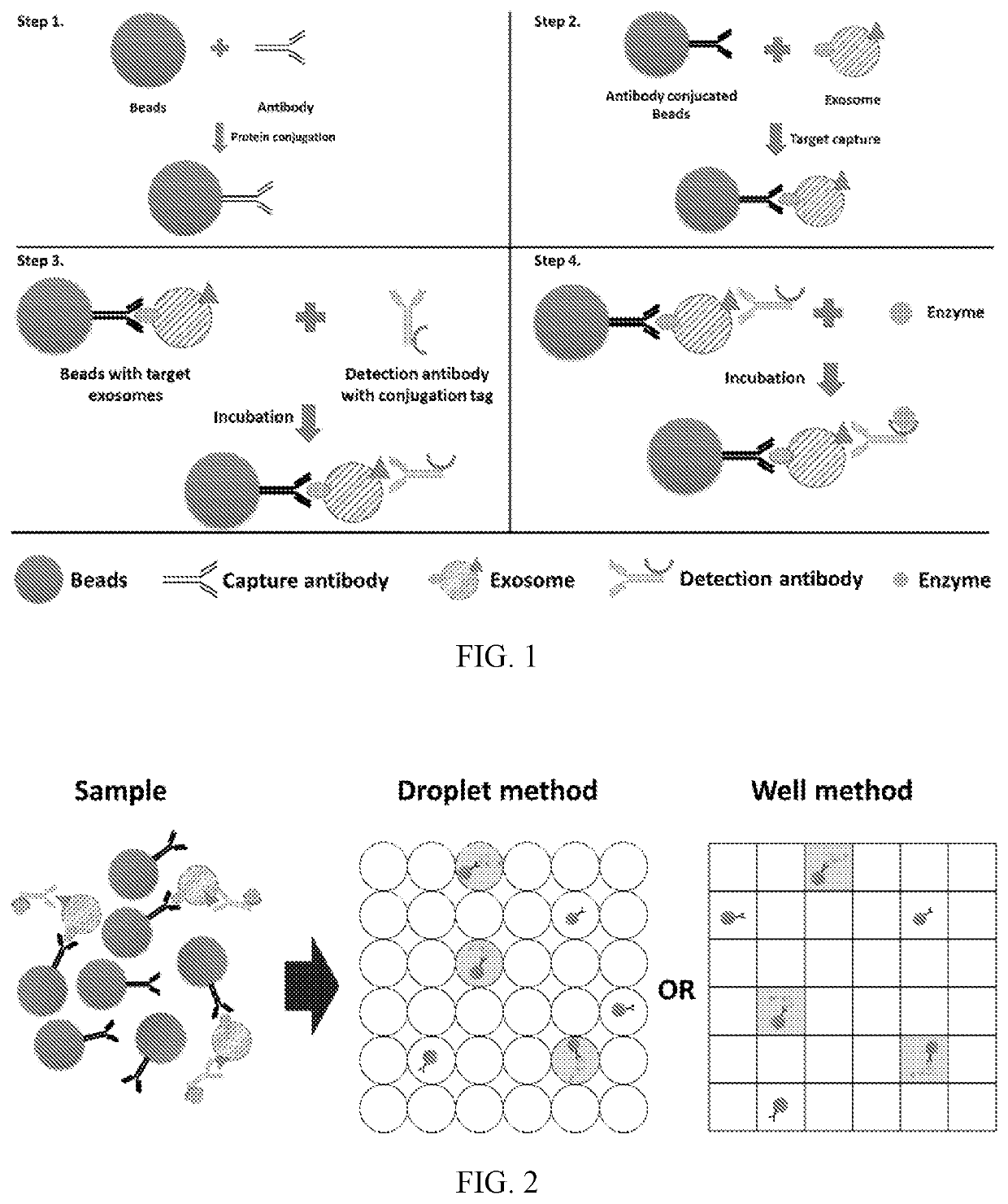
[0111]Digital enzyme-linked immunosorbent assays in various microfluidic platforms are demonstrated. Exosome solutions are obtained from biofluids and prepared through ultracentrifugation, ultrafiltration, density-gradient separation, and immunoaffinity capture methods. Since antigens exist on the surface of exosome, they can be recognized by the specific antibodies. One pair of antibodies which identify the exosome is constructed onto the bead in the form of an immunocomplex. The construction of immunocomplex onto the beads is shown in FIG. 1. The antibodies which can recognize the biomarkers (e.g. CD63) on the surface of exosomes are conjugated to the beads (e.g., Dynabeads™ or agarose beads). The beads are then incubated with an exosome solution. After incubation, the beads are collected by magnetic force or centrifugation. After thorough washing, the target exosomes conjugated on the beads are purified from the sample solution. Next, a second...

example 2

uantification of the Target Exosomes

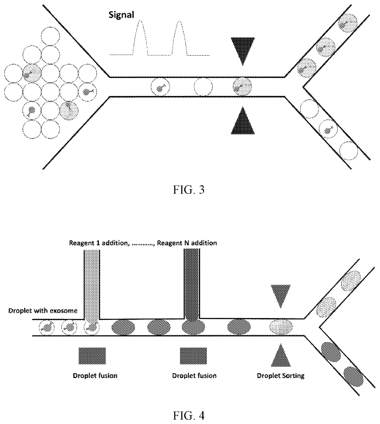
[0112]Digital quantification is carried out of the immunocomplex beads bound to the target exosomes via specific protein biomarkers. The immunocomplex constructed beads solution is flown into the channel to mix the solution with another channel of a substrate (e.g., FDG) flow and to form droplets of the mixtures. Instead of using droplets as the compartments, microwells fabricated on a flat chip can also be utilized to compartmentalize the sample solution. The sample with beads can be first dropped on the chip and be scraped into the wells. The substrate (e.g., FDG) solution is added into each compartment subsequently. The microwell chip is then sealed on the top to isolate each individual space for reaction. The microfluidic workflow is schematically shown in FIG. 2. After incubation, the droplets / wells with beads constructed immunocomplex emit color or fluorescent or electrochemical signal for detection. The signal can be detected by fluorescenc...

example 3

solation

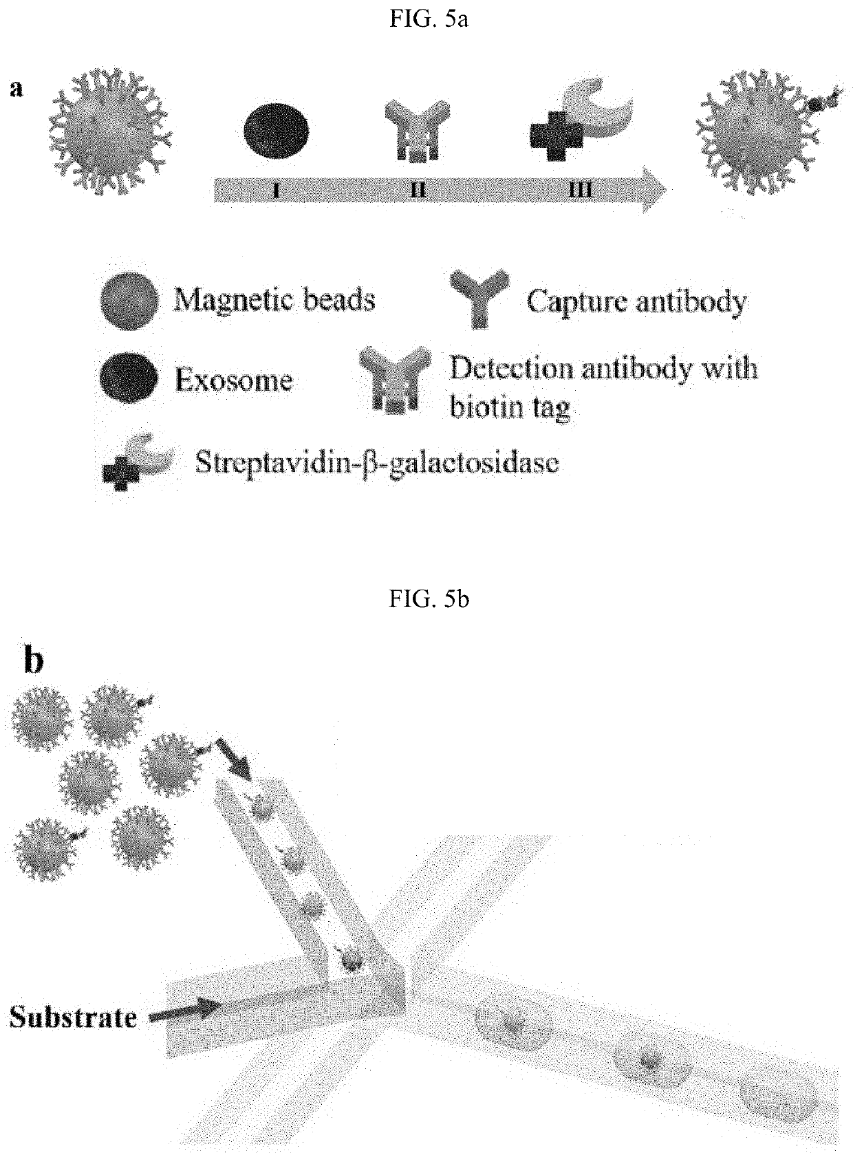
[0114]By constructing the immunocomplex on the beads and encapsulating them into droplets, the signal from labelled fluorescein or chemiluminescence can be used as a trigger for droplet sorting. The droplets that contain target exosomes can be separated through droplet sorting technology including electric sorting, mechanical sorting or acoustic sorting. FIG. 3 shows a schematic of the isolation of the fluorescent exosomes with desired information.


PUM


Abstract

Methods of quantification, isolation, and characterization of exosomes are provided. Exosomes can be quantified by contacting a sample with a capture bead comprising a bead and a first binding agent, and a second binding agent. The first binding agent binds to a first biomolecule in the exosomes to produce a first complex and the second binding agent binds to a second biomolecule in the exosomes of the first complex to produce a second complex. The first complexes and the second complexes are quantified based on a detectable signal conjugated to the second binding agent. A microwell or a droplet generation is utilized to quantify the first complexes and the second complexes. Quantifying the exosomes is used to diagnose a cancer in a subject. In such methods, the first and the second binding agents bind to cancer biomarkers present in the exosomes.

Description

CROSS-REFERENCE TO RELATED APPLICATION[0001]This application is the U.S. national stage application of International Patent Application No. PCT / CN2018 / 109760, filed Oct. 11, 2018; which claims the benefit of U.S. Provisional Patent Application Ser. No. 62 / 606,687, filed Oct. 5, 2017, the disclosure of each of which is hereby incorporated by reference in its entirety, including any figures, tables, or drawings.BACKGROUND OF THE INVENTION[0002]Exosomes are proposed as potent biomarkers for cancer diagnostics. Exosomes are non-uniform membranous particles with a diameter of 30-150 nm secreted from cells through plasma membrane fusion of multivesicular bodies (MVBs). Exosomes shed from tumor tissues carry numerous biomarkers such as transmembrane and cytosolic proteins (CD9, CD63, CD81, etc.), lipids, DNA and microRNA. Specific proteins, such as Glypican-1 (GPC1), Fibronectin (FN), Prostate-specific membrane antigen (PSMA), and functional nucleic acids, such as microRNA-145 have clinica...

Claims


Application Information

Patent Timeline
IPC IPC(8): G01N33/543G01N33/574
CPCG01N33/54326G01N33/57488G01N33/535G01N33/5436G01N33/574G01N2333/4722G16H50/20
Inventor YAO, SHUHUAIZHENG, LEIXU, XIAONANLIU, CHUNCHENHU, YU
Owner THE HONG KONG UNIV OF SCI & TECH
Features
  • R&D
  • Intellectual Property
  • Life Sciences
  • Materials
  • Tech Scout
  • Unparalleled Data Quality
  • Higher Quality Content
  • 60% Fewer Hallucinations
Learn More