Methods for determining blood gas on metabolic parameters
a metabolic parameter and blood gas technology, applied in the direction of instruments, material analysis, measurement devices, etc., can solve the problems of inability to measure the platelet count in heparinized blood, the formation of platelet aggregates, and the inability to measure the platelet coun
- Summary
- Abstract
- Description
- Claims
- Application Information
AI Technical Summary
Benefits of technology
Problems solved by technology
Method used
Image
Examples
example 1
Analysis of Platelet Aggregation Using Heparin and Different Anti-Platelet Agents
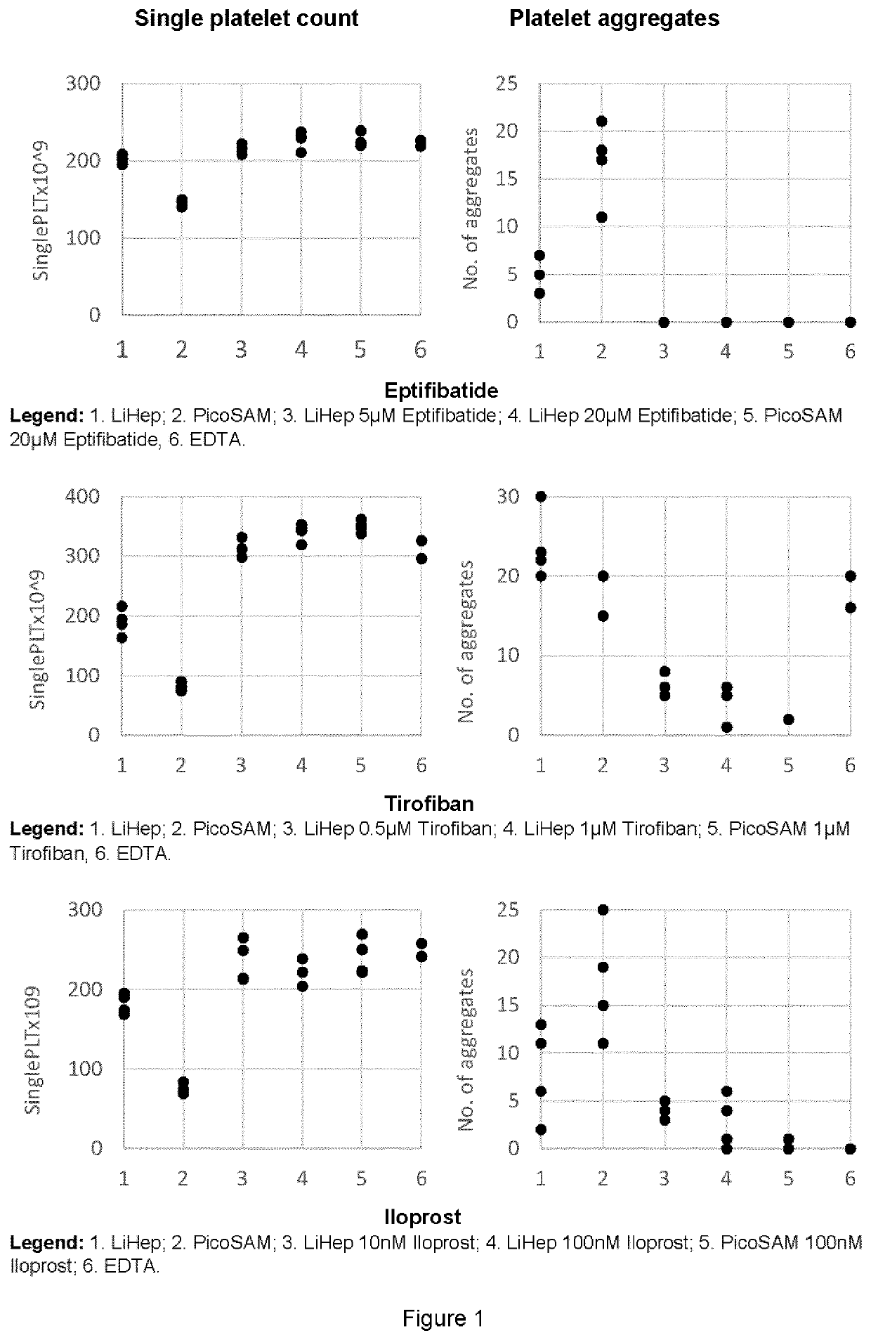
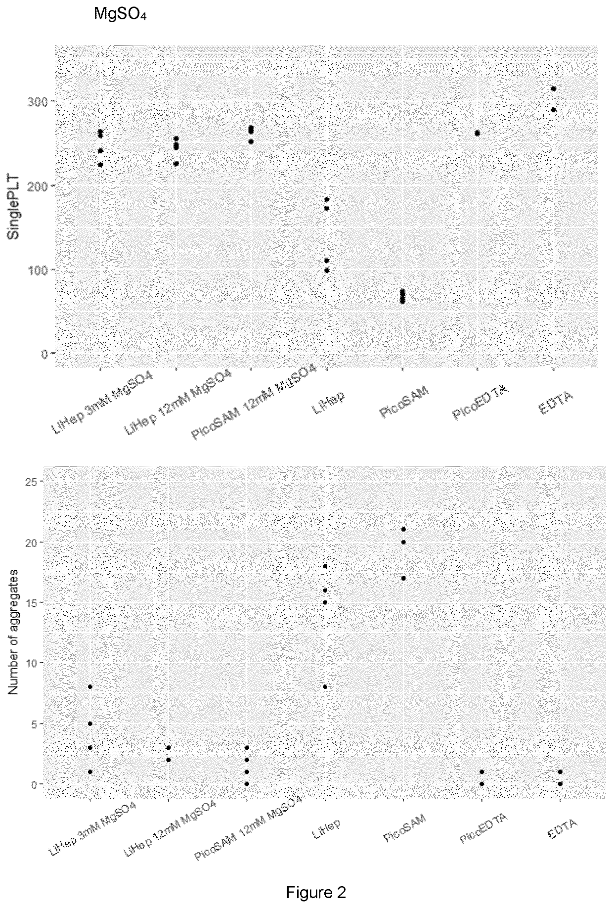
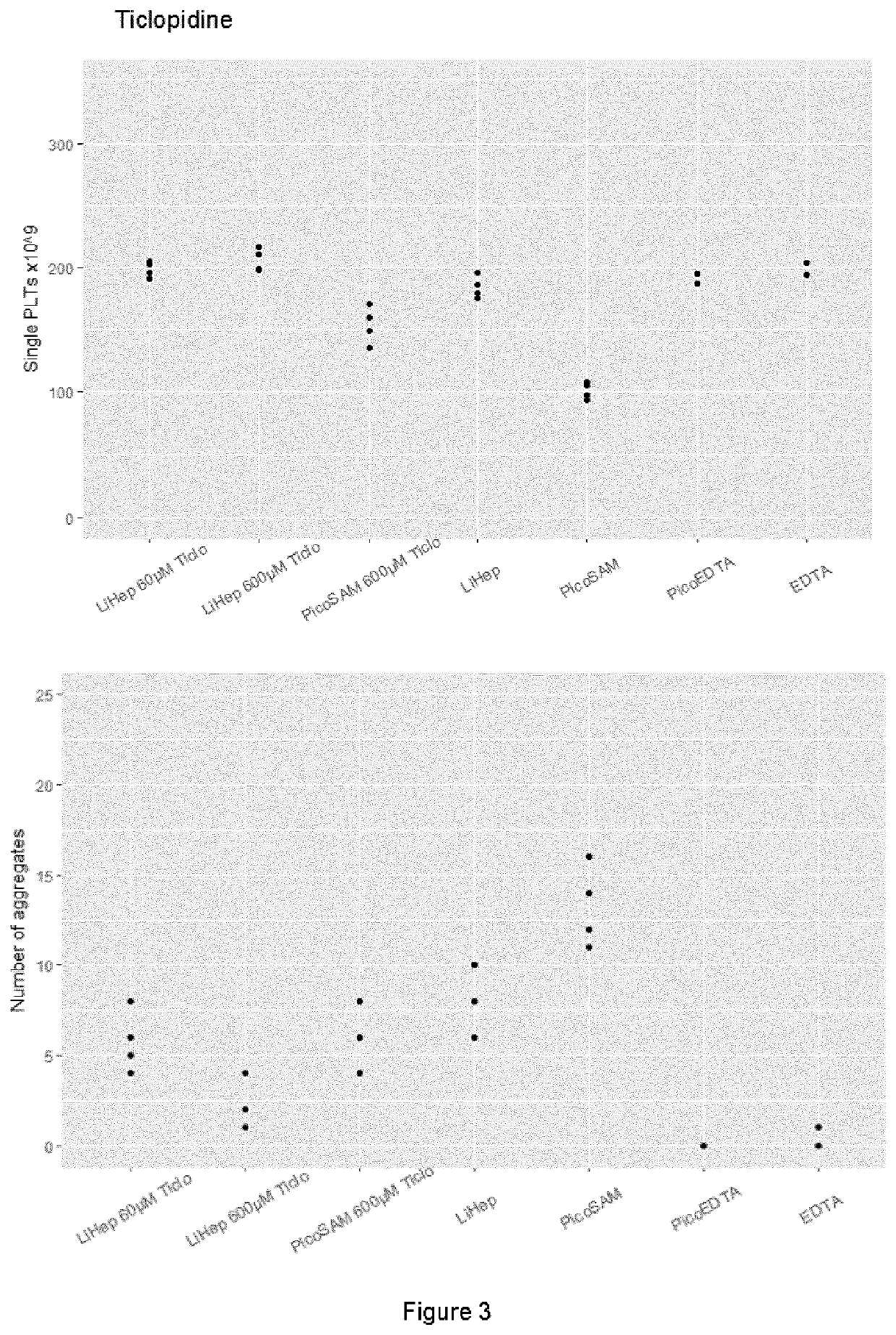
[0153]Each experiment was conducted on a separate day with blood from one voluntary donor testing one anti-platelet drug candidate. For each experiment PICO70 syringe samplers (Radiometer Medical ApS) were prepared just before sample drawing. For “LiHep” (Liquid Heparin) conditions, PICO70 samplers were emptied of the gold ball and heparin brick and 15 μl of aqueous liquid balanced heparin comprising heparin lithium (Celsus Laboratories) and heparin sodium (Celsus Laboratories) (final heparin conc. 60 IU / mL blood) and 15 μl of the solvent of the tested drug were added. For “PicoSAM” conditions, unmodified PICO70 samplers (with gold ball and heparin brick) were used and 15 μl of the solvent of the respective tested drug was added. For “LiHep xxx drug” conditions, PICO70 samplers were emptied as described for LiHep and 15 μl of liquid balanced heparin (final heparin conc. 60 IU / mL blood) and 15 μl of the ...
example 2
Platelet Aggregation Studies with Heparinized Blood and EDTA-Treated Blood
[0158]Venous blood samples drawn, mixed and fixed with 10% formalin solution, as described before in Example 1 were used to prepare wet mounts on a glass slide and covered with a glass coverslip. Wet mount samples of fixed blood cells were then imaged with a Leica 750 microscope using a 40× phase-contrast air objective. Aggregated platelets in heparinized blood versus non-aggregated single platelets in EDTA anti-coagulated blood and heparinized blood with e.g. eptifibatide are seen between abundant RBCs.
[0159]The results are shown in FIG. 6. Heparinized blood showed platelet aggregates which cannot be seen in blood samples prepared with EDTA or in a blood sample prepared with heparin and an antiplatelet drug such as eptifibatide.
example 3
Wet Mount Images of Stained Blood Samples
[0160]Venous blood samples were drawn and mixed as described before in Example 1 and used to prepare stained wet mounts images. Blood samples were mixed with a staining and hemolyzing agent (methylene blue and deoxycholic acid, respectively) and incubated at 47° C. in a water bath for 30 seconds. Stained and hemolyzed blood samples were then prepared in wet mount samples on a glass slide and covered with a glass coverslip. Images of stained samples were then acquired with a Leica 750 microscope using a 40× air objective in bright field mode. An image processing software (FIJI, ImageJ) was used to select regions of interest (ROIs) of representative stained WBCs from the images.
[0161]PicoSAM samples (heparinized blood without an anti-platelet drug) showed many aggregated platelets (small roundish cells).
[0162]PicoSAM samples with 1 μM tirofiban or 20 μM eptifibatide showed single platelets but also platelet satellinism, i.e. platelets binding t...
PUM
 Abstract
Description
Claims
Application Information
 - R&D
- Intellectual Property
- Life Sciences
- Materials
- Tech Scout
- Unparalleled Data Quality
- Higher Quality Content
- 60% Fewer Hallucinations
Browse by: Latest US Patents, China's latest patents, Technical Efficacy Thesaurus, Application Domain, Technology Topic, Popular Technical Reports.
© 2025 PatSnap. All rights reserved.Legal|Privacy policy|Modern Slavery Act Transparency Statement|Sitemap|About US| Contact US: help@patsnap.com


