

Method for restoration of normal phenotype in cancer cells
a cancer cell and normal phenotype technology, applied in the field of normal phenotype restoration in cancer cells, can solve problems such as cell apoptos
- Summary
- Abstract
- Description
- Claims
- Application Information
AI Technical Summary
Benefits of technology
Problems solved by technology
Method used
Image


Examples
example 1
Cell Proliferation Measurements
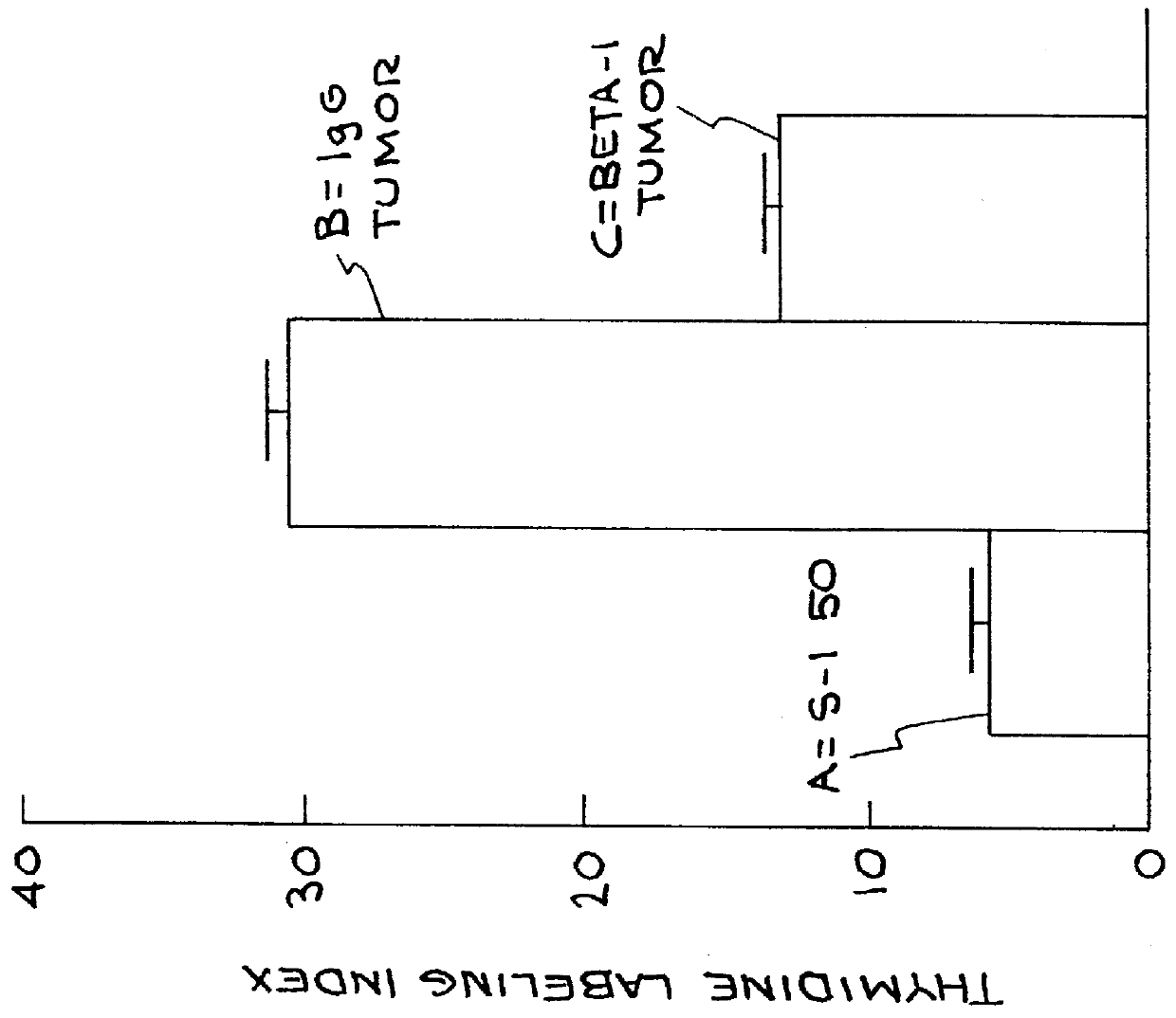
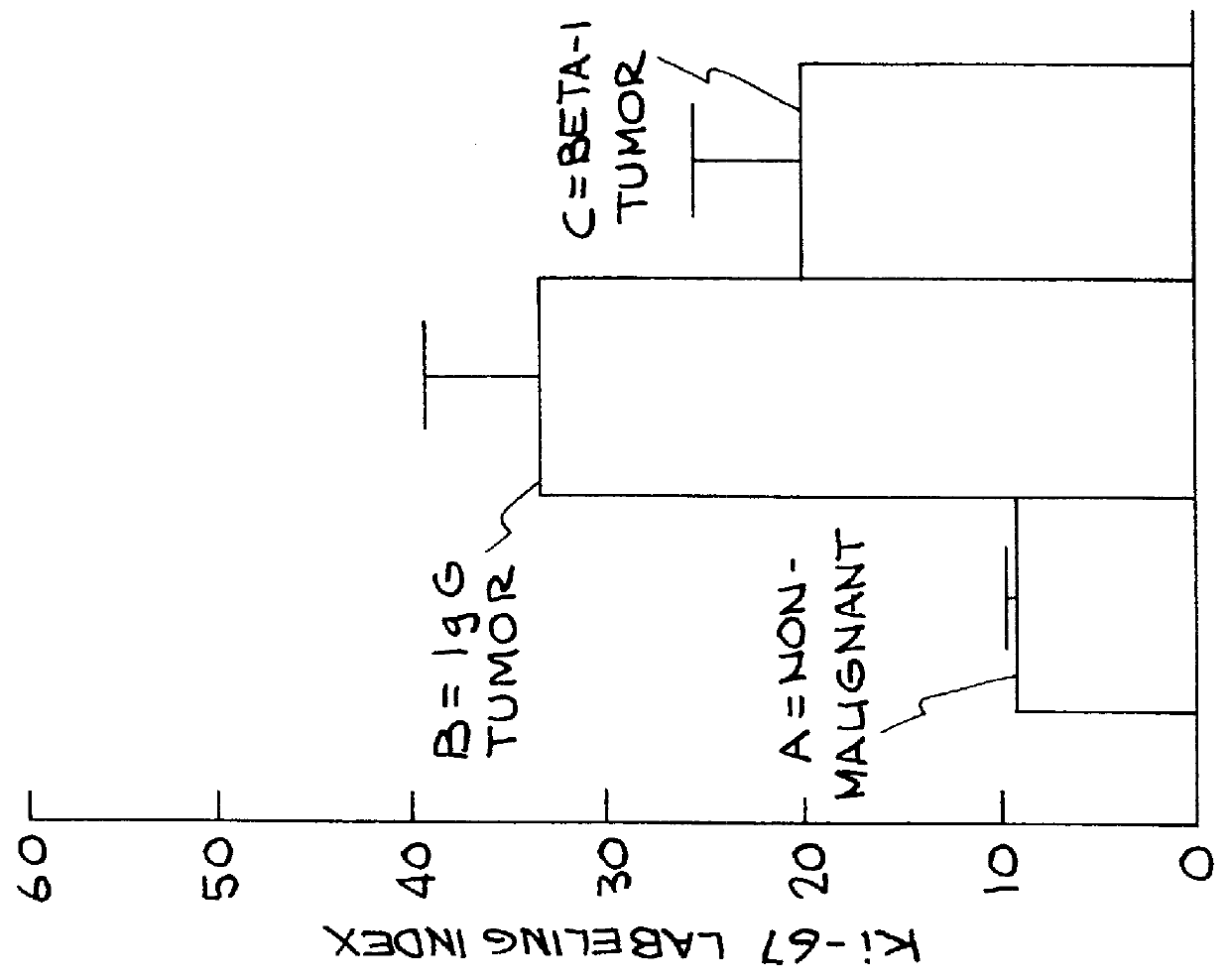
Cell proliferation was determined by 3 separate criteria: a) percentage of cells in S phase of the cell cycle (actively dividing) b) percentage of cells in the cell cycle and c) number of cells per spheroid.
When cultured within a 3 dimensional reconstituted basement membrane, non-malignant MECs undergo growth arrest after 6-10 days. In contrast MEC tumor cells fail to growth arrest and continue to grow. Restoration of non-malignant behavior by a treatment regime comprising application of .beta..sub.1 integrin function-blocking antibody to tumor cells resulted in the ability of the tumor cells to growth arrest after 6-10 days when grown in a 3-D basement membrane. This was assessed by .sup.3 H thymidine labeling studies of day 10-12 cultures which determines the percentage of cells in S phase of the cell cycle. Three-dimensional cell cultures were pulse labeled with .sup.3 H thymidine for 24 hours after which the cultures were washed, fixed and frozen. ...
example 2
Morphology
Morphology was assessed using four criteria: a) colony organization; b) actin microfilament organization; c) formation of adherens junctions; and d) formation of .beta..sub.1 catenin / E cadherin interactions.
FIG. 4 illustrates that non-malignant MECs undergo morphogenesis 8-10 days after culturing within a 3 dimensional basement membrane while tumor cells form large, disorganized colonies. Treatment with .beta..sub.1 integrin function-blocking antibody allowed the tumor cells to form organized spheroids. The ability of cells to form organized colonies was assessed by examining colony structure in live 3 dimensional, day 10-12, cultures using a phase contrast microscope with a 10.times. objective. Organization was determined by measuring colony size, length and width, using an eyepiece objective with a ruler. Colonies were deemed organized if they were uniformly spherical and had well defined, rounded structures. At least 50-100 colonies were assessed per group in living bas...
example 3
Responsiveness to a Basement Membrane
The ability cells to respond to the presence of an exogenous basement membrane was assessed by two criteria: a) the cells' ability to organize their own endogenous basement membranes and b) the cells' ability to repress apoptosis. Non-malignant MECs respond to an exogenous basement membrane by synthesizing and organizing an endogenous basement membrane around their acini and repressing apoptosis.
FIG. 8: The presence of an endogenous basement membrane was assessed by immunostaining frozen sections of day 10-12, 3-dimensional cultures for the presence of basally deposited collagen IV, using an anti-human collagen IV antibody (a known constituent of the endogenous basement membrane).
In FIG. 8, frame A shows the endogenous basement membrane indicated by collagen type IV or laminin of non-malignant S-1 50 control cells; frame B shows a schematic representation of endogenous basement membrane surrounding mock rat IgG antibody-treated tumor T-4 cells; f...
PUM
| Property | Measurement | Unit |
|---|---|---|
| Concentration | aaaaa | aaaaa |
| Concentration | aaaaa | aaaaa |
Abstract
Description
Claims
Application Information