

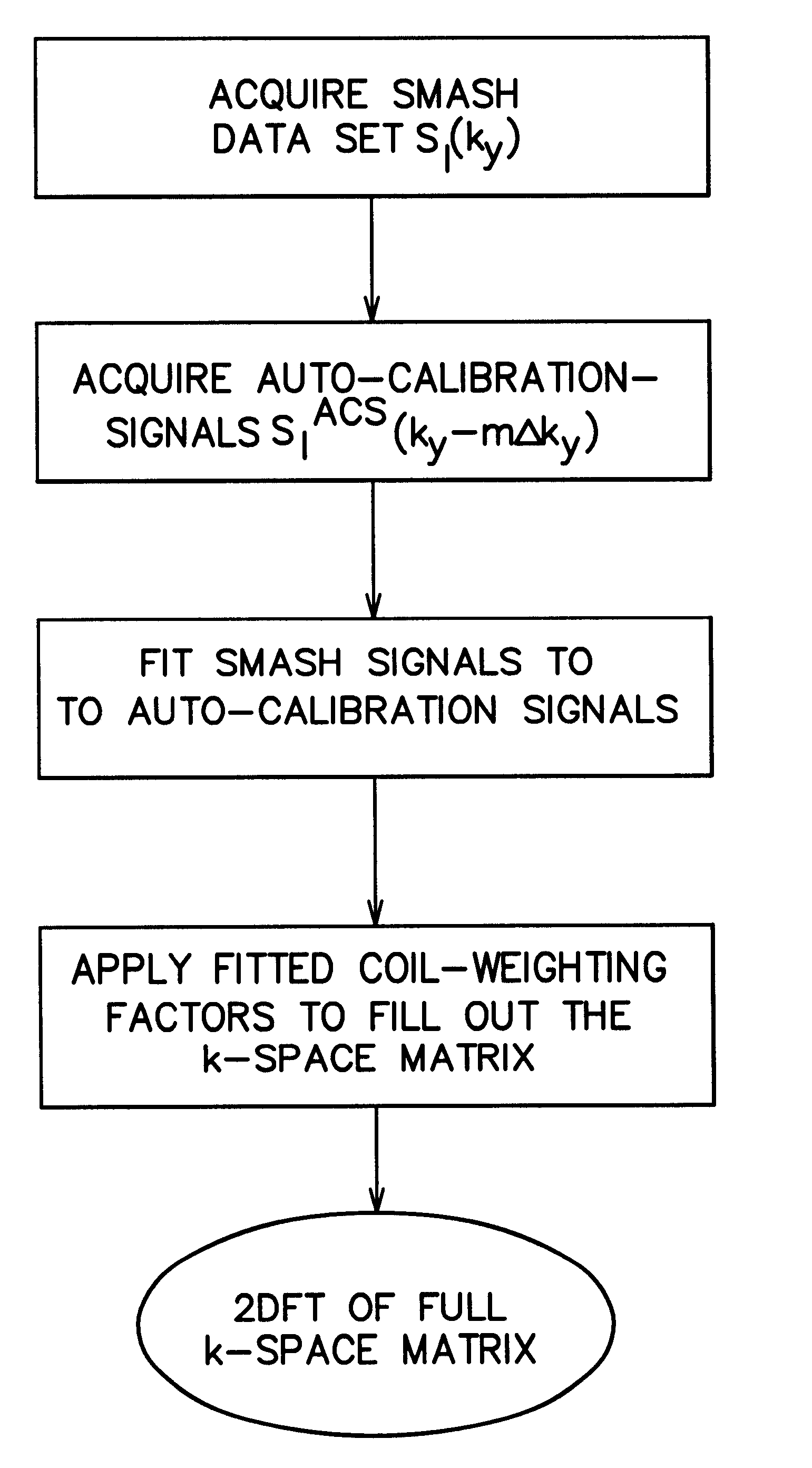
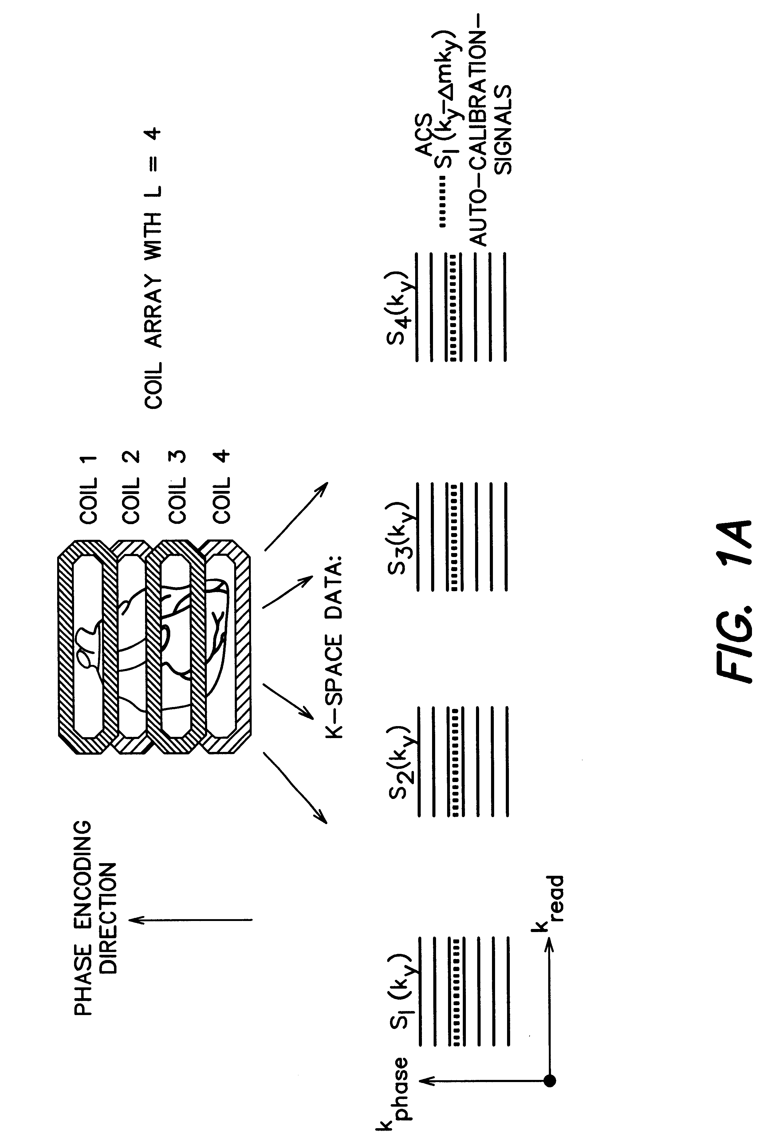
Coil array autocalibration MR imaging
a technology of autocalibration and mr imaging, applied in the direction of nmr measurement, diagnostic recording/measuring, instruments, etc., can solve the problems of blurred and distorted images, inability to achieve the resolution necessary for fast image acquisition in a shorter time, and trade-off between spatial resolution and blurred images
- Summary
- Abstract
- Description
- Claims
- Application Information
AI Technical Summary
Benefits of technology
Problems solved by technology
Method used
Image

Examples
example 1
In one test, to assess the performance of the AUTO-SMASH approach in a well-controlled experiment, a resolution phantom was imaged using the 4-element array. Images were acquired in an 8 mm thick coronal slice parallel to and approximately 60 mm above the plane of the array. A FLASH imaging sequence was used, with TE=6 ms, TR=12 ms, and flip angle=15.degree.. Phase encoding was performed in the direction of the array. The FOV was 320 mm.times.320 mm and matrix size was 128.times.128 for full-time reference images acquired with no omitted gradients. The reference image is shown in FIG. 4A. Reduced time data sets with either two (left column) or three (right column) times the phase encode step (interval), and hence one half or one third the FOV and matrix size in the phase encode direction, were used for both SMASH and AUTO-SMASH reconstruction. Reference component coil images were combined using a conventional sum of squares algorithm. SMASH reconstructions used coil sensitivity info...
example ii
A principal motivation for the development of both the SMASH and AUTO-SMASH procedures was to improve cardiac imaging. Since SMASH effectively allows multiple acquisitions to proceed simultaneously, it may be used to gather high resolution information in a given acquisition time, or else to acquire images of a given spatial resolution in a shorter acquisition time. Thus, the SMASH technique, when supplemented with AUTO-SMASH calibration, offers a possible remedy for the competing constraints of spatial versus temporal resolution in cardiac MRI. A methodical study of cardiac imaging with two different acquisition strategies was therefore undertaken in order to confirm the benefits of AUTO-SMASH for such imaging. These are referred to as strategy I and strategy II below.
STRATEGY I. Since a reduction in breath-hold times is particulary important for patients with cardiac disease for whom current long breath-hold times are impractical, the AUTO-SMASH strategy was used to reduce breath-h...
PUM
 Abstract
Description
Claims
Application Information