Diagnosing intrapulmonary infection and analyzing nasal sample

a technology for intrapulmonary infections and nasal samples, applied in the field of diagnosing intrapulmonary infection and analyzing nasal samples, can solve the problems of inability to present data to support these uses, time-consuming and resource-intensive process for diagnosing pulmonary infections in mammals such as humans, and achieve the effect of rapid detection of lung infections

Inactive Publication Date: 2002-10-08
THE TRUSTEES OF THE UNIV OF PENNSYLVANIA
View PDF12 Cites 26 Cited by
  • Summary
  • Abstract
  • Description
  • Claims
  • Application Information

AI Technical Summary

Benefits of technology

The present invention uses the Aromascan product or some other "electronic nose" for the purpose of analyzing gases exhaled or otherwise obtained from a patient for the purpose of rapidly determining the presence of pathologic processes in the lung or other parts of the patient resulting in alterations or deviations in the analyzed gases. A "pathologic process" as defined herein is necrosis, hemorrhaging, or an inflammatory process in the lung, such as an infection, along with other disease conditions for which a diagnosis is desired. The present invention is particularly useful in the rapid detection of lung infections and specifically differentiation between lung infiltrates (on chest x-rays) caused by infections versus lung collapse. Currently, such differentiation relies upon cultures which are expensive and time consuming (one to two days). With the present invention, such differentiation can be determined substantially immediately, and without requiring culturing or growing of a sample.

Problems solved by technology

Because of its subjectivity and the lack of correlative monitors, smell, as a diagnostic tool, has never achieved significant prominence in modern medicine.
No data is presented to support these uses.
The diagnosis of pulmonary infections in mammals such as humans is a time-consuming, resource intensive process and sometimes inaccurate process.
A chest x-ray does not necessarily provide an accurate indication of the presence or absence of an infection.
In such patients, CSF may leak or drain through a skull-base defect into the sinuses and then into the nose.
Although this test is very accurate, the test requires a relatively large amount of fluid.
It is sometimes difficult to obtain a sufficiently large amount of fluid to conduct the test.

Method used


Image

  • Diagnosing intrapulmonary infection and analyzing nasal sample
  • Diagnosing intrapulmonary infection and analyzing nasal sample
  • Diagnosing intrapulmonary infection and analyzing nasal sample

Examples


examples 2-6

FIGS. 4-8 provides additional patient data which shows distinguishable clusters for patient samples which have different types of lung infections and for uninfected patient samples. FIGS. 4-8 are self-explanatory and thus are not further described herein.

The samples collected in Examples 1-6 used a vapor form of expired air from the patients, and did not use condensate of the vapor form.

example 7

In example 7, testing was done on condensate from the patients since it is known that condensate contains volatiles from the expired air. Test equipment included a standard Aromascan A32S / CEM using a temperature controlled 25 ml sample vial and a carrier gas.

The protocol for obtaining and testing samples in Example 7 was as follows:

1. Collect 10 ml of condensate from a collection vessel inserted just before the ventilator catch pot using a plastic syringe.

2. Extract 250 .mu.l of condensate into the syringe.

3. A reference signal was set up for the Aromascan A32S / CEM by passing approximately 90 ml / min of carrier gas through a sample vial containing 2 ml of 10% sodium hydroxide (NaOH) held at 33.degree. C.

4. At 60 seconds into the run, the syringe containing 250 .mu.l of condensate is injected into the sample vial.

5. At 180 seconds into the run, the sample vial is replaced with a fresh vial of 2 ml of 10% NaOH to act as a wash and to become the next sample vial.

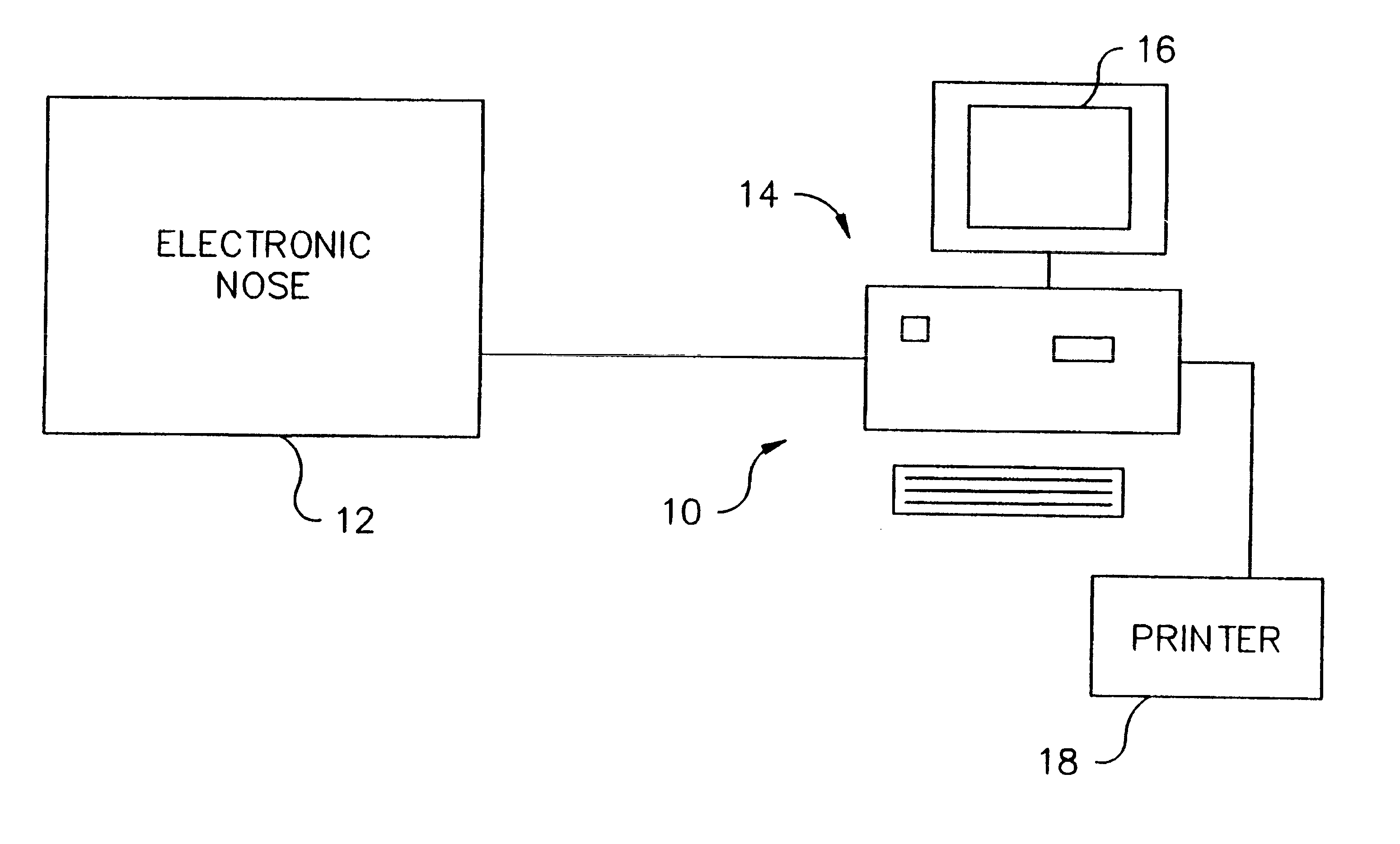
FIG. 9 is a schematic di...


PUM

PropertyMeasurementUnit
turnaround timeaaaaaaaaaa
timeaaaaaaaaaa
electrical propertyaaaaaaaaaa

Abstract

The presence of a pathologic process in a lung of a mammal is detected by applying exhaled gas of a mammal to an electronic nose (12). Data derived from the electronic nose (12) is used to determine whether a pathologic process is present in the lung of the mammal. The pathologic process may also be a lung infection such as pneumonia. Also, a mammalian fluid sample obtained from the sinus or nose is applied to an electronic nose (12) to determine if the fluid sample contains significant amounts of cebrospinal fluid.

Description

The sense of smell has long been used as a diagnostic tool by medical clinicians. Because of its subjectivity and the lack of correlative monitors, smell, as a diagnostic tool, has never achieved significant prominence in modern medicine."Electronic noses" or "electronic olfactory sensors" have recently been developed to provide objective measurements and analysis of aromas. One particular "electronic nose" which has achieved some commercial success, primarily in the area of quality control and environmental monitoring in particular industries such as the beverage, flavor, perfume and certain aspects of the food industries, is manufactured by Aromascan, Inc. of Hollis, N.H. The Aromascan product uses changes in an electrical property (specifically, impedance or resistance) of sensors in a sensor array made of a layer of a semi-conducting organic polymer, when exposed to particles in a gas for aroma analysis. The Aromascan product is disclosed in U.S. Pat. No. 4,887,455 (Payne et al....

Claims


Application Information

Patent Timeline
Patent Type & Authority Patents(United States)
IPC IPC(8): G01N33/00G01N33/497G01N33/483
CPCG01N33/0034G01N33/0047G01N33/497G01N2033/4975A61B5/082A61B5/7264G16H50/20Y02A90/10
Inventor HANSON, III, C. WILLIAMTHALER, ERICA R.
Owner THE TRUSTEES OF THE UNIV OF PENNSYLVANIA
Why Eureka
Try Eureka