Optical imaging of subsurface anatomical structures and biomolecules

a biomolecule and optical imaging technology, applied in the field of optical imaging and medical diagnosis, can solve the problems of deficient lack of effective means of imaging subsurface anatomical, use of ultraviolet, visible and infrared radiant electromagnetic energy for imaging, etc., and achieve enhanced imaging, enhanced contrast in medical imaging, and low cost

Inactive Publication Date: 2005-05-03
ROCKY MOUNTAIN BIOSYST
View PDF13 Cites 176 Cited by
  • Summary
  • Abstract
  • Description
  • Claims
  • Application Information

AI Technical Summary

Benefits of technology

[0008]The present invention provides various methods / systems of optical imaging of subsurface anatomical structures and biomolecules utilizing red and infrared radiant energy. Such methods enhance contrast in medical imaging, and are generally useful in non-invasive and relatively low cost imaging instruments. The techniques used include pulsatile enhanced imaging, confocal enhanced imaging, Raman enhanced imaging, laser speckle enhanced imaging, multiphoton interaction enhanced imaging, optical coherence tomography enhanced imaging, time correlated single photon counting enhanced imaging, and polarization enhanced imaging.

Problems solved by technology

However, there is a major limitation in using ultraviolet, visible and infrared radiant electromagnetic energy for imaging in that most tissues are highly scattering, and often strongly absorbing to such photons.
The prior art is deficient in the lack of effective means of imaging subsurface anatomical structures and biomolecules by using red and infrared radiant energy.

Method used


Image

  • Optical imaging of subsurface anatomical structures and biomolecules
  • Optical imaging of subsurface anatomical structures and biomolecules
  • Optical imaging of subsurface anatomical structures and biomolecules

Examples


example 1

Pulsatile Enhanced Imaging

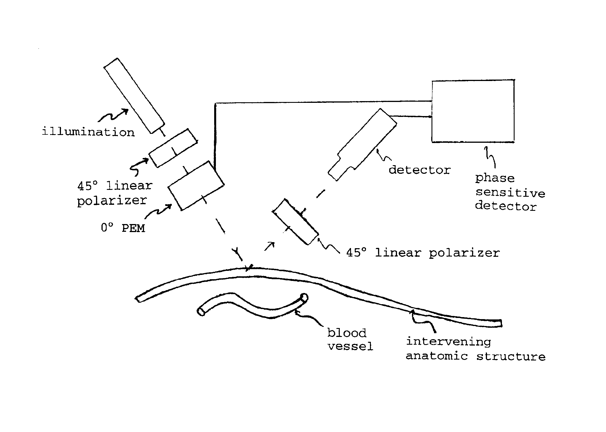
[0029]Pulse oximeters are relatively common devices used to measure the percent oxygen saturation of blood, wherein red (λ1≈660 nm) and near infrared (λ2≈940 nm) radiant energy are passed through tissue (typically the fingertip). Using this device, it is possible to obtain a measurement of oxygen saturation of the blood based on the relative signals transmitted through the fingertips at each wavelength, knowing the absorption characteristics of the pertinent absorbing chromophores (tissue, oxygenated hemoglobin or HbO2, deoxygenated hemoglobin or Hb), and after extensive calibration of the device with a direct measurement of blood oxygenation. One important operating characteristic that makes the pulse-oximeters useful is that they can discern between arterial and non-arterial absorption by discriminating between time-varying signals (due to the heart pumping and more evident in arterial blood) and non-varying signals (venous blood and tissue).

[0030]FIG. 1 ...

example 2

Confocal Enhanced Imaging

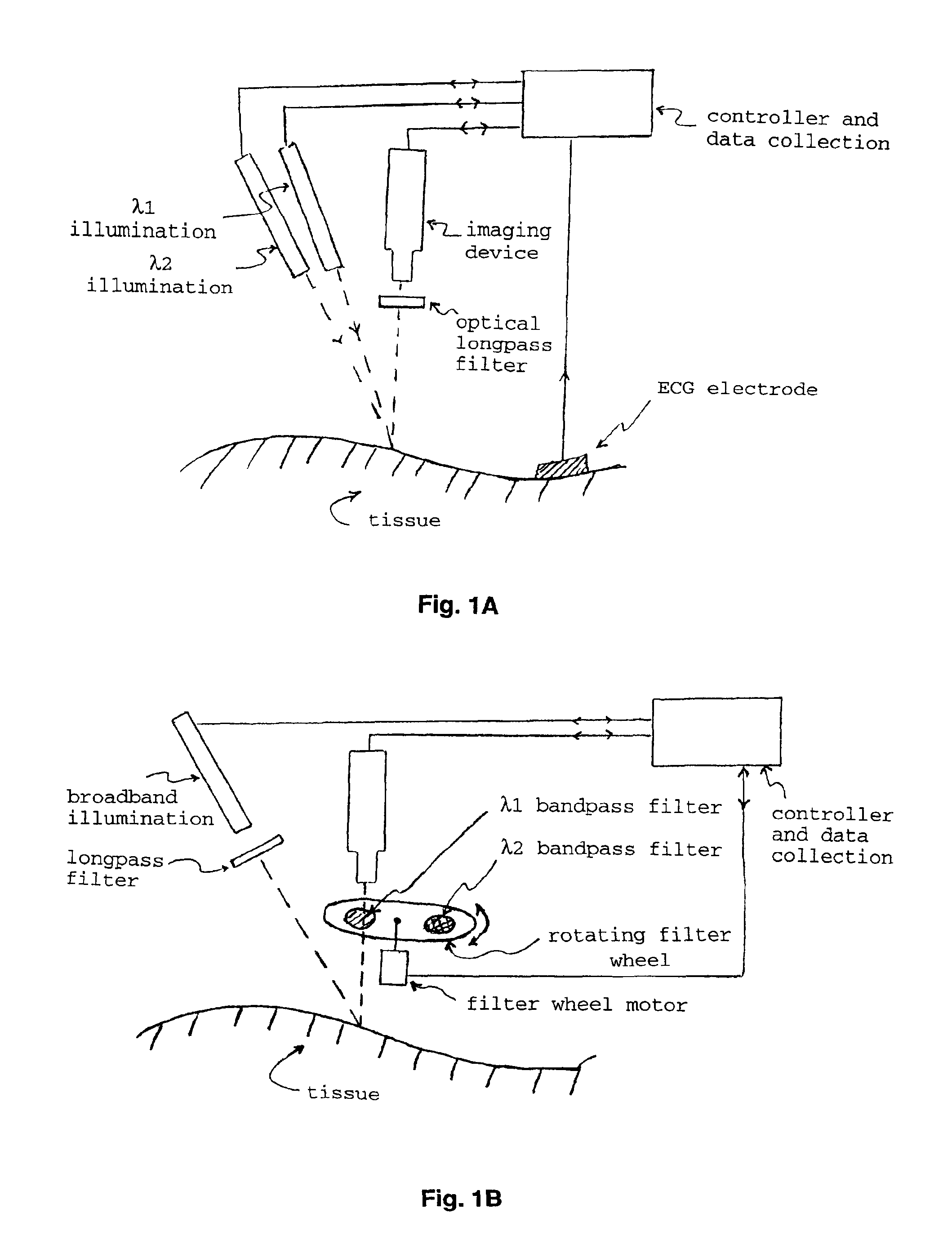
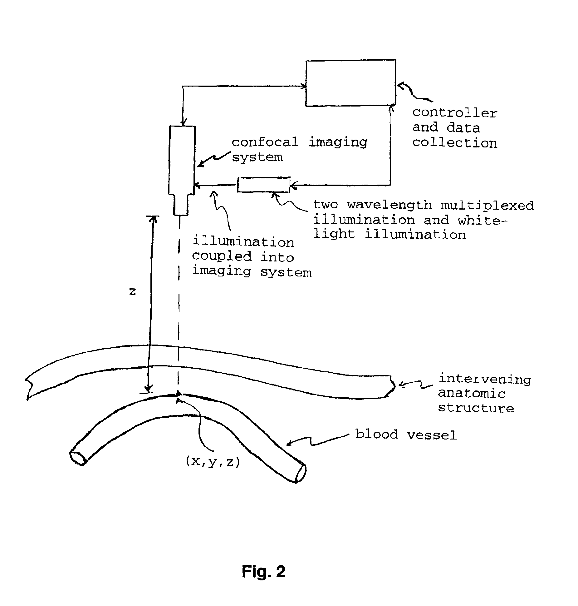
[0034]The concept of rejecting light scattered from locations other than the point being imaged (that is, with coordinates x,y,z), by using apertures in the imaging system, is referred to as confocal imaging. This concept has been used to “optically section” microscopic specimens being viewed with a microscope. Confocal microscopy normally uses white light illumination, or ultraviolet to green illumination to induce fluorescence in the sample. The former illumination results in significant chromatic aberrations in the final image, while the latter provides only a fluorescent image.

[0035]It would be beneficial, in certain cases, to use red and / or near infrared radiant energy in a confocal imaging optical system, along with white-light illumination. Narrow band illumination through the use of diode lasers or bandpass filtered broadband light sources would be optimal (FIG. 2). By alternately illuminating the region-of-interest (ROI), for example, 660 nm and 940...

example 3

Raman Enhanced Imaging

[0036]Raman spectroscopy is a light scattering technique that uses (typically) laser radiation to excite the sample, whereby the scattered radiation emitted by the sample is analyzed. Emission data has two main characteristics: the frequencies at which the sample emits the radiation (a small number of the incident photons, perhaps only 1 in 106, is emitted at frequencies different from that of the incident light), and the intensities of the emissions. Determining the frequencies allows identification of the sample's molecular makeup, since chemical functional groups are known to emit specific frequencies and emission intensity is related to the amount of the analyte present. Now, with highly sensitive imaging light detectors (e.g. CCDs) and efficient optics such as transmission holographic gratings and notch filters, Raman spectroscopy has seen a resurgence of interest in the medical field.

[0037]It would be useful to illuminate the anatomic structure of interes...


PUM


Abstract

The present invention provides various methods/systems of optical imaging of subsurface anatomical structures and biomolecules utilizing red and infrared radiant energy. Also provided are various applications of such methods/systems in medical diagnosis and treatment.

Description

CROSS-REFERENCE TO RELATED APPLICATION[0001]This non-provisional patent application claims benefit of provisional patent application U.S. Ser. No. 60 / 201,592, filed May 3, 2000, now abandoned.BACKGROUND OF THE INVENTION[0002]1. Field of the Invention[0003]The present invention relates generally to the fields of optical imaging and medical diagnosis. More specifically, the present invention relates to advanced methods of optical imaging of subsurface anatomical structures and biomolecules.[0004]2. Description of the Related Art[0005]The importance of imaging structures and biomolecules in humans is self-evident. Recently, many groups of investigators have been trying to develop optical imaging techniques in order to supercede, or reduce the requirements of using expensive and potentially harmful techniques including x-rays, ultrasound or magnetic and radio-frequency fields. Optical imaging techniques employing non-ionizing electromagnetic radiation are not harmful to biological tissu...

Claims


Application Information

Patent Timeline
Patent Type & Authority Patents(United States)
IPC IPC(8): A61B5/00A61B5/0456A61B5/026A61B5/0452A61B1/045A61B5/352
CPCA61B5/0066A61B5/489A61B5/0075A61B5/0071A61B1/043A61B1/045A61B5/0068A61B5/0086A61B5/0261A61B5/0456A61B5/352A61B2090/366
Inventor MARCHITTO, KEVIN S.FLOCK, STEPHEN T.
Owner ROCKY MOUNTAIN BIOSYST
Why Eureka
Try Eureka