Device and method of use for detection and characterization of pathogens and biological materials

a technology for pathogens and biological materials, applied in the detection and characterization field of pathogens, viruses, and other biological materials, can solve the problems of large number of deaths, economic hardship, and critical problems of pathogens, and achieve the effects of simple, rapid, sensitive and high throughput methods

Inactive Publication Date: 2005-05-24
BIOFORCE NANOSCI
View PDF118 Cites 35 Cited by
  • Summary
  • Abstract
  • Description
  • Claims
  • Application Information

AI Technical Summary

Benefits of technology

[0072]As may be appreciated, the invention can be applied to detection and characterization of a broad range of biological materials, such as, but not limited to, other viruses, bacteria, bacterial spores, prions, pathogenic microbes such as fungus, parasites, mold, and pollen spores.
[0073]The information and examples described herein are for illustrative purposes and are not meant to exclude any derivations or alternative methods that are within the conceptual context of the invention. It is contemplated that various deviations can be made to this embodiment without deviating from the scope of the present invention. Accordingly, it is intended that the scope of the present invention be dictated by the appended claims rather than by the foregoing description of this embodiment.

Problems solved by technology

Pathogens constitute a critical problem for human, animal and plant health.
Pathogens may cause infections that result in a variety of human illnesses and can lead to a large number of deaths.
Moreover, pathogens infect agriculturally important plant and animal species, resulting in economic hardship.
The use of animals in infectivity assays is costly, time consuming and subject to ethical dispute.
Serodiagnosis still exists in many-hospitals principally because there are no good alternatives for some infections.
Antibody based tests are popular, but are usually limited to a battery of individual tests in a macroscopic format (e.g., Enzyme Linked ImmunoSorbant Assay, or ELISA).
While precise in their verification of pathogen identity, these procedures are very slow.
PCR methods, however, are relatively costly and time consuming.
Furthermore, PCR tests are of a relatively limited applicability because of the requirement of enzyme activity and because of the frequency of false positives.

Method used


Image

  • Device and method of use for detection and characterization of pathogens and biological materials
  • Device and method of use for detection and characterization of pathogens and biological materials
  • Device and method of use for detection and characterization of pathogens and biological materials

Examples


example i

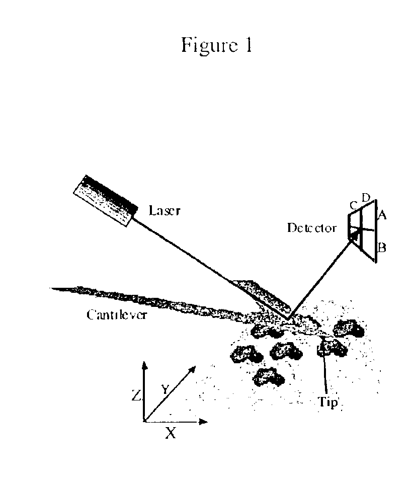
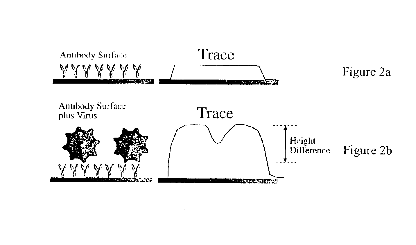
[0048]With reference to FIGS. 2a-b, 4a-c, 5a, 6a-c, and 7a-c, CPV is detected by constructing a chip with anti-canine parvovirus monoclonal antibody deposited thereon, exposing the chip to a target sample, and then reading the chip using the AFM to determine whether CPV is bound to the chip surface.

[0049]The viral particles were prepared at a stock concentration of 0.3 mg / ml. Purified monoclonal antibody which recognizes the CPV capsid, A3B10 (0.9 mg / ml) was utilized. In another test run, purified anti-canine parvovirus monoclonal antibody (2 mg / ml) was obtained from a separate source (Custom Monoclonal International, California) with no observable change in results.

[0050]A chip with a protein G coated surface was used for the deposition of antibodies against CPV at specific regions. The domains were formed using the above described microjet method. The antibodies of the present embodiment were diluted to 0.1 mg / ml in 1×PBS before being deposited onto the surface using the microjet....

example ii

[0062]Anti-vaccinia and anti-adenovirus (control) antibodies were obtained from Biodesign. Vaccinia virus strain WR (American Type Culture Collection (ATCC) 1354) was grown in HeLa S3 (ATCC CCL-2.2) cells maintained on RPMI 1640 supplemented with 7% v / v fetal bovine serum and penicillin, streptomycin and fungizone. Near confluent S3 cells in Blake bottles were infected at a multiplicity of infection (MOI) of 0.2. Cells were collected when detachment became apparent in about 3 days. Virus was purified from frozen and thawed cells by successive cycles of sedimentation velocity and equilibrium density centrifugation. Stocks of purified virus were titered by end-point dilution assays and stored at −80C. The virus had a stock concentration of 107 pfu / ml. The purified rabbit anti-vaccinia (B65101R lot 8304600) and goat anti adenovirus hexon (B65101G, lot 4A01901) antibodies were divided into aliquots and stored at −20 C. until use.

[0063]To construct the vaccinia chips, the process of acti...


PUM

PropertyMeasurementUnit
bar sizeaaaaaaaaaa
bar sizeaaaaaaaaaa
diameteraaaaaaaaaa

Abstract

The present invention includes a method and apparatus for the detection of a target material. The method and apparatus includes providing a substrate with a surface and forming a domains of deposited materials thereon. The deposited material can be placed on the surface and bound directly and non-specifically to the surface, or it may be specifically or non-specifically bound to the surface. The deposited material has an affinity for a specific target material. The domains thus created are termed affinity domains or deposition domains. Multiple affinity domains of deposited materials can be deposited on a single surface, creating a plurality of specific binding affinity domains for a plurality of target materials. Target materials may include, for example, pathogens or pathogenic markers such as viruses, bacteria, bacterial spores, parasites, prions, fungi, mold or pollen spores. The device thus created is incubated with a test solution, gas or other supporting environment suspected of containing one or more of the target materials. Specific binding interactions between the target materials and a particular affinity domain occurs and is detected by various methods.

Description

CROSS-REFERENCE TO RELATED APPLICATION(S)[0001]This application is a CIP of U.S. application Ser. No. 09 / 519,271, filed Mar. 7, 2000, U.S. Pat. No. 6,716,578 and is a CIP of U.S. application Ser. No. 09 / 574,519, filed May 18, 2000, U.S. Pat. No. 6,573,369.FIELD OF THE INVENTION[0002]The present invention relates to the detection and characterization of pathogens, viruses, and other biological materials.BACKGROUND OF THE INVENTION[0003]Pathogens constitute a critical problem for human, animal and plant health. Pathogens may cause infections that result in a variety of human illnesses and can lead to a large number of deaths. Such pathogens may include viruses, bacteria, prions, fungi, molds, eukaryotic microbes and parasites of many types. Moreover, pathogens infect agriculturally important plant and animal species, resulting in economic hardship. Detection and identification of pathogens in relevant materials (e.g., water, air, blood, tissues, organs, etc.) is essential to minimize ...

Claims


Application Information

Patent Timeline
Patent Type & Authority Patents(United States)
IPC IPC(8): B05D3/00C12Q1/70G01N33/569G01N33/554G02B26/10C12Q1/02C12Q1/68G01N33/552G01N33/553G01Q10/00G01Q30/20G01Q60/34
CPCG01N33/553G01N33/56983B82Y35/00Y10S977/853G01N2333/07C12Q1/70G01N33/53
Inventor HENDERSON, ERIC R.NETTIKADAN, SAJU R.MOSHER, CURTIS L.
Owner BIOFORCE NANOSCI
Why Eureka
Try Eureka