Method for measuring tissue morphology

a tissue morphology and morphology technology, applied in the field of tissue morphology, can solve the problems of inability to reliably diagnose dysplasia in-vivo, limited dysplastic changes, and huge sampling error potential, and achieve the effects of reducing the level of background light, increasing the nucleic acid density, and increasing the refractive index

Inactive Publication Date: 2005-07-26
MASSACHUSETTS INST OF TECH
View PDF47 Cites 81 Cited by
  • Summary
  • Abstract
  • Description
  • Claims
  • Application Information

AI Technical Summary

Benefits of technology

[0011]A preferred embodiment of the present invention relates to a system of measuring a fine structure component in backscattered light from mucosal tissue which is periodic in wavelength. This structure is ordinarily masked by a diffusive background, which must be removed to render it observable. The origin of this component is due to light which is Mie-scattered by surface epithelial cell nuclei. By analyzing the amplitude and frequency of the periodic structure, the density and size distribution of these nuclei can be extracted. These quantities are important indicators of neoplastic precancerous changes in biological tissue, and the technique can thus provide a useful tool for observing such changes in patients undergoing endoscopy.
[0012]The light that is incident on the thin layer at the tissue surface is not completely randomized. In this thin region the details of the elastic scattering process can be preserved. Mucosal tissues, which line the hollow organs of the body, generally consist of a thin surface layer of epithelial cells supported by underlying, relatively acellular connective tissue. In healthy tissues the epithelium often consists of a single, well-organized layer of cells with an endface diameter of about 10-20 μm and a height of about 25 μm. In cancerous and pre-cancerous (dysplastic) epithelium cells proliferate, the cellular layer often thickens and becomes more tightly packed, and the cell nuclei enlarge and appear darker (hyperchromatic) when stained. This may indicate increased nucleic acid density, hence increased refractive index.
[0013]A preferred embodiment of the invention utilizes a broadband light source to illuminate the region of interest in the tissue with optical radiation in the range between 350 and 700 nm. A fiber optic probe can be used to deliver and / or collect radiation from the tissue. The system can be used during endoscopy of a patient to optically survey a body lumen within the patient and thereby eliminate the need for removal of tissue for biopsy, or alternatively, can be used to aid in locating tissue suitable for biopsy.
[0014]Backscattered light is preferably collected over a small collection angle of between 2° and 12°, preferably in the range between 3° and 8°. When using an optical fiber system to collect the scattered light fibers having a numerical aperture between 0.05 and 0.22, and preferably between 0.07 and 0.1 can be used. Collection angles within this range reduce the level of background light without loss of the periodic component in the returning light.

Problems solved by technology

Despite extensive investigations, no reliable optical technique to diagnose dysplasia in-vivo is known.
One of the difficulties resides in the fact that dysplastic changes are limited to the uppermost epithelial layer, which can be as thin as 20 μm, a one cell layer that is nearly transparent to optical radiation.
Thus, there is tremendous potential for sampling error.

Method used


Image

  • Method for measuring tissue morphology
  • Method for measuring tissue morphology
  • Method for measuring tissue morphology

Examples


Embodiment Construction

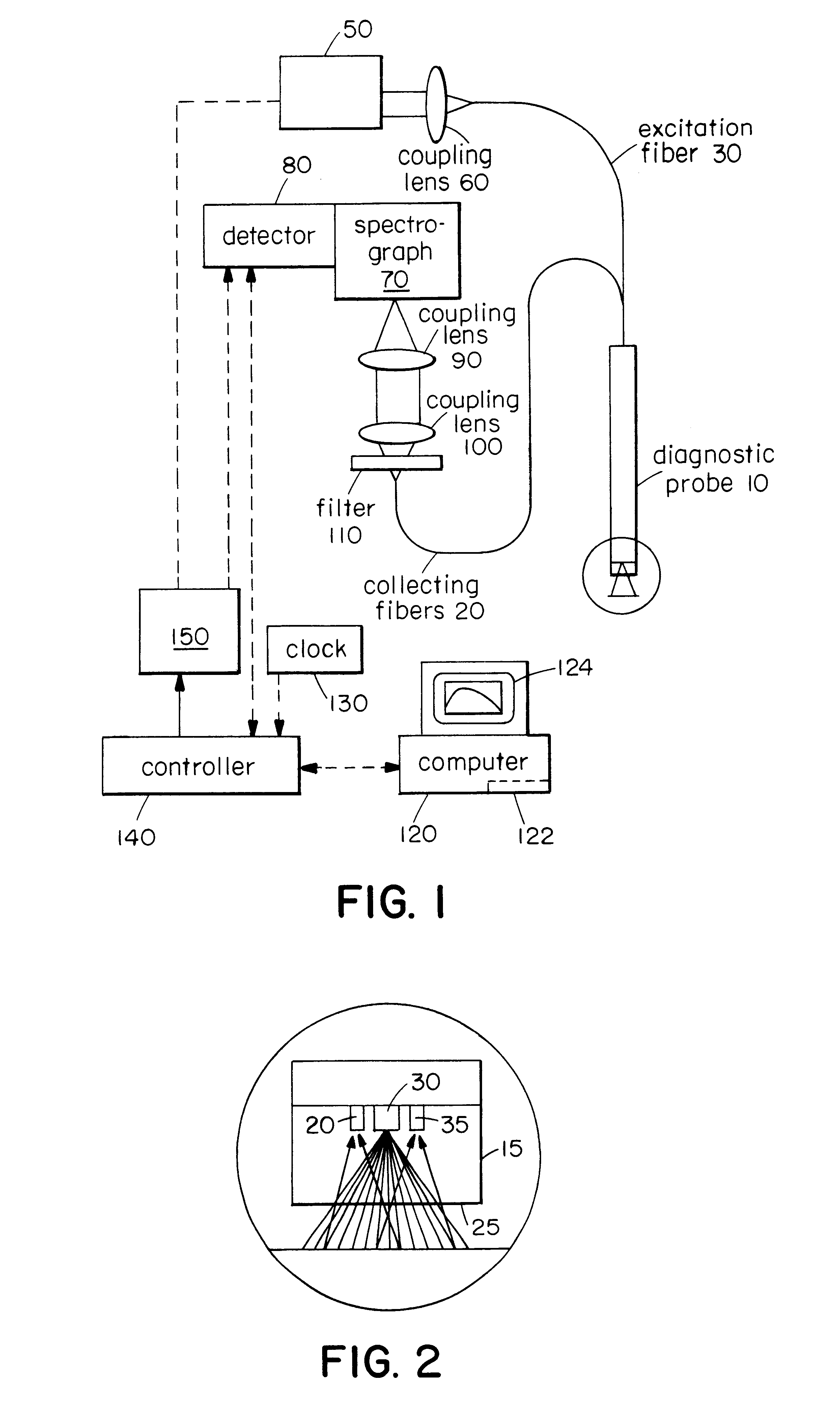
[0031]A preferred embodiment of the invention involves the use of a fiber optic system to deliver and collect light from a region of interest to measure one or more physical characteristics of a surface layer. Such a system is illustrated in FIG. 1. This system can include a light source 50 such as a broadband source, a fiber optic device for delivery and / or collection of light from the tissue, a detector system 80 that detects the scattered light from the tissue, a computer 120 having a memory 122 that analyzes and stores the detected spectra, and a display 124 that displays the results of the measurement. A lens 60 can be used to couple light from the source 50 into the excitation fiber 30 of the probe 10. A filter 110 and lens system 90, 100 can be used to efficiently couple collected light to a spectrograph 70. A controller 140 connected to the data processing system 120 can be connected to a clock and a pulser 150 that controls the light source 50.

[0032]The distal end 15 of the...


PUM

PropertyMeasurementUnit
wavelengthsaaaaaaaaaa
collection angleaaaaaaaaaa

Abstract

The present invention relates to systems and methods for measuring one or more physical characteristics of material such as tissue using optical radiation. The system can use light that is scattered by a tissue layer to determine, for example, the size of nuclei in the tissue layer to aid in the characterization of the tissue. These methods can include the use of fiber optic devices to deliver and collect light from a tissue region of interest to diagnose, for example, whether the tissue is normal or precancerous.

Description

RELATED APPLICATIONS[0001]This is a continuation application of International Application No. PCT / US98 / 21450 filed on Oct. 9, 1998 which is a continuation-in-part of U.S. Ser. No. 08 / 948,734 filed on Oct. 10, 1997, now U.S. Pat. No. 6,091,984, the entire contents of the above applications being incorporated herein by reference.GOVERNMENT SUPPORT[0002]The invention was supported, in whole or in part, by a Grants No. P41RR02594 and CA53717 from the National Institutes For Health. The Government has certain rights in the invention.BACKGROUND OF THE INVENTION[0003]Methods for diagnosis of cancer at an early stage are essential for cancer prevention and therapy. Many types of cancers grow from epithelial tissues, which cover inner and outer surfaces of the human body. Many of these, for example cancer in gastrointestinal tract, progress through the stage of dysplasia. Dysplasia can be defined as neoplastic tissue which is not malignant yet, but is considered to be a precursor of malignan...

Claims


Application Information

Patent Timeline
Patent Type & Authority Patents(United States)
IPC IPC(8): A61B5/00G01N21/17A61B10/00
CPCA61B5/0075A61B5/0084A61B5/0086
Inventor PERELMAN, LEV T.BACKMAN, VADIMFELD, MICHAEL S.ZONIOS, GEORGEITZKAN, IRVINGMANOHARAN, RAMASAMY
Owner MASSACHUSETTS INST OF TECH
Why Eureka
Try Eureka