Implantable medical device for measuring mechanical heart function

a medical device and mechanical heart technology, applied in the field of implantable medical devices, can solve the problems of insufficient cardiac output to sustain mild or moderate levels, inability to perform measurement procedures for even a resting patient, time-consuming measurement procedures, etc., and achieves the effects of not waste of pacing energy, prolonged battery life, and high strength

Inactive Publication Date: 2005-10-25
MEDTRONIC INC
View PDF39 Cites 171 Cited by
  • Summary
  • Abstract
  • Description
  • Claims
  • Application Information

AI Technical Summary

Benefits of technology

[0035]The present invention is preferably embodied in a multi-site, cardiac pacing system specifically implanted for treating heart failure having memory for storing data and wherein a pacing escape interval is timed out from a VS event or a ventricular pace (VP) pulse to trigger delivery of one or more bi-ventricular pacing therapy. VP pulses are selectively delivered to first and second ventricular sites synchronously within a V-V pace delay at a predetermined pacing rate so as to ensure that the synchronous depolarizations of the right and left ventricles is optimized to maximize cardiac output. The pacing system is preferably programmable to operate in one of a plurality of pacing modes.
[0044]The magnetic field responsive distance sensor of the present invention offers a number of advantages. The magnetic field permeates a lead body and any tissue encapsulation about it without any attenuation, so the magnetic field sensor can be reliably encased within the lead body and is not subject to drift caused by any ambient influences over time. The magnet can be made of a high strength material that is similarly encased securely within a lead body or a magnet housing and placed at a wide number of locations spaced across the ventricles or other heart chambers but close enough so that its magnetic field can be detected by the magnetic field sensor. The magnetic field sensor and the magnet are inexpensive, easy to position, and reliable over long periods of time.

Problems solved by technology

All these disease processes lead to insufficient cardiac output to sustain mild or moderate levels of exercise and proper function of other body organs, and progressive worsening eventually results in cardiogenic shock, arrhythmias, electromechanical dissociation, and death.
However, the measurement procedure is time consuming to perform for even a resting patient and cannot be practically performed replicating a range of metabolic conditions.
Typically, the echocardiography procedure is performed infrequently and months or years may lapse between successive tests, resulting in a poor understanding of the progress of the disease or whether or not intervening drug therapies have been efficacious.
Moreover, in many cases, diseased hearts exhibiting LVD and CHF also have conduction defects wherein cardiac depolarizations that naturally occur in one upper or lower heart chamber are not always conducted in a timely fashion either within the heart chamber or to the other upper or lower heart chamber.
In such cases, the right and left heart chambers do not contract in optimum synchrony with each other, and cardiac output suffers due to the conduction defects.
In addition, spontaneous depolarizations of the left atrium or left ventricle occur at ectopic foci in these left heart chambers, and the natural activation sequence is grossly disturbed.
Cardiac output deteriorates because the contractions of the right and left heart chambers are not synchronized sufficiently to eject the maximal blood volume.
Furthermore, significant conduction disturbances between the right and left atria can result in left atrial flutter or fibrillation.
Moreover, the changed conductive patterns or a heart in heart failure are manifested by other changes in the PQRST waveforms, particularly an abnormally wide or long duration of the ventricular depolarization signal, or QRS.
However, fixed or physiologic sensor driven rate responsive pacing in such patients does not always lead to improvement in cardiac output and alleviation of the symptoms attendant to such disease processes because it is difficult to assess the degree of compromise of cardiac output caused by CHF and to determine the pacing parameters that are optimal for maximizing cardiac output, particularly the AV delay.
Moreover, conventional DDD and DDDR pacemakers pace and sense only in the right atrium and right ventricle and cannot alleviate or alter IAB, LBBB, RBBB and QT interval widening.
Consequently, while some improvement has been reported in certain patients receiving two-chamber DDD or DDDR AV sequential pacemakers, the efficacy of the treatment is not established for larger patient populations.
For example, if conduction paths in the left ventricle are impaired, delivering a pacing stimulus to the left ventricle at precisely the same time as to the right ventricle may nonetheless result in left ventricular contraction being slightly delayed with respect to the right ventricular contraction.
However, it is not always easy to determine just how to program the CDW duration to optimize the hemodynamics of the heart.
Moreover, while such AV sequential, three or four-chamber pacing systems can be programmed to at least initially restore right and left and upper and lower heart synchrony in the clinical setting, they are not always able to maintain that synchrony over a range of heart rates and as the patient is exposed to other conditions of daily life including stress and exercise.
While measurement and storage of this group of parameters of cardiac function and contractile state can provide valuable information about the state of heart failure, the sensors are not always easy to implant so that they perform reliably chronically and under the range of conditions encountered by the patient and resulting from progression of the heart failure.
The proposed systems employing locally disposed accelerometers at one or more location in the heart or distributed impedance measuring electrodes to detect and measure heart motion and to derive the above-described parameters are difficult to implement and subject to outside influences that distort the signals.
The sensitive semiconductor or miniature beam elements of accelerometers are subject to damage or change in sensitivity over time.
Implantable pressure sensors that employ a diaphragm that is deflected by contacting blood have proven to be problematic.

Method used


Image

  • Implantable medical device for measuring mechanical heart function
  • Implantable medical device for measuring mechanical heart function
  • Implantable medical device for measuring mechanical heart function

Examples


Embodiment Construction

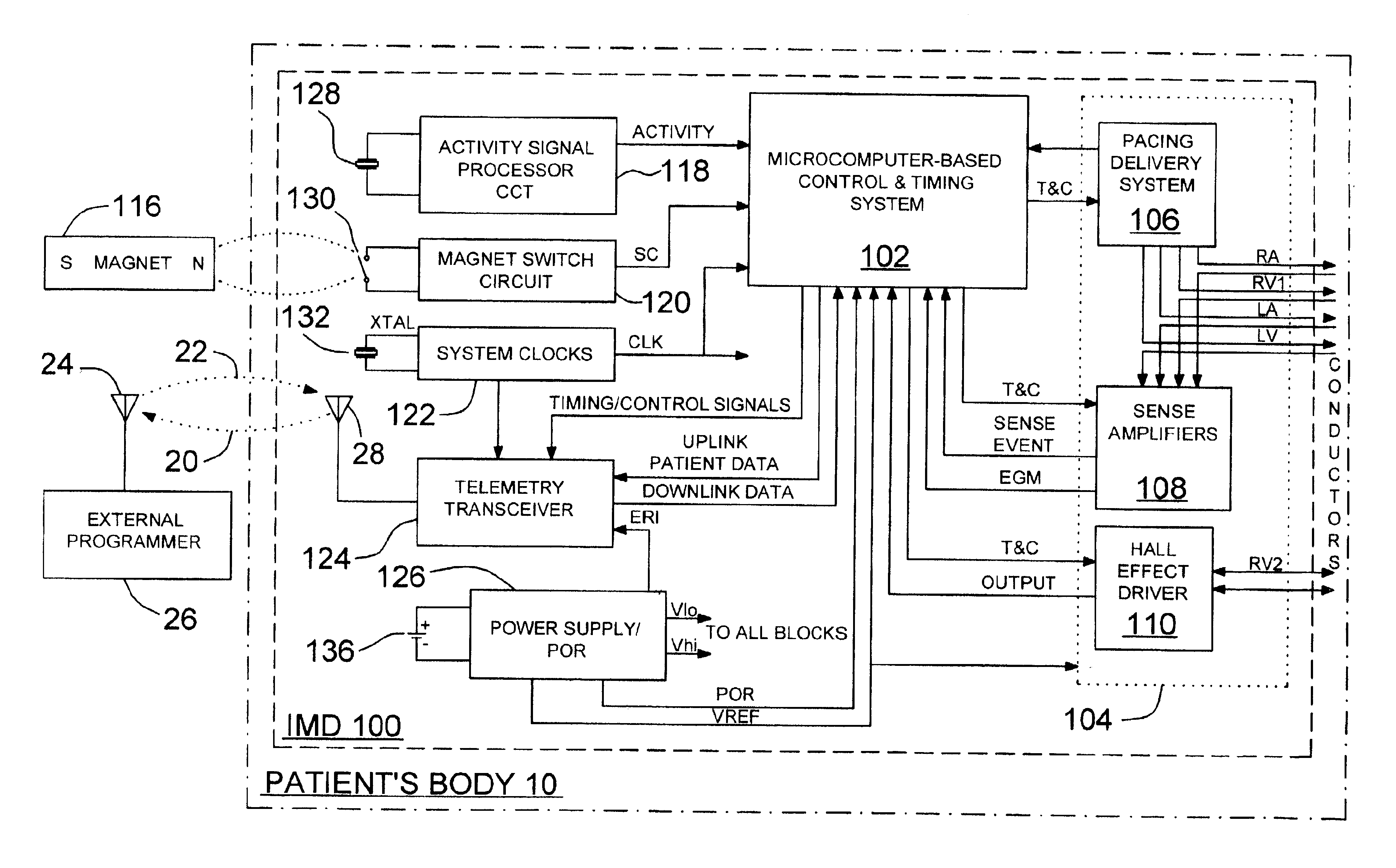
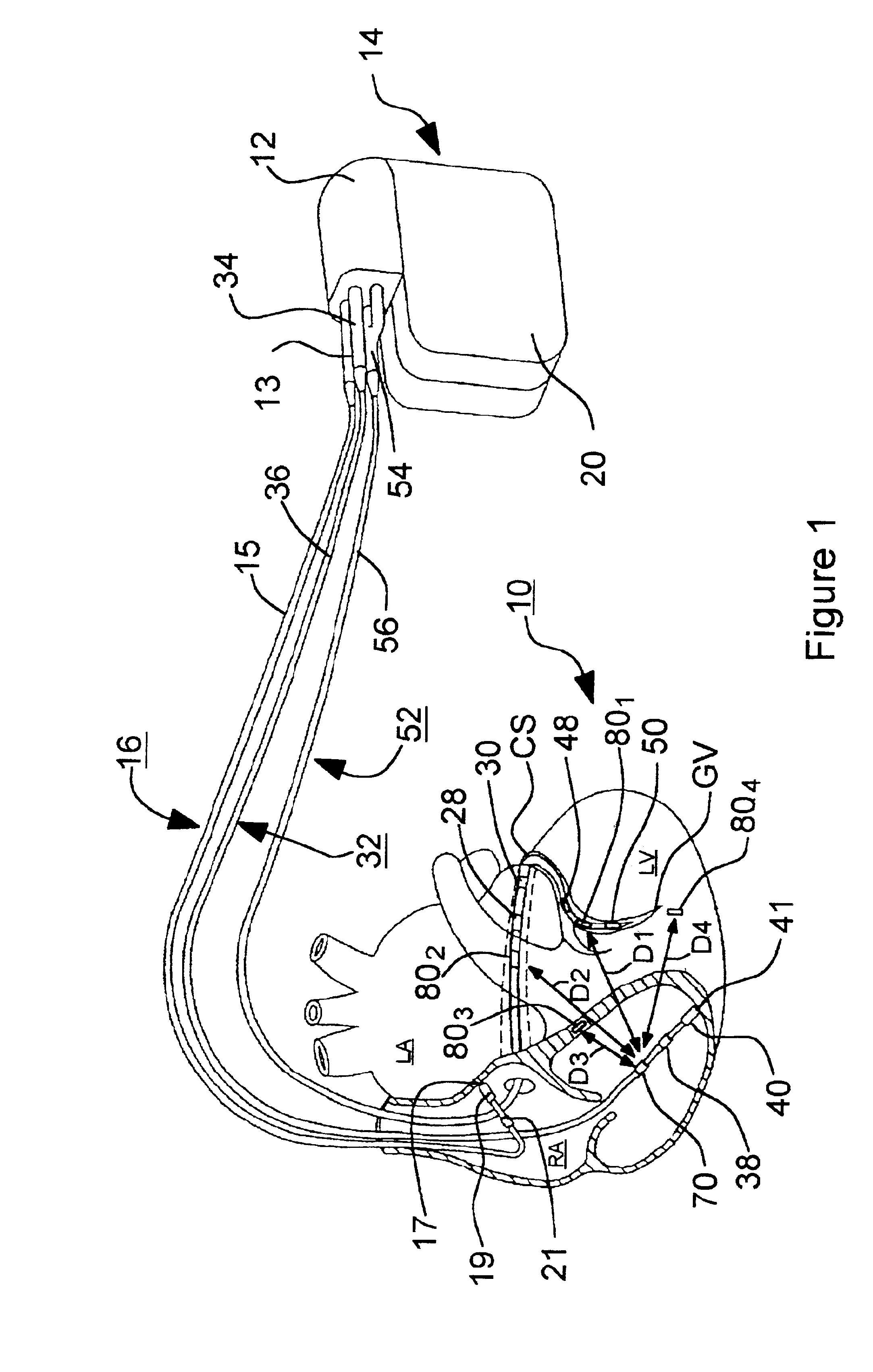
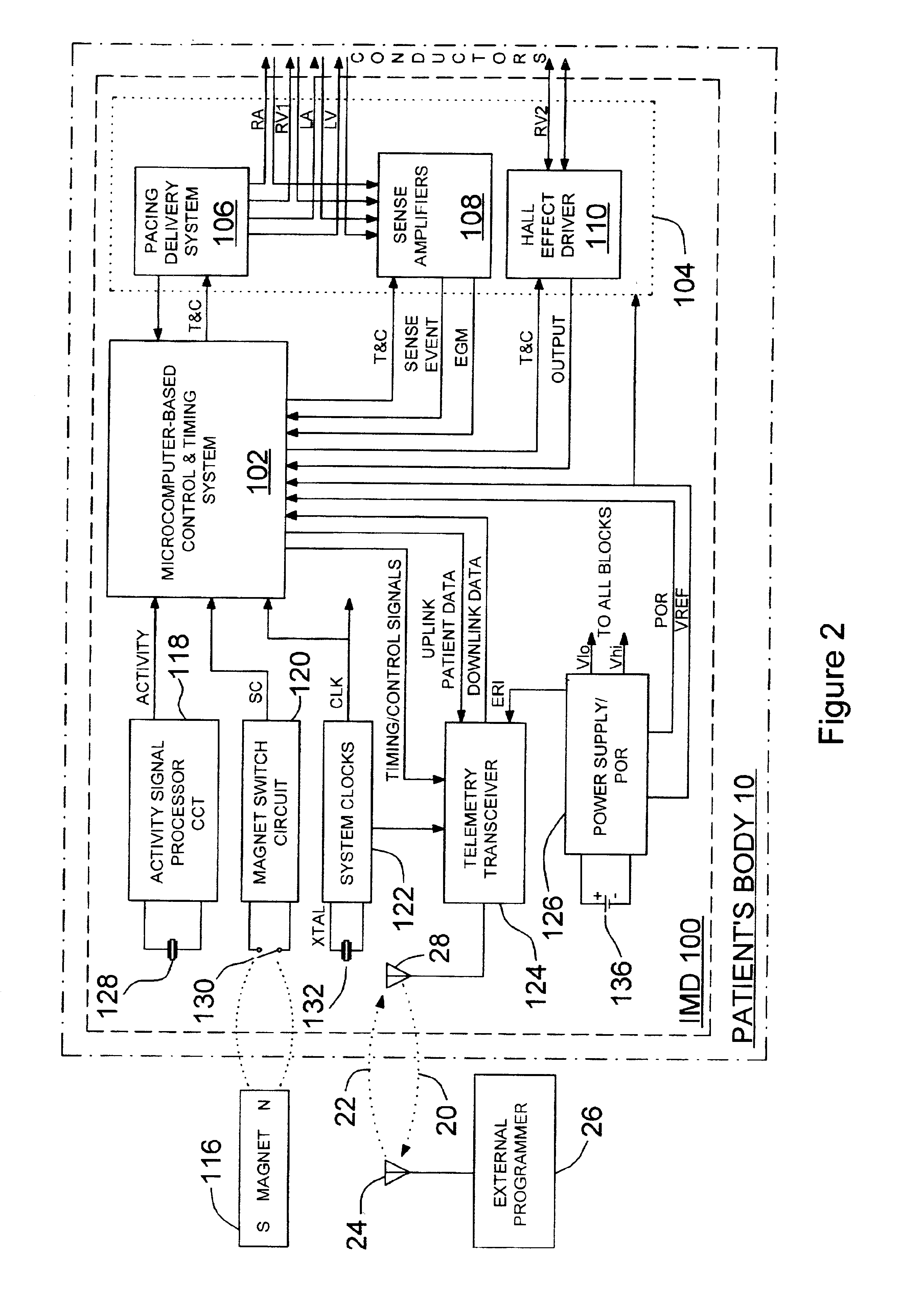
[0055]In the following detailed description, references are made to illustrative embodiments for carrying out the invention. It is understood that other embodiments may be utilized without departing from the scope of the invention. For example, the invention is disclosed in detail herein in the context of an AV sequential, three chamber or four chamber, pacing system operating in demand, atrial tracking, and triggered pacing modes for restoring synchrony in depolarizations and contraction of left and right ventricles in synchronization with atrial sensed and paced events for treating heart failure and / or bradycardia in those chambers. This embodiment of the invention is programmable to operate as a three or four chamber pacing system having an AV synchronous operating mode for restoring upper and lower heart chamber synchronization and right and left atrial and / or ventricular chamber depolarization synchrony. Or, simply bi-ventricular pacing may be applied to the right and left vent...


PUM


Abstract

An implantable device for measuring mechanical heart function of selected heart chambers using a heart contraction detection system that includes a magnetic field sensor. The system may be used for monitoring signs of acute or chronic cardiac heart failure, to enable diagnosis of the condition of the heart, to prescribe appropriate therapies, and to assess delivered pacing therapies. Distance measurements within the heart are made using the magnetic field sensor which is implanted at a sensor site in or on one of the right or left ventricle. A magnet implanted at a site relative to the other of the left or right heart ventricle is sufficiently spaced at a distance that fluctuates with expansion and contraction of the ventricles. The magnetic field sensor provides a sensor output signal having a signal magnitude proportional to the magnetic field strength of the magnet, and which is indicative of changing cardiac dimensions.

Description

FIELD OF THE INVENTION[0001]The present invention relates generally to implantable medical devices (IMDs) for measuring mechanical heart function of selected heart chambers using a magnetic field sensor based, heart contraction detection and measuring system.BACKGROUND OF THE INVENTION[0002]Patients suffering from chronic CHF manifest an elevation of left ventricular end-diastolic pressure, according to the well-known heterometric autoregulation principles espoused by Frank and Starling. This may occur while left ventricular end-diastolic volume remains normal due to a decrease in left ventricular compliance concomitant with increased ventricular wall stiffness. CHF due to chronic hypertension, ischemia, infarct or idiopathic cardiomyopathy is associated with compromised systolic and diastolic function involving decreased atrial and ventricular muscle compliance. These may be conditions associated with chronic disease processes or complications from cardiac surgery with or without s...

Claims


Application Information

Patent Timeline
Patent Type & Authority Patents(United States)
IPC IPC(8): A61N1/365A61N1/362
CPCA61N1/36514A61N1/3627
Inventor PAPE, FORREST C. M.HUELSKAMP, PAUL J.
Owner MEDTRONIC INC
Why Eureka
Try Eureka