Cell fusion assays using fluorescence resonance energy transfer
a cell fusion and energy transfer technology, applied in the field of using fluorescence resonance energy transfer to assay, can solve the problems of insufficient methods for identifying such inhibitors, inability to use fluorescence resonance energy transfer, and inability to simplify the picture, so as to prevent or ameliorate the effects of hiv-1 infection
- Summary
- Abstract
- Description
- Claims
- Application Information
AI Technical Summary
Benefits of technology
Problems solved by technology
Method used
Image
Examples
example 1
A CHO Cell / Jurkat Cell Embodiment of the Present Invention
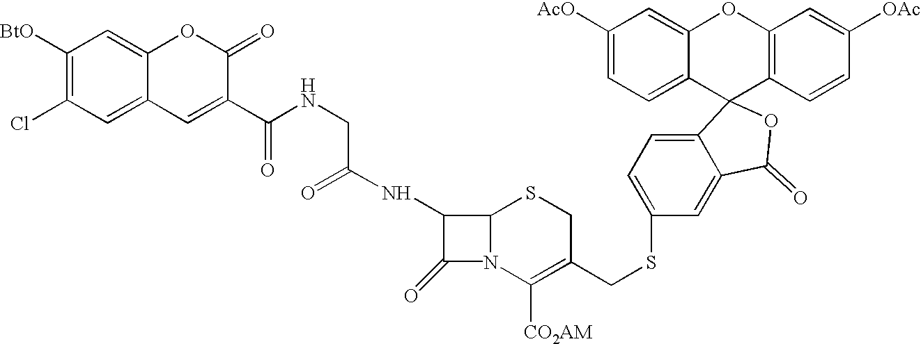
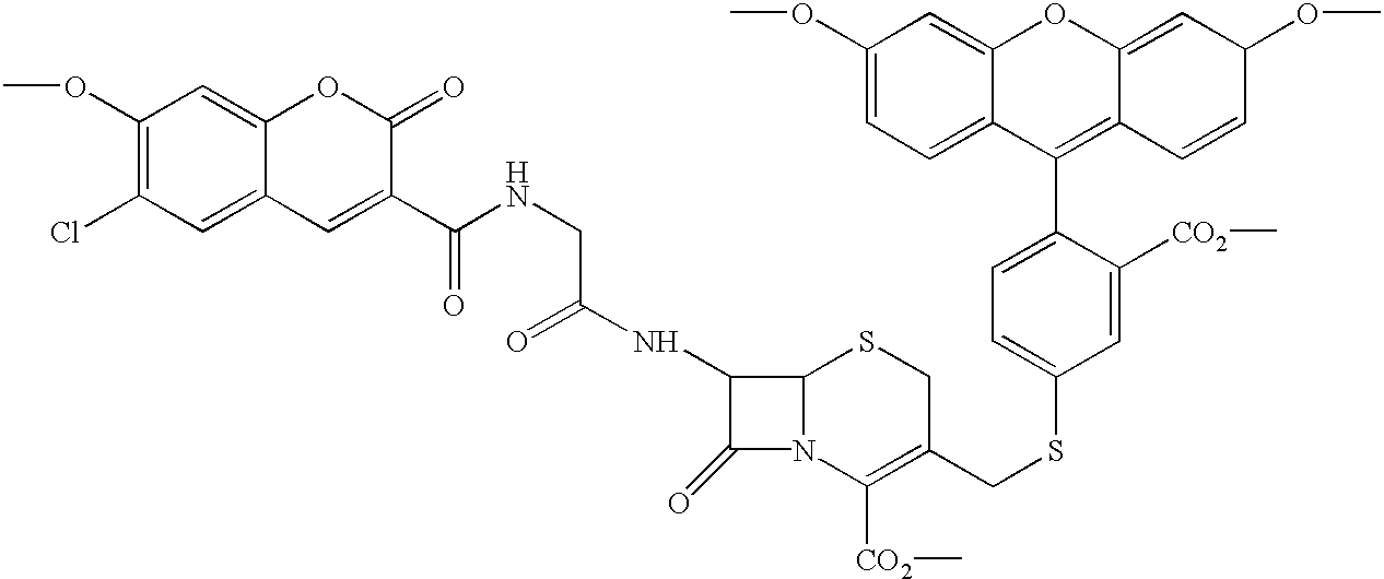
[0183]A particular embodiment of the invention makes use of Chinese hamster ovary (CHO) cells (an adherent cell line) that have been transfected so as to express an HIV Env protein and Jurkat T cells (a suspension cell line) that have been transfected so as to express β-lactamase. In a particular version of this embodiment, a CHO cell line expressing a truncated version of a T-tropic Env (gp143 / HXB2) was plated in 96-well, black, clear bottom microtiter plates and incubated overnight at 37° C. to allow the cells to attach. Jurkat cells expressing β-lactamase (and which naturally express CXCR4 and CD4) were added to the CHO cells and incubated at 37° C. for a period sufficient to allow fusion to occur. The non-fused cells were removed by washing and the fluorogenic 1β-lactamase substrate CCF2 / AM was added to the remaining cells. After a 1 hour incubation at room temperature, cell fusion was measured by fluorescence on a fluore...
example 2
A 293T Cell / Jurkat Cell Embodiment of the Present Invention
[0197]The assay was carried out in 6-well polyD-lysine microtiter plates. 293T cells (0.7×10⁶ cells per well; about 70% confluent) were transiently transfected with 2 μg pVIJneo / gp143HXB2 per well using a Gibco / BRL LIPOFECTAMINE PLUS® kit and 24 hours later were overlayed for 1 hour at 37° C. with 2×10⁶ Jurkat cells per well that constitutively express β-lactamase. Non-attached Jurkat cells were washed 3–4 times with tissue culture media and the 293T cells and remaining Jurkat cells were allowed to fuse for 3–4 hours at 37° C. The cells were then loaded with CCF2 / AM (2 μM per well, final concentration). As a control, this experiment was repeated with 293T cells that were mock transfected and thus did not express gp143. Results were visualized by fluorescence microscopy and documented by photography. This showed that the mock transfected control cells fluoresced green, indicating intact CCF2 and thus lack of fusion. In contra...
example 3
Construction of the Env Expression Vector pVIJneo / gp143HXB2
[0201]The expression vector pV1J is described in International Patent Publication WO 94 / 21797. pV1Jneo was derived from pV1J by removing the ampicillin gene (ampr) from pV1J and replacing it with the neomycin resistance gene. The ampr gene from the pUC backbone of pV1J was removed by digestion with SspI and Eam 1105I restriction enzymes. The remaining plasmid was purified by agarose gel electrophoresis, blunt-ended with T4 DNA polymerase, and then treated with calf intestinal alkaline phosphatase. The commercially available kanr gene, derived from transposon 903 and contained within the pUC4K plasmid (Pharmacia), was excised using the PstI restriction enzyme, purified by agarose gel electrophoresis, and blunt-ended with T4 DNA polymerase. This fragment was ligated with the pV1J backbone and plasmids with the kanr gene in either orientation were derived which were designated as pV1Jneo #'s 1 and 3. Each of these plasmids was ...
PUM
| Property | Measurement | Unit |
|---|---|---|
| Ratio | aaaaa | aaaaa |
Abstract
Description
Claims
Application Information
 - R&D
- Intellectual Property
- Life Sciences
- Materials
- Tech Scout
- Unparalleled Data Quality
- Higher Quality Content
- 60% Fewer Hallucinations
Browse by: Latest US Patents, China's latest patents, Technical Efficacy Thesaurus, Application Domain, Technology Topic, Popular Technical Reports.
© 2025 PatSnap. All rights reserved.Legal|Privacy policy|Modern Slavery Act Transparency Statement|Sitemap|About US| Contact US: help@patsnap.com


