Non-invasive gene targeting to ocular cells

a gene and ocular cell technology, applied in the field of non-invasive gene targeting to ocular cells, can solve the problems of inability to stabilize the electrostatic interaction between dna and polycations, high invasive routes of administration, and inability to invasively apply techniques, etc., to achieve the effect of increasing the plasma bioavailability of dna and increasing the stability of dna located within the immunoliposom

Inactive Publication Date: 2006-08-15
RGT UNIV OF CALIFORNIA
View PDF6 Cites 30 Cited by
  • Summary
  • Abstract
  • Description
  • Claims
  • Application Information

AI Technical Summary

Benefits of technology

[0015]The liposomes of the present invention include a neutral liposome having an exterior surface and an internal compartment in which the therapeutic gene is located. The surface of the liposome is decorated with several thousand strands of polyethyleneglycol (PEG), a process called “pegylation.” The PEG strands make the surface of the liposome “hairy,” and this prevents the rapid absorption of blood proteins to the surface of the liposome, which is what accelerates the rapid removal from blood of unprotected liposomes. In contrast, the pegylated liposomes are protected and are removed from blood at a much slower rate. The PEG strands also act as conjugation agents for attachment of transportable peptide targeting agents to the surface of the pegylated liposome. The transportable peptides are any kind of ligand that triggers the RMT of the complex through the BBB or BRB. In addition, the same or different transportable peptides also trigger RME through the BCM (or the plasma membrane of ocular cells) in vivo.
[0018]It was found in accordance with the present invention that the use of liposomes in which the polyethyleneglycol is conjugated to the liposome surface results in an increase in the plasma bioavailability of the DNA incorporated within the interior of the immunoliposome. It was also found that the stability of the DNA located within the immunoliposome is increased during in vivo use. Further, in addition to achieving expression of an exogenous gene in the brain or eye, it is also possible to achieve, in parallel, gene expression in other organs which contain or express high levels of the receptor targeted by the blood-brain barrier targeting agent. The pattern of organ expression of the therapeutic gene in the body can be controlled by changing the promoter controlling the expression of the exogenous gene. The promoter is placed at the 5′-end of the therapeutic gene within the non-viral expression plasmid, that is encapsulated within the liposomes. In the present case, differential patterns of organ gene expression are obtained depending on whether a widely expressed promoter, such as the SV40 promoter, is used, or a promoter specific to neural cells is used. The neural cell specific promoter is taken from the 5′-flanking sequence of the gene for human glial fibrillary acidic protein (GFAP). When the β-galactosidase expression plasmid is driven by the SV40 promoter, this construct is called the SV40 / β-galactosidase gene. Conversely, when the β-galactosidase gene is under the influence of the GFAP promoter, the plasmid is called the GFAP / β-galactosidase gene.

Problems solved by technology

However, in either case, highly invasive routes of administration are required.
Such invasive techniques are needed because of the failure of either viruses or cationic liposomes to cross the brain capillary wall, which forms the blood-brain barrier (BBB) in vivo.
However, electrostatic interactions between DNA and polycations may not be stable in blood, and highly polycationic proteins such as histone or polylysine exert toxic effects at the BBB and cause generalized BBB permeability changes in vivo (18).
Rather, the rate-limiting problem is how to target the therapeutic gene throughout the entire retina.
There are at least two problems with the existing forms of gene therapy of retinal diseases.
The development of these anti-viral neutralizing antibodies sets the stage for inhibiting the therapeutic effect of the subsequent gene treatments, and may also cause local inflammation within the eye.
The second problem with the existing forms of retinal gene therapy relates to the fact that the virus does not cross the blood-retinal barrier (BRB).
Therefore, the virus cannot access the retina following non-invasive (intravenous, subcutaneous) forms of administration.
This would be an invasive procedure that may not be widely adapted to hundreds of thousands of patients with RP, and would also create an aberrant ‘multiple pin-hole’ field of vision for the patient.

Method used


Image

  • Non-invasive gene targeting to ocular cells
  • Non-invasive gene targeting to ocular cells
  • Non-invasive gene targeting to ocular cells

Examples


Embodiment Construction

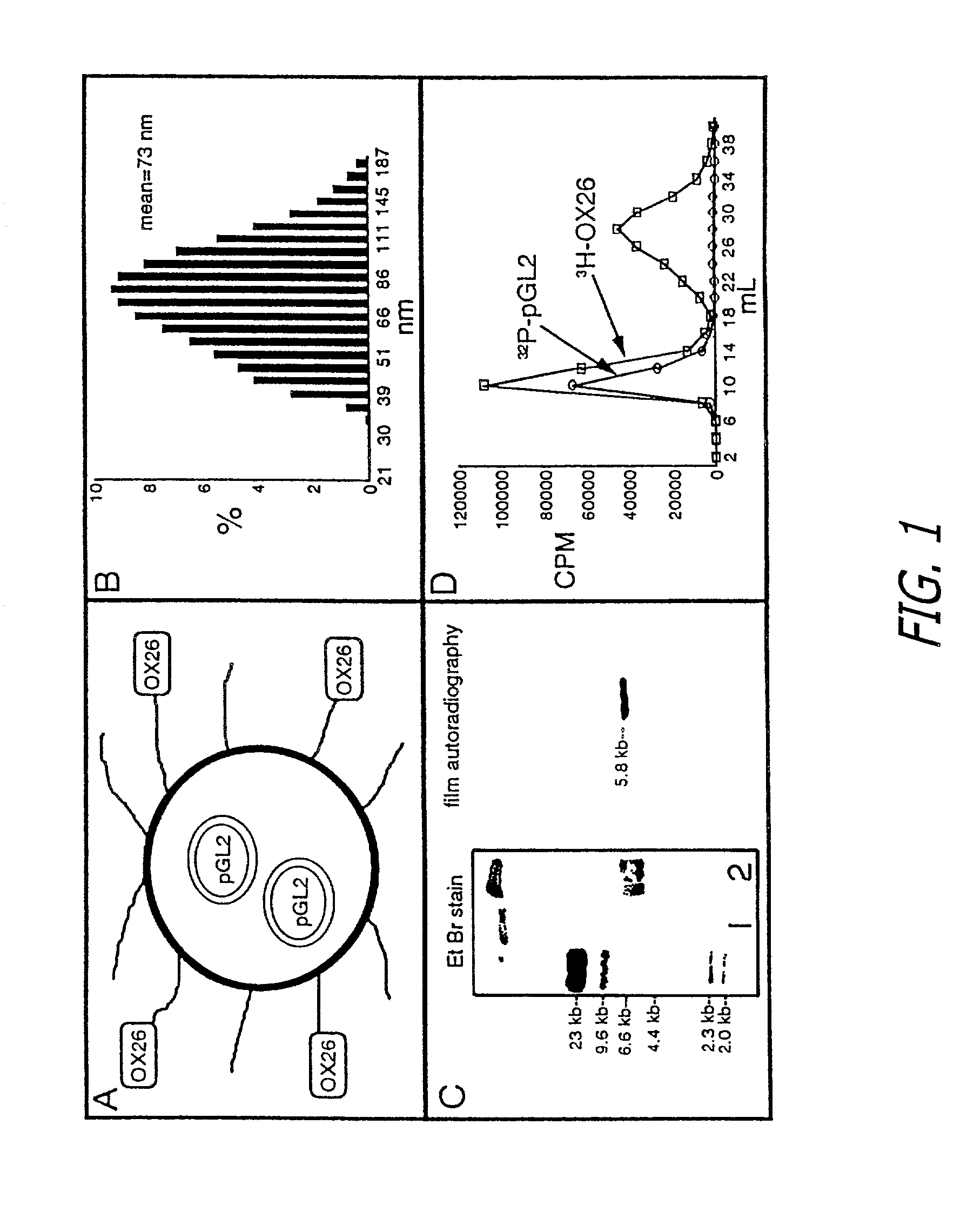
[0041]The immunoliposomes in accordance with the present invention are designed for delivering therapeutic genes across the blood-brain barrier followed by expression in the brain of the therapeutic agents encoded by the gene. The liposomes are a form of nanocontainer and nanocontainers, such as nanoparticles or liposomes, are commonly used for encapsulation of drugs. The liposomes preferably have diameters of less than 200 manometers. Liposomes having diameters of between 50 and 150 nanometer are preferred. Especially preferred are liposomes or other nanocontainers having external diameters of about 80 nanometers. Suitable types of liposomes are made with neutral phospholipids such as 1-palmitoyl-2-oleoyl-sn-glycerol-3-phosphocholine (POPC), diphosphatidy phosphocholine, distearoylphosphatidylethanolamine (DSPE), or cholesterol, along with a small amount (1%) of cationic lipid, such as didodecyldimethylammonium bromide (DDAB) to stabilize the anionic DNA within the liposome.

[0042]T...


PUM

PropertyMeasurementUnit
diameteraaaaaaaaaa
thicknessaaaaaaaaaa
diameteraaaaaaaaaa

Abstract

Liposomes containing therapeutic genes are conjugated to multiple targeting agents to provide transport of the encapsulated gene across the blood-retinal barrier and the plasma membrane of ocular cells. Once across the blood-retinal barrier and ocular cell membrane, the encapsulated gene expresses the encoded therapeutic agent within the ocular cells to provide diagnosis and / or treatment of disease.

Description

[0001]This is a continuation-in-part of application Ser. No. 09 / 557,349 which was filed on Apr. 25, 2000 and issued as U.S. Pat. No. 6,372,250 on Apr. 16, 2001.BACKGROUND OF THE INVENTION[0002]1. Field of the Invention[0003]The present invention relates generally to the delivery of gene medicines to the brain and the eye. More particularly the present invention involves the combination of liposome technology, blood-brain barrier (BBB) / blood-retinal barrier (BRB) receptor technology, pegylation technology, and therapeutic gene technology to provide formulations which are useful in the non-invasive delivery of genes to the brain and the eye.[0004]2. Description of Related Art[0005]The publications and other reference materials referred to herein to describe the background of the invention and to provide additional detail regarding its practice are hereby incorporated by reference. For convenience, the reference materials are numerically referenced and grouped in the appended bibliogra...

Claims


Application Information

Patent Timeline
Patent Type & Authority Patents(United States)
IPC IPC(8): A61K9/127A61K9/51A61K31/7088A61K38/00A61K38/16A61K38/28A61K39/395A61K47/48A61K48/00A61P5/48A61P25/16A61P35/00C12N15/88
CPCA61K9/0019A61K9/1271A61K47/48823A61K48/00A61K48/0041C12N15/88A61K9/1272Y10S977/907C12N2810/50C12N2810/80C12N2810/851C12N2810/854C12N2810/858C12N2810/859A61K47/6913A61P25/16A61P35/00A61P5/48
Inventor PARDRIDGE, WILLIAM M.
Owner RGT UNIV OF CALIFORNIA
Why Eureka
Try Eureka