Contrast enhancing solution for use in confocal microscopy

a confocal microscopy and contrast enhancement technology, applied in the field of optical detection of tumors, can solve the problems of limiting the depth to which the confocal microscope can optically penetrate to permit in-vivo observation in real-time, and sacrificing some non-involved or otherwise healthy tissue. , to achieve the effect of enhancing the contrast of a tumor and/or lesion, enhancing the optical characteristics

Inactive Publication Date: 2006-10-31
THE GENERAL HOSPITAL CORP
View PDF7 Cites 30 Cited by
  • Summary
  • Abstract
  • Description
  • Claims
  • Application Information

AI Technical Summary

Benefits of technology

[0008]The present invention provides a method of enhancing optical characteristics of at least one cell anomaly associated with a tumor. With this arrangement, a surgeon or medical technician can efficiently identify and demarcate the at least one cell anomaly associated with the tumor for subsequent removal. While the invention is primarily shown and described in conjunction with enhancing the contrast of a tumor and / or lesion, it is understood that the invention is applicable to other cell anomalies in general in which contrast enhancement is desirable.
[0009]In one aspect of the invention, a method of enhancing optical characteristics of at least one cell anomaly associated with a tumor includes applying a predetermined contrasting solution to an in-vivo defect area associated with the tumor for optically enhancing the at least one cell anomaly associated with the tumor. The method further includes imaging at least a portion of the in-vivo defect area associated with the tumor using a first optical imaging system, such as a confocal microscope, to provide an in-vivo enhanced tumor image. The in-vivo enhanced tumor image includes the at least one cell anomaly having enhanced attributes, which aids a surgeon or medical tecnician in identifying and demarcating the at least one cell anomaly associated with the tumor. In the exemplary embodiment, applying the predetermined contrasting solution to the in-vivo defect area includes applying an AlCl solution having a predetermined concentration of Al and a predetermined concentration of Cl.

Problems solved by technology

The depth to which the confocal microscope can optically penetrate to permit in-vivo observation in real-time is limited by the light penetration into the tissue and the reflective properties of the structures being observed.
Therefore, Mohs surgery can be a tedious, time-consuming and costly surgical procedure requiring correlating the positive histology with actual skin margins.
Although MMS is a tissue-sparing surgical technique, it can still result in the sacrifice of some noninvolved or otherwise healthy tissue.

Method used


Image

  • Contrast enhancing solution for use in confocal microscopy
  • Contrast enhancing solution for use in confocal microscopy
  • Contrast enhancing solution for use in confocal microscopy

Examples


Embodiment Construction

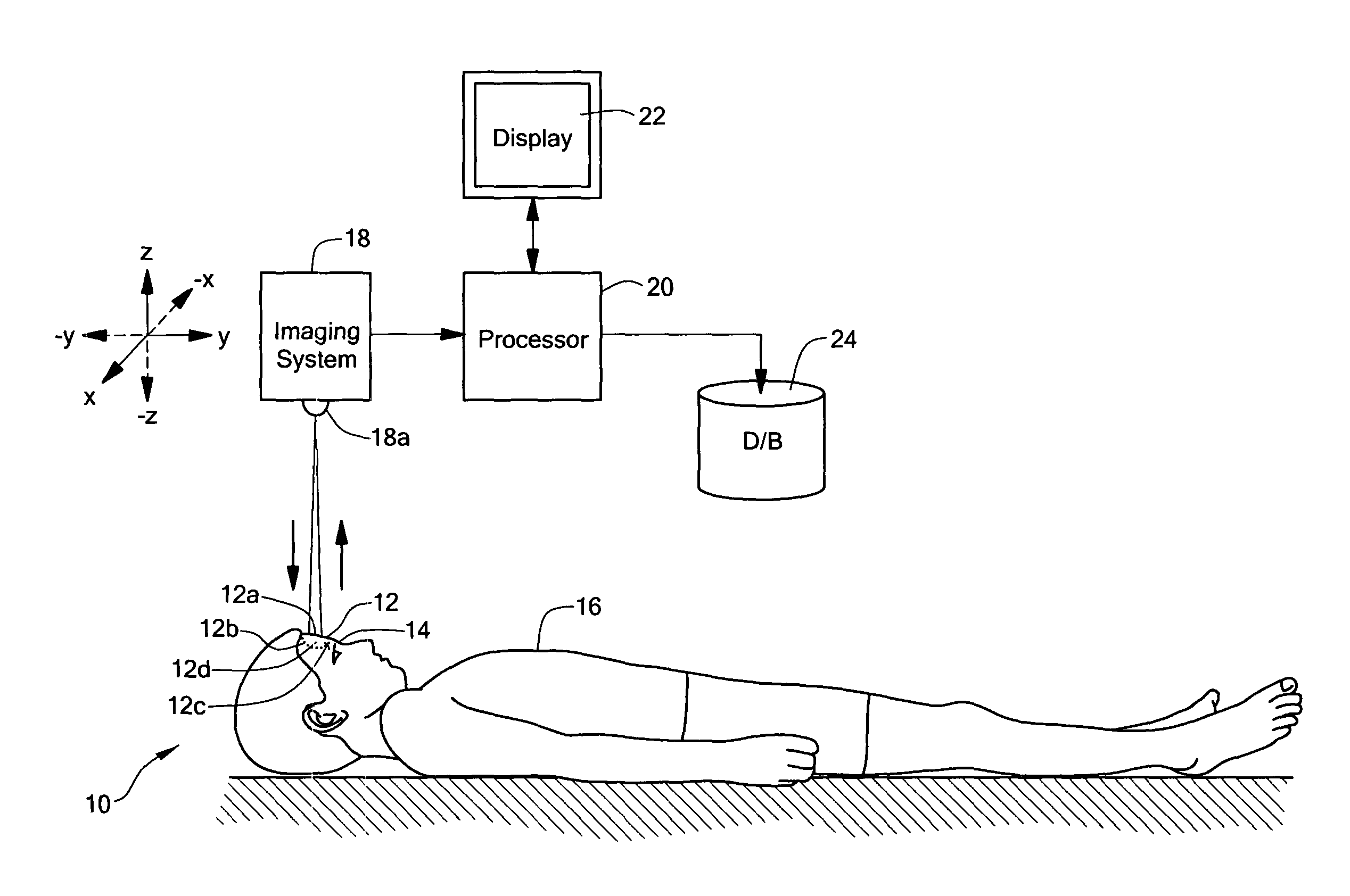
[0018]The present invention provides a system and method for enhancing the optical detection of tumors during surgery. The system and method assists a surgeon in optically locating the margins or boundaries of tumors and / or lesions, so that the surgeon can surgically remove the tumors and / or lesions while at the same time minimizing the unnecessary removal of healthy tissue.

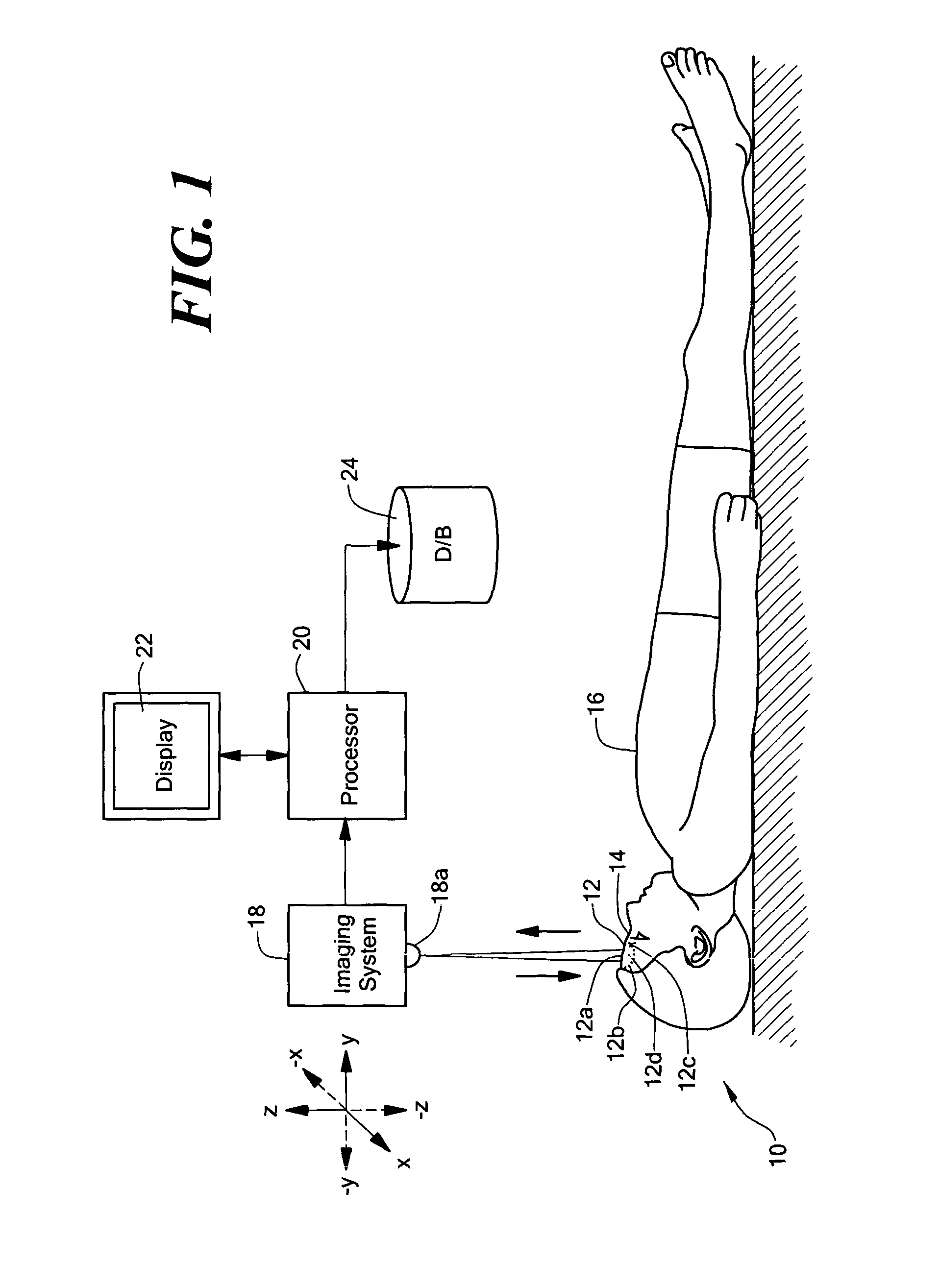
[0019]Referring now to FIG. 1, shown is an exemplary embodiment of a system 10 for enhancing the optical detection of a tumor and / or lesion 12 located on a patient 16 during surgery. The system 10 includes an imaging system 18 coupled to a processor 20. The processor 20 is coupled to a display 22 and to a database 24. In an embodiment, the tumor and / or lesion 12 (hereinafter referred to as “tumor 12”) is located on a forehead region 14 of the patient 16.

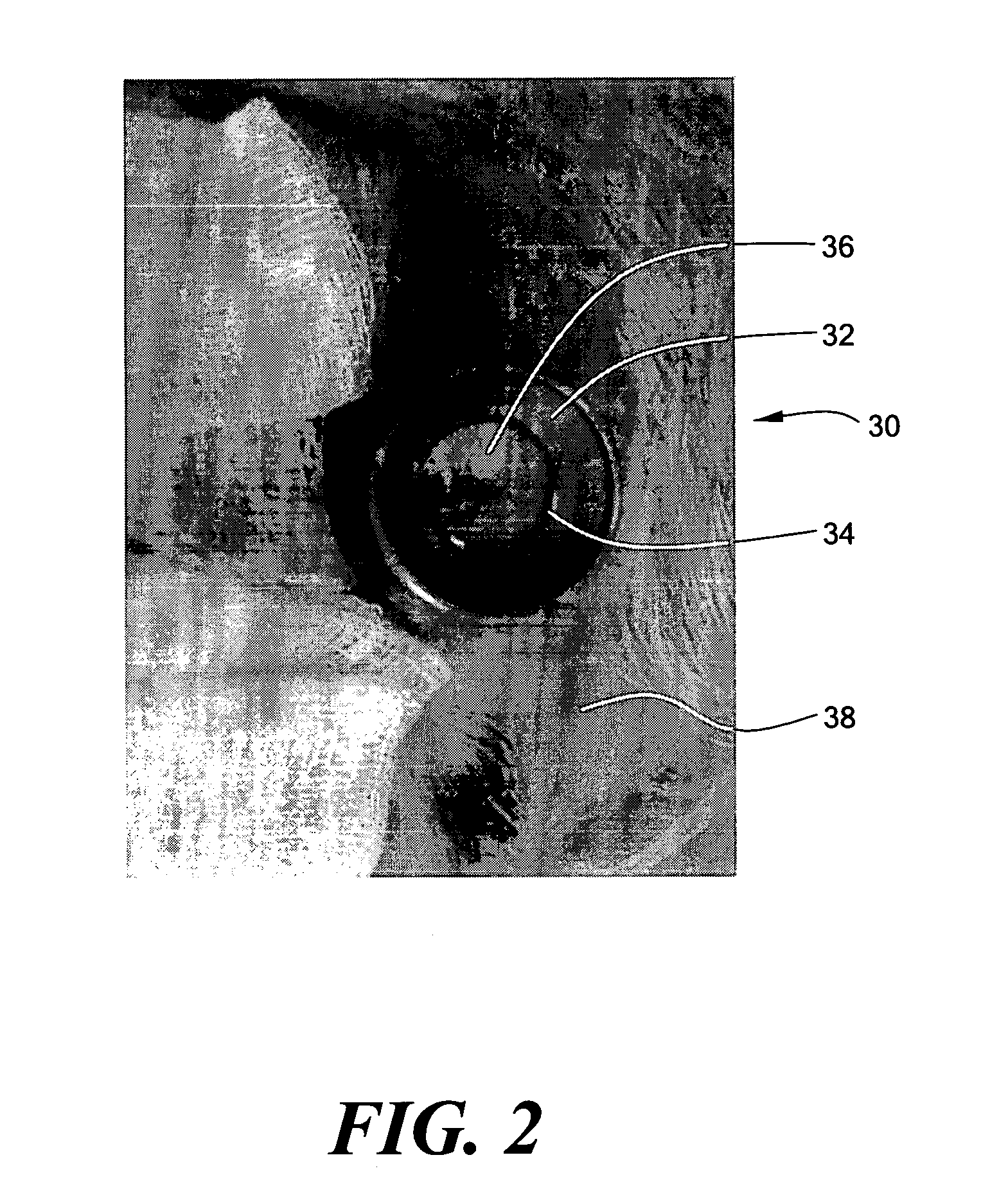
[0020]As part of preliminary preparation of the patient 16 for surgery, a surgeon or medical technician can mark a first test point 12a on a central region of the...


PUM


Abstract

A method of optically detecting a tumor during surgery. The method includes imaging at least one test point defined on the tumor using a first optical imaging system to provide a first tumor image. The method further includes excising a first predetermined layer of the tumor for forming an in-vivo defect area. A predetermined contrast enhancing solution is disposed on the in-vivo defect area, which is adapted to interact with at least one cell anomaly, such as basal cell carcinoma, located on the in-vivo defect area for optically enhancing the cell anomaly. Thereafter the defect area can be optically imaged to provide a clear and bright representation of the cell anomaly to aid a surgeon while surgically removing the cell anomaly.

Description

CROSS REFERENCE TO RELATED APPLICATIONS[0001]This application claims benefit of U.S. Provisional Patent Application No. 60 / 392,515, filed Jun. 27, 2002, entitled, Aluminum Chloride As Contrast Enhancer for In-Vivo Optical Detection of Tumors During Surgery, which application is hereby incorporated by reference in its entirety.STATEMENTS REGARDING FEDERALLY SPONSORED RESEARCH[0002]This invention was made with Government support under Grant No. DE-FG02-91ER61228 awarded by the U.S. Department of Energy. The Government may have certain rights to this invention.FIELD OF THE INVENTION[0003]The present invention relates generally to the optical detection of tumors during surgery and, more particularly, to enhancing the optical detection of tumors.BACKGROUND OF THE INVENTION[0004]As is known in the art, confocal microscopy is an imaging technique that can optically image biological objects using confocal microscopes. Confocal microscopes vary in configuration and location of components, bu...

Claims


Application Information

Patent Timeline
Patent Type & Authority Patents(United States)
IPC IPC(8): A61K49/00
CPCA61K49/0013
Inventor TANNOUS, ZEINATORRES, ABELGONZÁLEZ, SALVADOR
Owner THE GENERAL HOSPITAL CORP
Why Eureka
Try Eureka