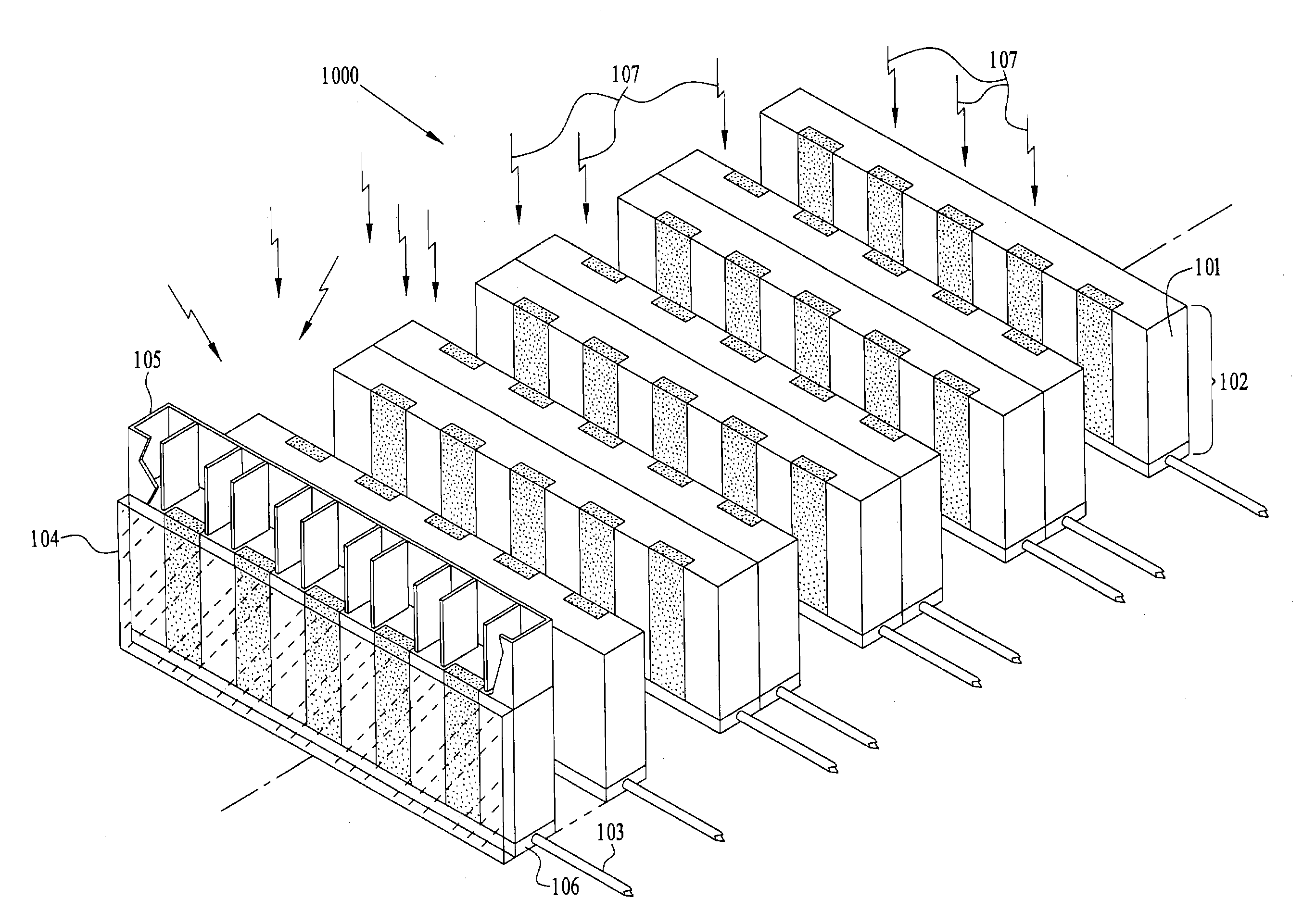
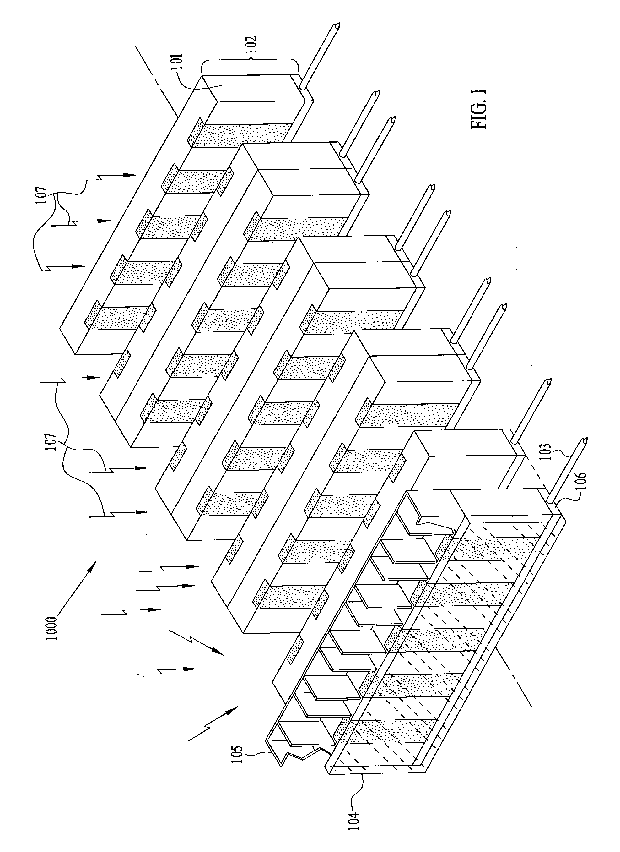
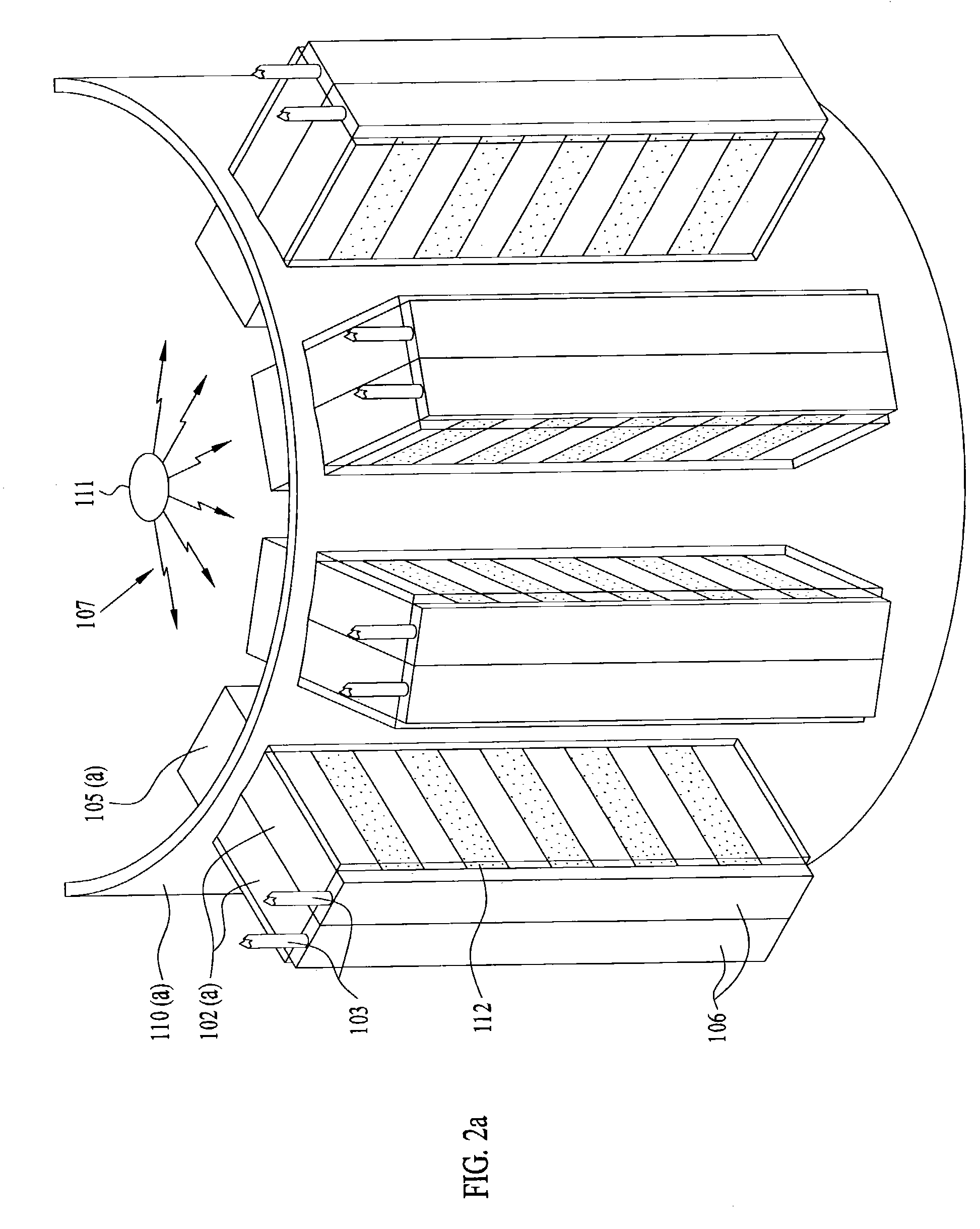
[0005]In accordance with the present invention, a radiation detection apparatus is provided for radiographic imaging and material
composition analysis in which the apparatus can dynamically configure its array geometry and radiation detector parameters for a specific imaging task or it can use an existing radiation detection geometry and settings. This invention is particularly suited for x-ray and
gamma ray imaging in nuclear
medicine, including scintimammography, and x-ray
radiography, specifically, x-ray mammography. There are several advantages inherent to this invention. Superior detectors in cost-effective formats can be utilized and detectors with different properties, including materials, resolution,
response time and
noise characteristics, can be used within an array. One or more radiation detectors are incorporated into a detector module and one or more modules make up a detector module array. The detector modules transmit detected
photon image data and relevant module parameters to a computer system which utilizes this information to electronically-control the modules and in some cases attached collimators. This system is implemented using detector sub-arrays, comprised of one or more detector modules, and detector arrays in order to enhance
image quality or analysis capability. Conventional attenuating or rigid geometry collimators, including ones characterized by coded apertures, and unconventional, including x-ray optic, configurable (adaptive), and Compton scatter module, collimators can be employed to improve the energy and / or spatial resolution for the
photon radiation detection system. In a similar manner additional types of radiation optic collimators such as
neutron optic collimators or
electron optic collimators capable of focusing electric or magnetic fields, can be used with neutrons or charged particles, respectively.
[0007]A typical
nuclear medicine imaging session begins with an operator selecting from a computer display menu a
specific detection system with pre-defined array geometry,
collimator, and module settings appropriate for the desired imaging task. The
detector array configuration can already exist or it can be set up by the computer system. Once a baseline detection system is established, an operator can then adjust and fine tune the
detector array position and settings or leave the
detector array adjustments and tuning under
computer control. While under
computer control electronic instructions can be issued dynamically in response to detector module parameter values and detected radiation data that is transferred to the computer system for
processing, display, and storage during
image acquisition or
adaptive imaging. Electronic commands can be used to control the array geometry and motion, detector module parameters, and some types of collimators. Thus an
information feedback loop can be implemented as a means of tuning detection
system parameters. For some imaging or analysis applications it will be sufficient to configure the detector array based on either a standard geometry, such as line, plane, open box, wedge, ring, cylinder,
ellipse,
ellipsoid, or sphere, or a contoured geometry, in order to compensate for the
radionuclide distribution within the subject and / or the shape of the subject at the
region of interest. For example, configurations may be based on the breast size of a woman or on the
head size,
waist size, or chest size of an adult, child, or infant. A versatile design allows at least a subset of these detector array geometries to be generated “
on the fly”. A less-versatile design still utilizes modules, but the modules are fixed within a specific detector array geometry or they are constrained to move to specific positions, for example, along a track, within a specific detector array geometry. Less-versatile designs reduce the mechanical complexity of the detection system and may be sufficient for specific imaging tasks. An optional capability is to allow the entire array to undergo discrete or uniform motion. The simplest example of this capability would be to scan a radiation source with a detector array comprised of a single detector module.
[0011]The system of the present invention may utilize devices detailed in prior inventions for slit-scan or slot-scan radiographic x-ray imaging in which photons are detected directly using edge-on array detectors; small, 2-D
semiconductor array detectors; or
semiconductor array detectors coupled to scintillators. This
new device can also use thick, linear
semiconductor array detectors and thick, small, 2-D semiconductor array detectors in addition to other types of detectors. Manufacturing costs for these detectors are much less than those associated with large-area or moderate-area, thick, planar, 2-D semiconductor array detectors made from materials such as, but not limited to, CdZnTe, CdTe, GaAs, Ge, Si, SiC, or HgI2. The detector format is also compatible with detectors such as thin, linear semiconductor arrays or thin, small, 2-D semiconductor arrays coupled to scintillators. For example, thin, linear semiconductor arrays of avalanche photodiodes coupled to scintillators can be used as radiation detectors. This approach can be extended to include scintillators coupled to integrated photoemissive cathodes or small PMTs; small, gas microcapillary detector assemblies; or small superconducting array detectors. Consider a
scenario in which radiation is incident upon a planar edge-on detector. The detector thickness (height) now defines the maximum detector entrance aperture while the length or width of the detector area now defines the maximum attenuation distance for edge-on radiation detector designs including semiconductor drift chamber, single-sided strip, and double-sided strip detectors, including micro-strip detector versions. Strip widths can be tapered or curved, in the case of drift chamber detectors, if focusing is desired. In the case of double-sided parallel strip detectors in which opposing strips are parallel, both electrons and holes can be collected to provide 2-D position information across the aperture. If strips on one side run perpendicular to those on the other side, then depth-of-interaction information can be obtained. If strips are segmented in either a single-sided or double-sided parallel strip detector then depth-of-interaction information can be obtained and readout rates can be improved.

