Multivalent constructs for therapeutic and diagnostic applications

a multi-functional construct and construct technology, applied in the field of therapeutic and diagnostic applications, can solve the problems of unacceptable levels of antigenicity, natural antibodies are difficult to produce in recombinant form, natural antibodies also present pharmacokinetic problems, etc., to improve the ability to inhibit the function of receptor tyrosine kinase, improve the ability to bind the target molecule, and improve the effect of anti-cancer

Inactive Publication Date: 2007-05-01
BRACCO SUISSE SA +1
View PDF170 Cites 57 Cited by
  • Summary
  • Abstract
  • Description
  • Claims
  • Application Information

AI Technical Summary

Benefits of technology

[0059]Therefore, the present invention is drawn to constructs comprising two or more binding polypeptides. The multimeric constructs of the present invention comprise two or more binding polypeptides, such that at least two of the binding polypeptides in the construct are specific for different epitopes of a target, for example, KDR and/or KDR/VEGF and cMet and/or cMet/HGF. These constructs are also referred to herein as “heteromeric constructs,”“hetermultimers” and/or “heteromultimeric constructs.” The constructs of the present invention can also include unrelated, or control peptide. The constructs can include two or more, three or more, or four or more binding poly

Problems solved by technology

However, use of such antibodies present certain disadvantages, such as unacceptable levels of antigenicity—even for humanized antibodies.
In addition, natural antibodies are difficult to produce in recombinant form, due to the number of chains, disulfide bonds, and glycosylation.
Natural antibodies also present pharmacokinetic problems.
Antibodies pose significant problems in imaging and radiotherapeutic applications because, due to their large size, accumulation in extravascular target tissue and clearance from the vascular system are both slow.
This problem is especially critical when dealing with solid tumors, which present additional barriers to the ingress of large blood born compounds.
Similar problems occur with ant

Method used


Image

  • Multivalent constructs for therapeutic and diagnostic applications
  • Multivalent constructs for therapeutic and diagnostic applications
  • Multivalent constructs for therapeutic and diagnostic applications

Examples


example 1

Peptide Synthesis and Fluorescein Labelling

[0295]Selected KDR or VEGF / KDR binding peptides corresponding to positive phage isolates were synthesized on solid phase using 9-fluorenylmethoxycarbonyl protocols and purified by reverse phase chromatography. Peptide masses were confirmed by electrospray mass spectrometry, and peptides were quantified by absorbance at 280 nm. For synthesis, two N-terminal and two C-terminal amino acids from the phage vector sequence from which the peptide was excised were retained and a -Gly-Gly-Gly-Lys-NH2 linker was added to the C-terminus of each peptide. Peptides with selected lysine residues were protected with 1-(4,4-dimethyl-2,6-dioxocyclohex-1-ylidene)-3-methybutyl (ivDde), which allows selective coupling to the C-terminal lysine, is not removed during peptide cleavage, and can be removed after coupling with 2% hydrazine in DMF or 0.5 M hydroxylamine, pH 8, in water.

[0296]Each peptide was labeled with fluorescein on the C-terminal lysine using Fluo...

example 2

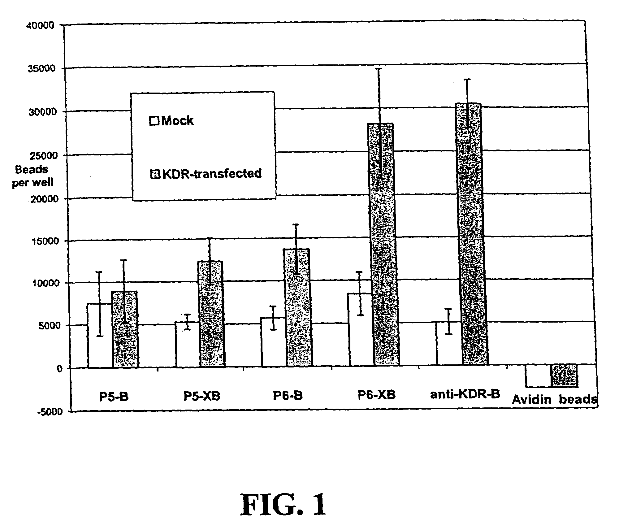
Bead-binding Assay to Confirm Ability of Peptides Identified by Phage Display to Bind KDR-expressing Cells

[0333]The following experiments were performed to assess the ability of KDR-binding peptides to bind to KDR-expressing cells. In this experiment, KDR-binding peptides P5-B and P5-XB and P6-B and P6-XB were conjugated to fluorescent beads and their ability to bind to KDR-expressing 293H cells was assessed. The experiments show that both peptide sequences can be used to bind particles such as beads to KDR-expressing sites. In general, the P6 peptides exhibited better binding to the KDR expressing cells than P5. However, the binding of both peptides improved with the addition of a spacer.

Biotinylation of an Anti-KDR Antibody

[0334]Anti-KDR from Sigma (V-9134), as ascites fluid, was biotinylated using a kit from Molecular Probes (F-6347) according to the manufacturer's instructions.

Preparation of Peptide-conjugated Fluorescent Beads

[0335]0.1 mL of a 0.2 mM stock solution of each biot...

example 3

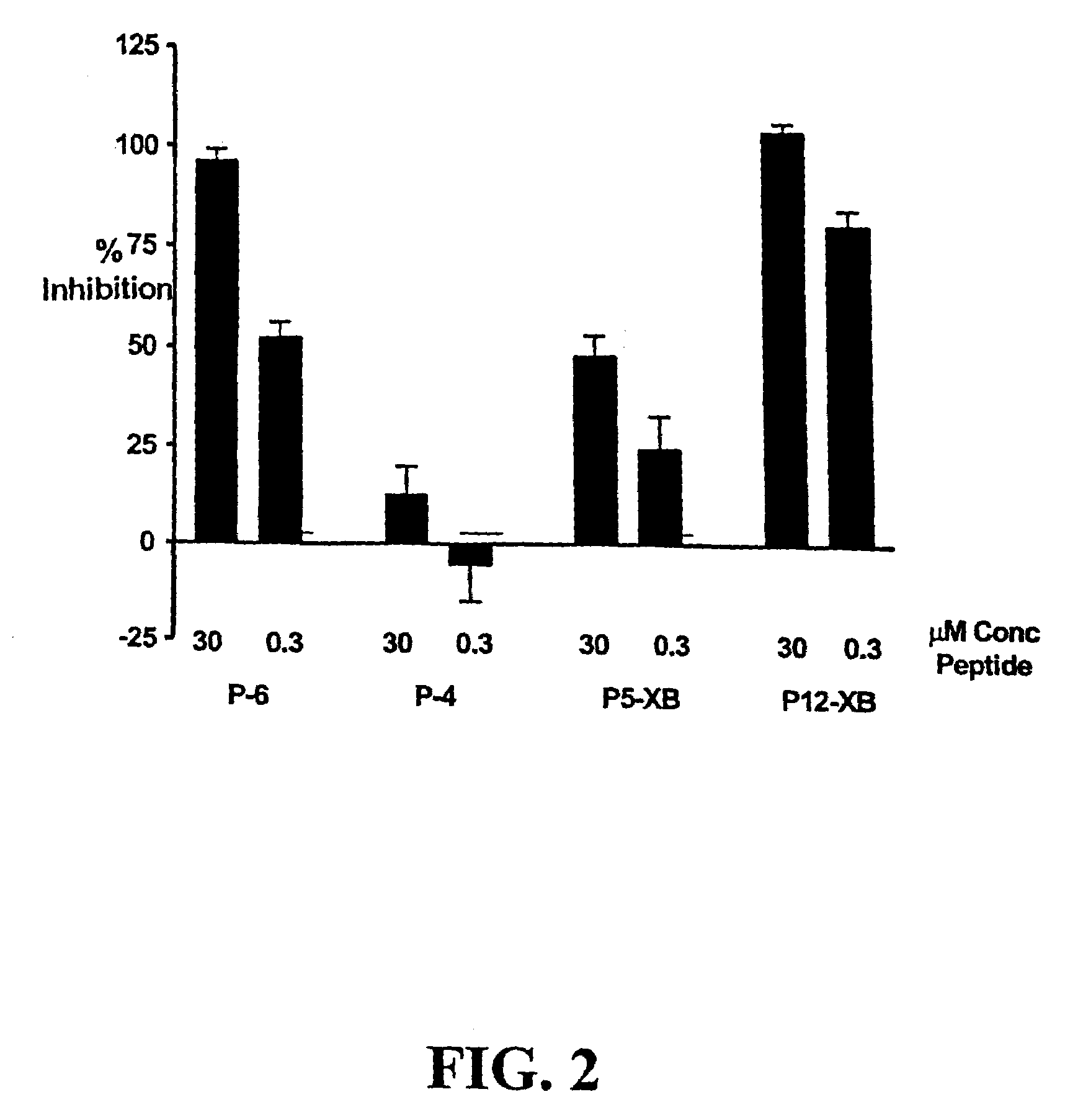
Competition of KDR Binding Peptides and 125I-labeled VEGF for Binding to KDR-transfected 293H Cells

[0340]The following experiment assesses the ability of KDR-binding peptides to compete with 125I-labeled VEGF for binding to KDR expressed by transfected 293H cells. While KDR-binding polypeptide P4 did not compete significantly with 125I-labeled VEGF, P5-XB, P6 and P12-XB competed very well with 125I-labeled VEGF, inhibiting 96.29±2.97% and 104.48±2.07% of 125I-labeled VEGF binding.

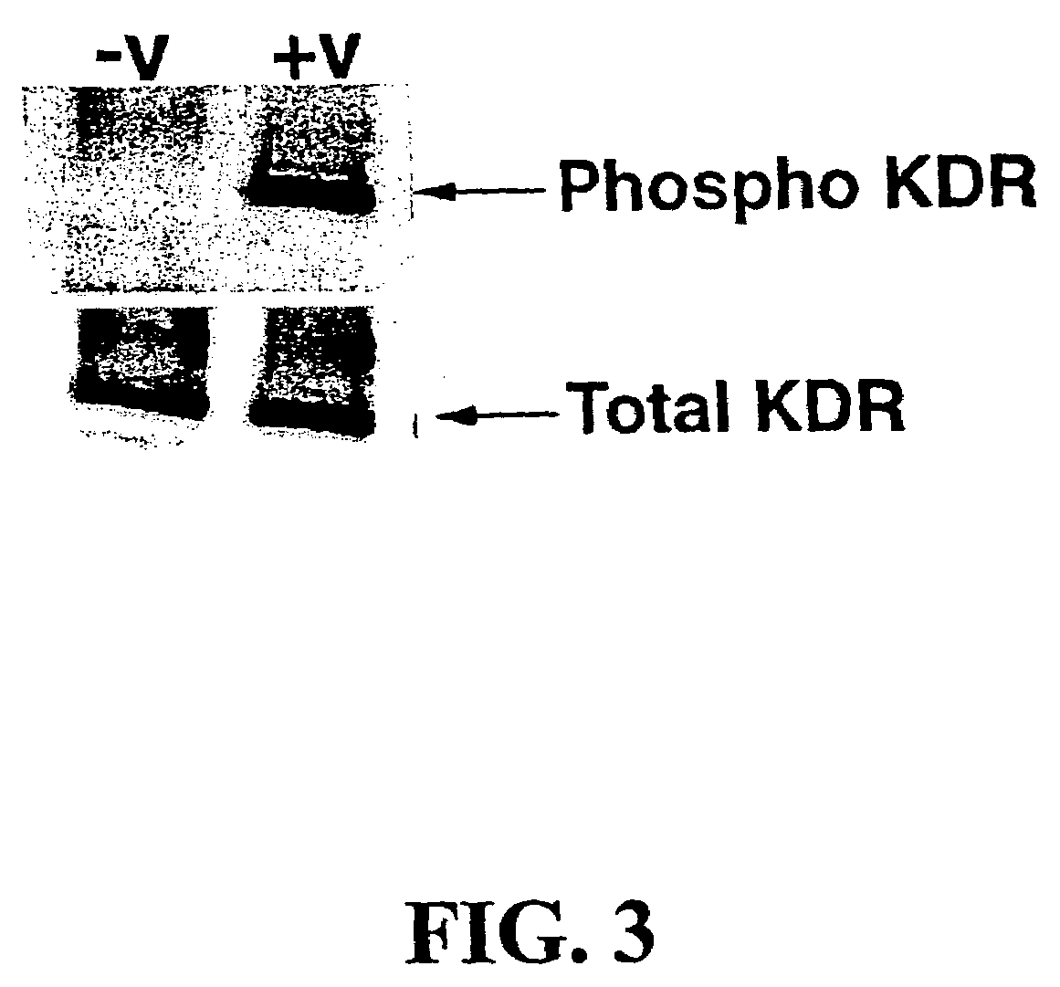
Transfection of 293H Cells

[0341]293H cells were transfected using the protocol described in Example 6. Transfection was done in black / clear 96-well plates (Becton Dickinson, cat. # 354640). The cells in one half of the plate (48 wells) were mock-transfected (with no DNA) and those in the other half of the plate were transfected with KDR cDNA. The cells were 80–90% confluent at the time of transfection and completely confluent the next day, at the time of the assay; otherwise the assay was aborted.

Preparatio...


PUM

PropertyMeasurementUnit
Lengthaaaaaaaaaa
Lengthaaaaaaaaaa
Currentaaaaaaaaaa

Abstract

The invention provides compositions and methods for therapeutic and diagnostic applications.

Description

RELATED APPLICATIONS[0001]This application claims priority from U.S. Provisional Application Ser. No. 60 / 440,201 filed on Jan. 15, 2003, and U.S. Provisional application Ser. No. 60 / 360,821, filed on Mar. 1, 2002, both of which are incorporated by referenceFIELD OF THE INVENTION[0002]The invention relates to compositions and methods for therapeutic and diagnostic applications.BACKGROUND OF THE INVENTION[0003]Researchers have long been attempting to exploit the ability of targeting moieties or ligands to bind to specific cells (via receptors or otherwise) to target compositions such as detectable labels or therapeutic agents to particular tissues of an animal (especially a human). In such situations, the ability of the targeting moiety to bind to the target (e.g., affinity, avidity, and / or specificity) significantly impacts the ability to successfully target the desired tissues.[0004]Numerous attempts have been made to use natural (e.g. polyclonal) and monoclonal antibodies, as targe...

Claims


Application Information

Patent Timeline
IPC IPC(8): A61K49/00A61K47/48C07K7/08C07K7/64
CPCA61K47/48215A61K47/48246C07K7/08C07K7/64A61K47/60A61K47/64A61P35/00Y02A50/30
Inventor ARBOGAST, CHRISTOPHEBUSSAT, PHILIPPEFAN, HONGLINDER, KAREN E.MARINELLI, EDMUND R.NANJAPPAN, PALANIAPPANUNN, ADRIANPILLAI, RADHAKRISHNAPOCHON, SYBILLERAMALINGAM, KONDAREDDIARSHRIVASTAVA, AJAYSONG, BOSWENSON, ROLF E.VON WRONSKI, MATHEW A.SATO, AARONWALKER, SHARON MICHELEDRANSFIELD, DANIEL T.
Owner BRACCO SUISSE SA
Why Eureka
Try Eureka