

High speed microscope with three-dimensional laser beam scanning including acousto-optic deflector for controlling the lateral position and collimation of the light beam
a three-dimensional laser beam and scanning microscope technology, applied in the direction of discharge tube/lamp details, instruments, optical elements, etc., can solve the problems of limiting the speed of such systems, raster scanning nor random access to multiple sites of interest at the required rate, and reducing resolution. , the effect of removing the time dependence of the deflection angl
- Summary
- Abstract
- Description
- Claims
- Application Information
AI Technical Summary
Benefits of technology
Problems solved by technology
Method used
Image

Examples
Embodiment Construction
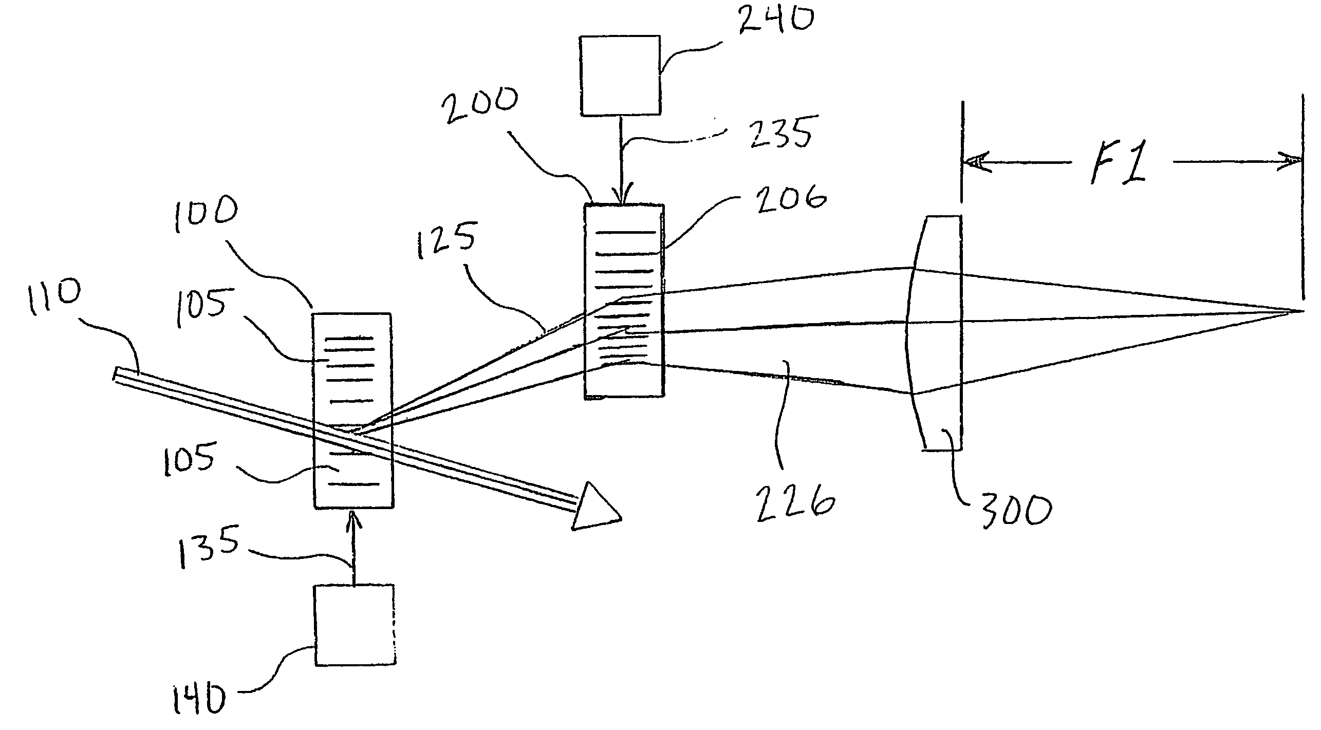
[0014]Preferred embodiments of the present invention comprise a scanning device to direct the focus of a light beam (such as a laser) to multiple predefined positions within a given volume. Preferred embodiments comprise an acousto-optically controlled light diffraction to independently change both collimation and direction of a laser beam. This inertia-free mechanism allows for very fast (microsecond range) three-dimensional positioning of the focus spot.
[0015]Embodiments of the present invention comprise a novel instrument for both structural and functional imaging studies of specimens such as living brain tissue. Certain embodiments use four AODs together with a commercial objective lens to deterministically and quickly (˜30 μs) position the focus spot in a microscopic 3D volume via a remote focusing strategy, while other embodiments may use fewer AODs with multiple acoustic waves in an individual AOD. Embodiments of the present invention utilize counter-propagating acoustic wave...
PUM
 Abstract
Description
Claims
Application Information