Labeled cell sets for use as functional controls in rare cell detection assays

a technology of functional controls and labeled cell sets, which is applied in the field of labeled cell sets for use as functional controls in rare cell detection assays, can solve the problems of difficult detection and elimination of colonies, inability to treat all colonies successfully, and a bit of futility in efforts, so as to facilitate the practice of methods and increase the severity of the disease sta

Inactive Publication Date: 2007-10-16
MENARINI SILICON BIOSYSTEMS SPA
View PDF22 Cites 54 Cited by
  • Summary
  • Abstract
  • Description
  • Claims
  • Application Information

AI Technical Summary

Benefits of technology

[0042]Also in accordance with the present invention are improved methods for isolating and enumerating rare cells, increasing numbers of rare cells being indicative of a disease state. In a particularly preferred embodiment, the rare cell is a cancer cell and the disease state is cancer. An exemplary method of the invention includes the following steps: i) obtaining a blood sample from a test subject, the sample comprising a mixed cell population suspected of containing said rare cells; ii) preparing an immunomagnetic sample wherein the blood sample is mixed with magnetic particles coupled to a ligand which reacts specifically with a determinant of the rare cells, to the substantial exclusion of other sample components; iii) contacting the immunomagnetic sample with at least one reagent which labels a determinant of the rare cells; and iv) analyzing the labeled rare cells to determine the presence and number of any rare cells in the immunomagnetic sample, the greater the number of rare cells present in said sample, the greater the severity of the disease state, the improvement comprising the addition of two fluorescently distinct sets of stabilized cell populations for use as internal or external control cells in said method, wherein the membrane of said control cell is

Problems solved by technology

Unfortunately, the metastatic colonies are harder to detect and eliminate and it is often impossible to treat all of them successfully.
These efforts have proven to be somewhat futile as the appearance of such antigens in a test sample have not been generally predictive and are often detected when there is little hope for the patient.
Despite the success of PSA testing, the test leaves much to be desired.
This is being done through the laborious procedure of isolating all of the mRNA from a blood sample and performing RT-PCR.
It is noteworthy that PCR is difficult, if not impossible in many situations, to perform quantitatively, i.e., to determine the number of tumor cells per unit volume of biological sample.
Additionally, false positives are often observed using this technique.
An added drawback is the finite and practical limit to the sensitivity of this technique based on the sample size examined.
As a further potential complication, tumor cells are often genetically unstable.
Further, there are very considerable doubts in the medical literature regarding the existence of such cells and the potential of such information.
The general view is that tumors are initially well confined and hence there will be few if any circulating cells in early stages of disease, and that early detection of cancer cells in circulation, even if feasible, would be unlikely to yield any useful information.
The current sample preparation procedures in which blood samples are incubated with fluorescently-labeled antibodies followed by addition of an erythrocyte lysing agent dilutes the sample ten-fold and is thus not suitable for detection of rare cells.
These procedures lead to variable cell losses and are difficult to standardize between laboratories.
Moreover, no significant enrichment of the target cells is obtained.
While the detection of circulating tumor cells in one's blood is frightening to the patient, a negative test result has not yet been proved to be an indication that a patient is free of cancer.
Even worse would be a false-negative result for circulating blood cells.
Reagent failure, instrument failure and operator errors can all lead to erroneous negative results.
As the cancer cells in blood are rare, (often less than 1 cell/ml of blood) the blood volumes needed to perform the test are restrictively large.
The requirement for such a large volume of blood prohibits the use of additional blood samples for traditional external control purposes.
With 0 cells detected, one has no way of knowing whether the reagents and/or process are working.
Use of a genuine ‘isotopic dilution’ protocol is not

Method used


Image

  • Labeled cell sets for use as functional controls in rare cell detection assays
  • Labeled cell sets for use as functional controls in rare cell detection assays
  • Labeled cell sets for use as functional controls in rare cell detection assays

Examples


example 1

Preparation of Stabilized Pre-Labeled Control Cells

[0091]The positive control cells can be pre-labeled with diverse markers. One method entails labeling cells using a lipophilic, membrane-specific fluorescent dye. There are numerous types of membrane dyes known in the art which are available commercially. Carbocyanines are among the most strongly absorbing membrane dyes known. Membrane dyes label cells by binding to membrane lipids. It is important that this labeling does not prevent the antibody binding to specific epithelial antigens. The binding of the dye to cells should be essentially non-reversible, and no leakage should occur during storage and test procedures. Another approach is to label cells using a fluorescent-antibody conjugate specific for cell surface antigens. A further approach entails labeling cellular components with fluorescent dyes. Examples of this approach include, without limitation, DAPI and Hoechst 33342 for double stranded DNA, acridine orange for DNA and ...

example 2

Preparation of Pre-Labeled Control Cells Using an Antibody Conjugated to a Fluorescent Dye

[0098]In this example, the SKBR-3 cultured tumor cells described in Example 1 were again used. However, the cells were pre-labeled with a Her2neu antibody conjugated to a cyanine dye. Anti-Her2neu specifically binds a surface antigen present on certain tumor cells including SKBR-3. The Her2neu MAb was conjugated to a Cy2™ dye using a N-hydroxysuccinimide ester of Cy2™ dye (Amersham catalog # PA22000) and following the manufacturer's recommendations.

[0099]SKBR-3 cells, adhered to the flask, were released with trypsin and washed twice with PBS by centrifugation. The cells were resuspended in permeabilization solution and stained with Her2neu-Cy2® dye for labeling. Permeabilization reagent did not have any effect on staining of cells with antibody. The final concentration of antibody during staining was 2 μg / ml and the concentration of cells was 1×10⁶ cells / ml. The staining and permeabilization we...

example 3

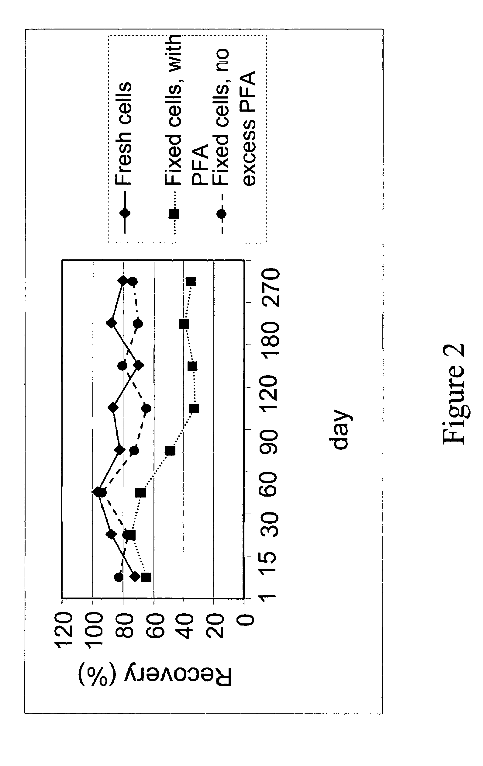
Stability of Pre-Labeled Control Cells

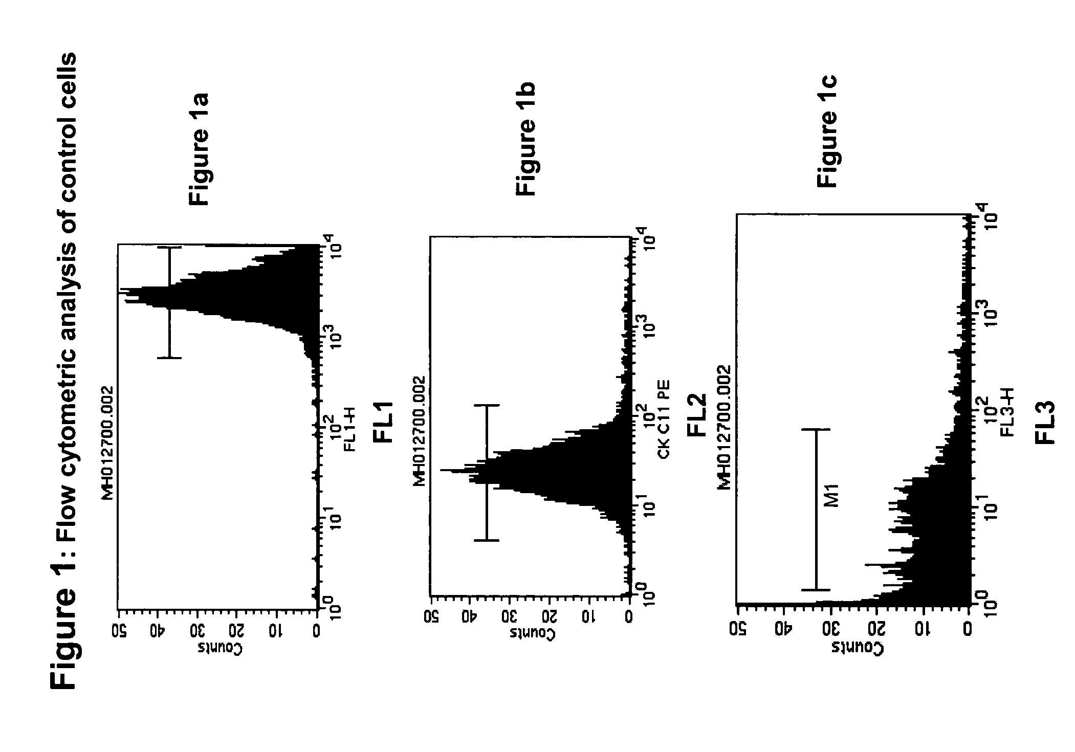
[0101]Fresh cells are generally stable for only one or two days. After this time, the antigens begin to shed and soon the cells disintegrated, causing cell number to decrease drastically. The pre-labeled control cells described in Examples 1 and 2 remained stablity for much longer periods. Two important criteria were used to follow stability: physical stability and biological stability. Physical stability is defined as the presence of an intact cell in a suspension. Biological stability is defined as the preservation of antigens present on cell surfaces and inside cells. Both physical and biological stability are important indicators of functional stability of control cells.

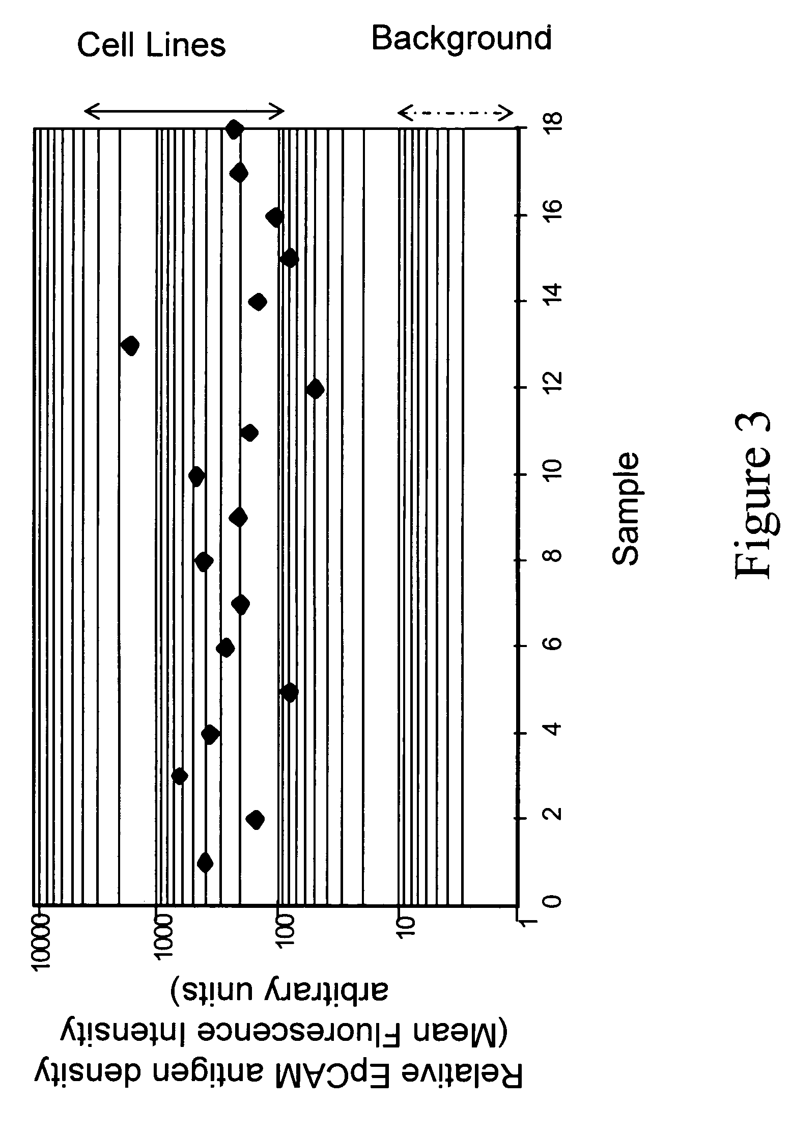
[0102]The physical stability of control cells was observed as a function of time by determining the number of cells present in suspension using flow cytometry for cell size, presence of a nucleus, and integrity of antigens. Two antigens were checked for integrity, which are i...


PUM


Abstract

Methods and kits for used as an internal or external control in rare cell analysis are disclosed. A plurality of fluorescently distinct sets of cells is used to define a range to assess the isolation of rare target cells from a sample. Thus, a known number of cells, expressing the surface and intracellular antigens present in the targeted rare cells, are stabilized and modified in such a way that they can be discriminated from the targeted rare cells. In addition, these cells are separated into at least two sets based upon the number. These sets are detectably distinct from each other and provide an upper and lower indication of the detection ability of the rare cell assay.

Description

CROSS REFERENCE TO RELATED APPLICATIONS[0001]This application is a continuation-in-part of U.S. application Ser. No. 09 / 801,471, filed Mar. 8, 2001, which is a continuation-in-part of U.S. application Ser. No. 09 / 248,388 (now U.S. Pat. No. 6,365,362), filed Feb. 12, 1999, which is a non-provisional U.S. application of U.S. Provisional Application Ser. No. 60 / 074,535, filed on Feb. 12, 1998, and which is a non-provisional U.S. application of U.S. application Ser. No. 60 / 110,279, filed on Nov. 30, 1998, and which is a non-provisional U.S. application of U.S. Provisional Application Ser. No. 60 / 110,202, filed on Nov. 30, 1998.FIELD OF THE INVENTION[0002]This invention relates to the use of pre-labeled cells as an external or internal functional control in cell selection and analysis procedures. The invention provides a range for controlling such diverse parameters as magnetic labeling, magnetic selection, viscosity, temperature, reagent addition, reagent activity, and operator error in...

Claims


Application Information

Patent Timeline
IPC IPC(8): C12N5/06G01N1/30B03C1/01G01N15/14G01N33/574H01F1/00
CPCB03C1/01B82Y25/00G01N15/14H01F1/0054G01N33/57484G01N33/57492G01N33/574
Inventor RAO, GALLA CHANDRARUTNER, HERMAN
Owner MENARINI SILICON BIOSYSTEMS SPA
Why Eureka
Try Eureka