
Membrane receptor reagent and assay

Inactive Publication Date: 2007-10-30
ZWEIG STEPHEN ELIOT
View PDF15 Cites 27 Cited by
  • Summary
  • Abstract
  • Description
  • Claims
  • Application Information

AI Technical Summary

Benefits of technology

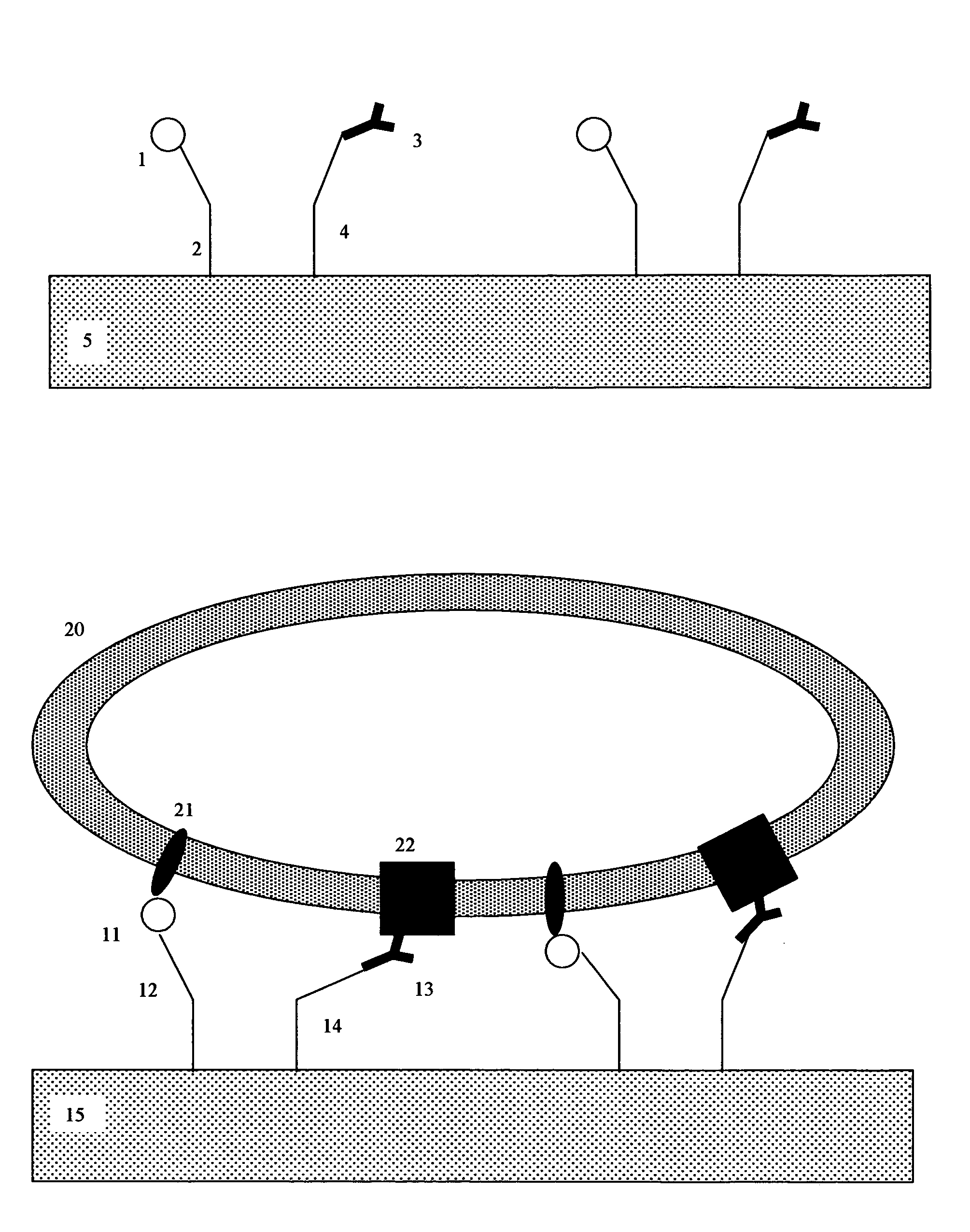
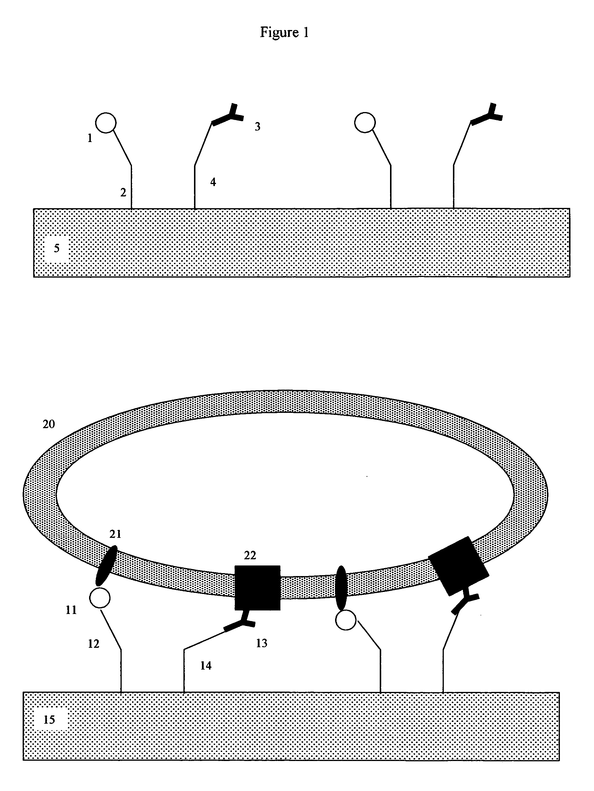
[0031]Here, a novel type of membrane-receptor reagent, suitable for high performance membrane receptor microarrays, is taught. As will be discussed in later sections, this type of microarray may have high utility for HTS drug discovery and development. For example, a comprehensive GPCR microarray, containing a large number of different GPCR families and common GPCR variants, would detect unwanted cross-reactions at the earliest stage of the drug discovery process. This would greatly facilitate the development of highly specific and well-targeted new drugs.
[0046]This reagent and method exhibits a number of distinct advantages over the prior art. One distinct advantage is that all of the chemistry required to perform the desired analysis is present on the support as a single homogeneous unit. Thus for each test, no additional chemistry or processing steps (such as washing or centrifugation) are required. Because the reagent contains all necessary detection means, the test-ligands do not need to be artificially labeled in any way. The assay chemistry is inherently reusable, and, by simply applying a wash step between assays, may be reused for multiple assays with different test-ligands. The reagent and method uses several orders of magnitude less reagents than previous art. Since the reagents used in this type of test are typically quite expensive, the economic savings are considerable.

Problems solved by technology

As a result, one of the key challenges going forward is to utilize this new knowledge to aid in the development of next generation drugs.
At present, however, membrane HTS screening methods are sub-optimal.
Such faithful recreations of native membrane structures are difficult to reproduce in a synthetic, in-vitro, environment.
Although the use of natural membranes has strong physiological merit, it is slow, expensive, and cumbersome.
At present, HTS methods are typically restricted to large, well-financed, commercial organizations.
This is because the present methodologies require the use of large quantities of expensive membrane receptors, expensive synthetic drug candidates, and expensive automation.
Although useful for demonstration purposes, one fundamental critique of this approach is that due to the close (1 nm) association between the inner leaflet of the artificial membrane lipid bilayer, and the solid phase support, the physiology of the artificial membrane is highly distorted.
In particular, larger membrane proteins, such as GPCR proteins, are large enough that they can interact with the solid phase support, resulting in distorted conformations and inactivity.
Additionally, the narrow aqueous layer is too thin to enable GPCR proteins to interact with other cytoplasmic side cofactors, which play a key role in the proper function of the receptor.
A second critique of this approach is that FRAP detection techniques are too crude to spot many interesting types of interactions.
This is because the binding of the relatively small (low molecular weight) drug candidates to relatively large (high molecular weight) membrane receptors will have a negligible impact on the FRAP mobility of the receptors, and thus will be poorly detected by FRAP techniques.
Additional drawbacks are the relatively slow throughput of the single element flow-cell design, and a large number of false positives produced by the mass-spectrometer detection methodology.
Evanescence biosensor techniques are known in the art, but have not been generally used for membrane bound analytes.

Method used


Image

  • Membrane receptor reagent and assay
  • Membrane receptor reagent and assay
  • Membrane receptor reagent and assay

Examples


example 1

Evanescent Wave Calculations

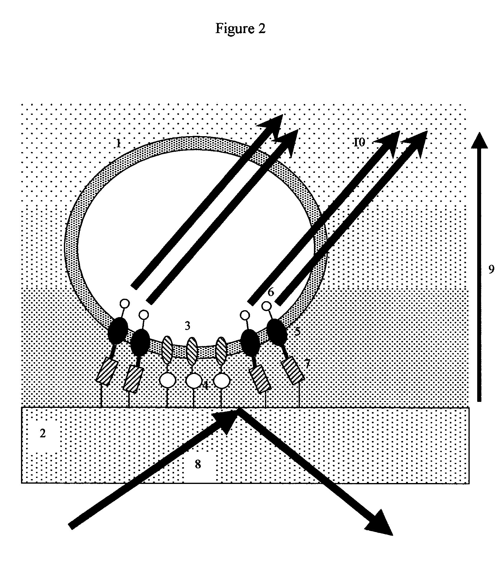
[0109]Upon hitting a boundary between two media with different indexes of refraction, there is a critical angle in which light waves can either pass from the first media to the second media, or bounce back into the first media. At angles where light bounces back, part of the wave energy passes into the second media for a very short distance. This is called the evanescent wave. Evanescent wave techniques are commonly used in microscopy to help visualize regions where cultured cell membranes adhere to transparent supports.

[0110]In the common situation where light is passing from a glass or transparent support into an aqueous media, evanescent waves typically penetrate several hundred nanometers into the aqueous media. The wave decays in intensity at a rate of

Intensity=Ioe(−z / d)

Where Io is the illumination intensity at the support surface, z is the distance in nanometers above the support surface, and d is a constant (approximately 277 nanometers). (For thes...

example 2

Signal Processing

[0118]As previously discussed, one of the most important signal enhancing techniques is signal normalization, using concurrent normal fluorescence (epifluorescent) illumination to provide a second reference, signal. Here the microarray spots are sequentially visualized by both evanescent illumination and epifluorescence illumination, and the evanescent signal is normalized by the epifluorescent signal. Since the epifluorescent signal is relatively unaffected by the position of the membrane proteins on the liposome, epifluorescence normalization corrects for many variables, including liposome spot density, labeling efficiency, and GPCR receptor concentration.

[0119]As an example of the issues that are involved in signal processing, consider the following model:

[0120]Any given microarray spot will have a variable number of liposomes “x”, and a background signal “b”, where b may be due to contaminating lysed lyposomes, autofluorescence, or other impurities in the reagen...

example 3

Studies with Model GPCR Target Membrane Receptors

[0135]Not all GPCR receptors bind drug ligands. Some, such as bacteriorhodopsin, act as sensors. Bacteriorhodopsin is a 7-transmembrane protein with well-understood properties. It is available in low cost and large quantities from a variety of commercial sources. It is easy to work with, and is often used for exploratory biophysical research. Here, methods to construct prototype membrane sensors using liposomes with fluorescent bacteriorhodopsin are described.

Preparation of Large Liposomes Containing Fluorescent Bacteriorhodopsin:

[0136]Bacteriorhodopsin from commercial sources can be labeled with the Alexa Fluor 488 fluorophore (a high efficiency fluorescent moiety), and incorporated into giant (5 micron) phospholipid vesicles (liposomes) following the methods of Kahya et. al (Kahya N, Pecheur E, de Boeij, W, Wiersma D, Hoekstra D, “Reconstitution of Membrane Proteins into Giant Unilamellar Vesicles via Peptide-Induced Fusion”, Biophy...


PUM


Abstract

A membrane receptor reagent and assay is disclosed in which liposomes are bound to an evanescent wave emitting surface. Membrane receptors on the liposome's fluid lipid bilayer membrane are labeled with a fluorescent or luminescent moiety. These membrane receptors are free to diffuse randomly throughout the liposome surface, and thus tend to redistribute according to externally applied forces. The evanescent wave-emitting surface additionally contains reagents that reversibly bind to the membrane receptors, tending to bring them closer to region of high evanescent wave intensity. Test analytes that disrupt or promote the association between the membrane receptors and the surface reagents act to change the average distance between the membrane receptors and the evanescent wave emitting surface, resulting in a change in the fluorescent or luminescent signal. This reagent and assay system functions with physiologically important membrane receptors such as GPCR receptors, other 7-tm receptors, drug transport proteins, cytochrome P450 membrane proteins and other clinically important membrane components. The reagent and assay methods may be incorporated into microarrays, capillaries, flow cells and other devices, and used for drug discovery, ADMET, and other biomedically important assays.

Description

[0001]This patent is a continuation in part of patent application Ser. No. 10 / 444,390 “Membrane receptor reagent and assay”, filed May 23, 2003 now U.S. Pat. No. 6,790,632. Application Ser. No. 10 / 444,390 claimed priority benefit of provisional patent applications 60 / 389,679; and 60 / 400,396 both entitled “Tethered receptor-ligand reagent and assay”, filed Jun. 17, 2002 and Jul. 31, 2002; and provisional patent application 60 / 428,137; entitled “Membrane receptor reagent and assay”, filed Nov. 21, 2002.BACKGROUND OF THE INVENTION[0002]1. Field of the Invention[0003]The invention generally concerns reagents and methods useful for membrane receptor-ligand binding assays.[0004]2. Description of the Related Art[0005]Much of modern pharmacology and biochemistry is focused on the interactions between different types of biological signaling molecules and their corresponding membrane receptors. Structurally, these membrane receptors are often G-Protein Coupled Receptors (GPCR) from the 7-tran...

Claims


Application Information

Patent Timeline
IPC IPC(8): C12Q1/00G01N33/543
CPCG01N33/5432G01N33/54373Y10S435/968G01N33/543
Inventor ZWEIG, STEPHEN ELIOT
Owner ZWEIG STEPHEN ELIOT
Learn More