
Retroviral vectors comprising a functional splice donor site and a functional splice acceptor site

a technology of retroviral particles and acceptor sites, which is applied in the field of vectors, can solve the problems of reducing the potential for production of replication-competent viruses, unable to bring about encapsidation, and defective replication of retroviral vectors

Inactive Publication Date: 2007-12-04
OXFORD BIOMEDICA (UK) LTD
View PDF5 Cites 140 Cited by
  • Summary
  • Abstract
  • Description
  • Claims
  • Application Information

AI Technical Summary

Benefits of technology

[0088]In particular, the present invention seeks to provide a novel retroviral vector capable of providing efficient expression of a NOI—or even a plurality of NOIs—at one or more desired target sites.
[0259]The invention also permits the production of retroviral vectors such as MMLV-based vectors in non-dividing and slowly-dividing cells in vivo. It had previously been possible to produce MMLV-based retroviral vectors only in rapidly dividing cells such as tissue culture-adapted cells proliferating in vitro or rapidly dividing tumour cells in vivo. Extending the range of cell types capable of producing retroviral vectors is advantageous for delivery of genes to the cells of solid tumours, many of which are dividing slowly, and for the use of non-dividing cells such as endothelial cells and cells of various haematopoietic lineages as endogenous factories for the production of therapeutic protein products.

Problems solved by technology

In some cases, this specificity may restrict the transduction potential of a recombinant retroviral vector.
This makes the retroviral vector replication-defective.
The packaging cell line produces the proteins required for packaging retroviral DNA but it cannot bring about encapsidation due to the lack of a psi region.
This reduces the potential for production of a replication-competent virus.
If the vector encodes toxic genes or genes that interfere with the replication of the host cell, such as inhibitors of the cell cycle or genes that induce apotosis, it may be difficult to generate stable vector-producing cell lines, but transient transfection can be used to produce the vector before the cells die.
In view of the toxicity of some HIV proteins—which can make it difficult to generate stable HIV-based packaging cells—HIV vectors are usually made by transient transfection of vector and helper virus.
With regard to vector titre, the practical uses of retroviral vectors have been limited largely by the titres of transducing particles which can be attained in in vitro culture (typically not more than 108 particles / ml) and the sensitivity of many enveloped viruses to traditional biochemical and physicochemical techniques for concentrating and purifying viruses.
Although a 20-fold increase in viral titre can be achieved, the relative fragility of retroviral Env protein limits the ability to concentrate retroviral vectors and concentrating the virus usually results in a poor recovery of infectious virions.
While this problem can be overcome by substitution of the retroviral Env protein with the more stable VSV-G protein, as described above, which allows for more effective vector concentration with better yields, it suffers from the drawback that the VSV-G protein is quite toxic to cells.
In these cases, it is not sufficient simply to expose the cells to a larger volume of virus to compensate for a low virus titre.
However, when the inserted gene is not selectable, this approach has the disadvantage that it is more difficult to generate cells that produce a high titre vector stock.
In addition it is usually more difficult to determine the titre of the vector.
One drawback of this strategy is that foreign sequences are inserted into the intron of the spliced gene.
Because these effects are unpredictable, they can affect the production of the encoded genes.
These products then result in viral packaging and release of virions.
However, certain second generation vectors appear not to give longer-term gene expression, even though the DNA seems to be maintained.
The “gutted” or “gutless” viruses can be grown to high titres with a first generation helper virus in the 293 cell line but it has been difficult to separate the “gutted” vector from the helper virus.
Thus, in normal nontumour cells, in the absence of E1b, the virus is unable to block apoptosis and is thus unable to produce infectious virus and spread.
However, current adenoviral vectors suffer from some major limitations for in vivo therapeutic use.
These include: (i) transient gene expression—the adenoviral vector generally remains episomal and does not replicate so that it is not passed onto subsequent progeny (ii) because of its inability to replicate, target cell proliferation can lead to dilution of the vector (iii) an immunological response raised against the adenoviral proteins so that cells expressing adenoviral proteins, even at a low level, are destroyed and (iv) an inability to achieve an effective therapeutic index since in vivo delivery leads to an uptake of the vector and expression of the delivered genes in only a proportion of target cells.

Method used


Image

  • Retroviral vectors comprising a functional splice donor site and a functional splice acceptor site
  • Retroviral vectors comprising a functional splice donor site and a functional splice acceptor site
  • Retroviral vectors comprising a functional splice donor site and a functional splice acceptor site

Examples


example 1

Construction of a Split-intron MLV Vector

(i) Addition of Small-T Splice Donor:

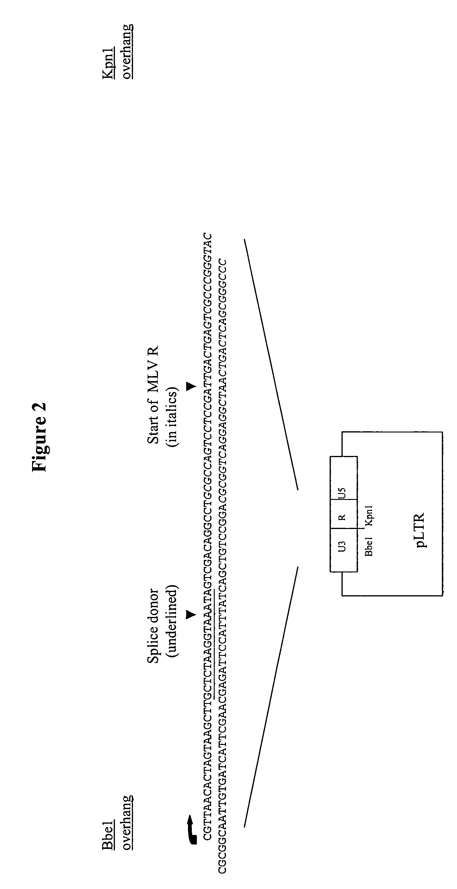
[0320]The starting plasmid for this construct is pLXSN (Miller el al 1989 ibid); Firstly this construct is digested with Nhe1 and the backbone re-ligated to create an LTR (U3-R-U5) plasmid. Into this plasmid is then inserted an oligonucleotide containing the splice donor sequence between the Kpn1-Bbe1 sites. Also contained within this oligonucleotide, downstream of the splice donor is the MLV R sequence up to the Kpn1. The resulting plasmid is named 3′LTR-SD (see FIG. 2).

(ii) Addition of Splice Acceptor:

[0321]The splice acceptor sequence used in this construct (including the branch point—an A residue between 20 and 40 bases upstream of the splice acceptor involved in intron lariat formation (Aebi et al 1987 Trends in Genetics 3: 102-107) is derived from an immunoglobulin heavy chain variable region mRNA (Bothwell et al 1981 Cell 24: 625-637) but with a consensus / optimised acceptor site. Such a sequence sig...

example 2

Construction of a Split-intron Lentivector

Construction of Initial EIAV Lentiviral Expression Vector (Also see Patent Application GB 9727135.7)

[0324]For the construction of a split-function lentiviral vector the starting point is the vector named pEGASUS-1 (see patent application GB 9727135.7). This vector is derived from infectious proviral EIAV clone pSPEIAV19 (accession number: U01866; Payne et al 1994). Its construction is outlined as follows: First; the EIAV LTR, amplified by PCR, is cloned into pBluescript II KS+ (Stratagene). The MluI / MluI (216 / 8124) fragment of pSEIAV19 is then inserted to generate a wild-type proviral clone (pONY2) in pBluescript II KS+ (FIG. 1). The env region is then deleted by removal of the Hind III / Hind III fragment to generate pONY2-H. In addition, a BglII / NcoI fragment within pol (1901 / 4949) is deleted and a β-galactosidase gene driven by the HCMV IE enhancer / promoter inserted in its place. This is designated pONY2.10nlsLacZ. To reduce EIAV sequence t...

example 3

Construction of an MMLV Amphotropic env Gene with Minimal Homology to the pol Gene and a gag-pol Transcription Cassette

[0349]In the Moloney murine leukaemia virus (MMLV), the first approximately 60 bps of the env coding sequence overlap with sequences at the 3′ end of the pol gene. The region of homology between these two genes was removed to prevent the possibility of recombination between them in cells expressing both genes.

[0350]The DNA sequence of the first 60 bps of the coding sequence of env was changed while retaining the amino acid sequence of the encoded protein as follows. A synthetic oligonucleotide was constructed to alter the codon usage of the 5′-end of env (See FIG. 15) and inserted into the remainder of env as follows.

[0351]The starting plasmid for re-construction of the 5′ end of the 4070A gene was the pCI plasmid (Promega) into which had previously been cloned the Xba1-Xba1 fragment containing the 4070A gene from pHIT456 (Soneoka et al 1995 ibid) to form pCI-4070A....


PUM


Abstract

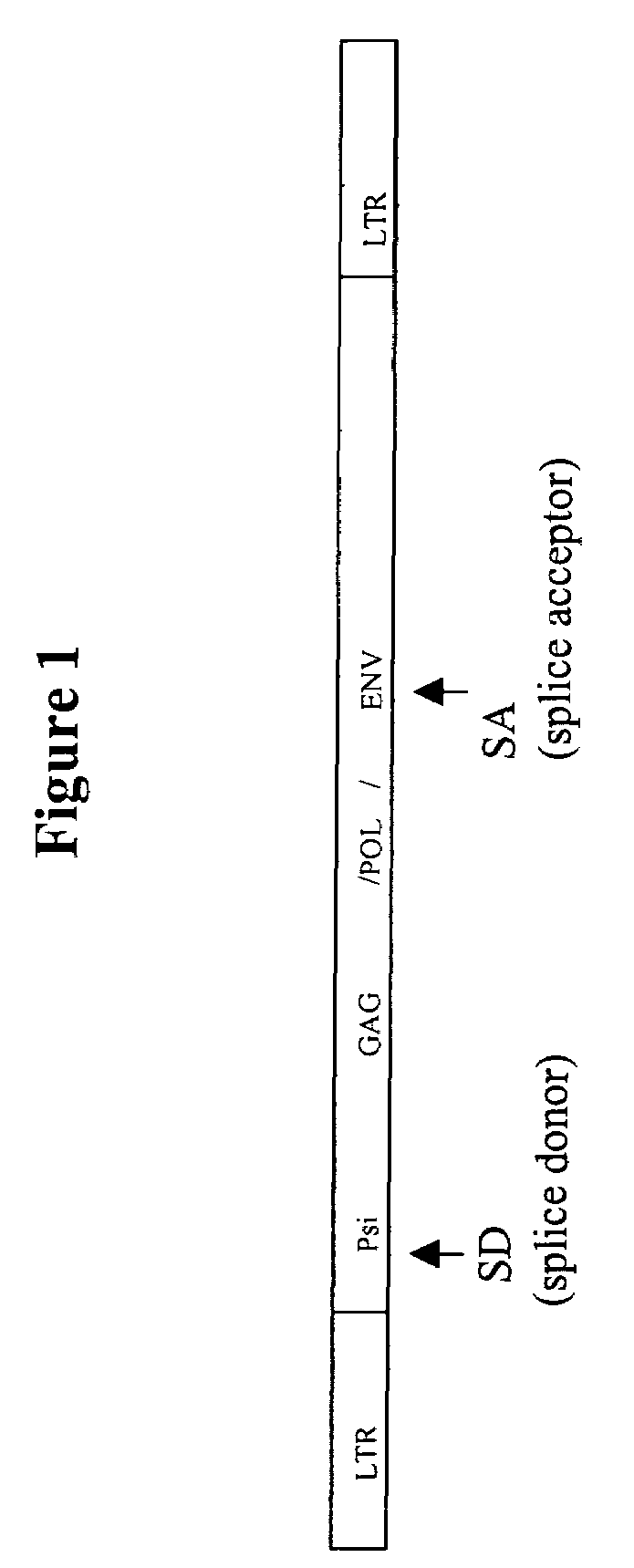
A retroviral vector is described. The retroviral vector comprises a functional splice donor site and a functional splice acceptor site; wherein the functional splice donor site and the functional splice acceptor site flank a first nucleotide sequence of interest (“NOI”); wherein the functional splice donor site is upstream of the functional splice acceptor site; wherein the retroviral vector is derived from a retroviral pro-vector; wherein the retroviral pro-vector comprises a first nucleotide sequence (“NS”) capable of yielding the functional splice donor site and a second NS capable of yielding the functional splice acceptor site; wherein the first NS is downstream of the second NS; such that the retroviral vector is formed as a result of reverse transcription of the retroviral pro-vector.

Description

CROSS-REFERENCE TO RELATED APPLICATIONS[0001]This is a continuation of U.S. application Ser. No. 09 / 508,516, filed on Jun. 8, 2000 now U.S. Pat. No. 6,808,922, which is a 371 of PCT / GB98 / 02867, filed on Sept. 23, 1998, claiming priority to UK application 9720465.5, filed on Sep. 25, 1997.FIELD OF THE INVENTION[0002]The present invention relates to a vector.[0003]In particular, the present invention relates to a novel system for packaging and expressing genetic material in a retroviral particle.[0004]More in particular, the present invention relates to a novel system capable of expressing a retroviral particle that is capable of delivering a nucleotide sequence of interest (hereinafter abbreviated as “NOI”)—or even a plurality of NOIs—to one or more target sites.[0005]In addition, the present invention relates to inter alia a novel retroviral vector useful in gene therapy.BACKGROUND OF THE INVENTION[0006]Gene therapy may include any one or more of: the addition, the replacement, the ...

Claims


Application Information

Patent Timeline
IPC IPC(8): C12N15/63C12N15/09A61K35/76A61K48/00C07K14/145C07K14/155C12N1/15C12N1/19C12N1/21C12N5/10C12N7/00C12N7/01C12N15/861C12N15/867
CPCC12N15/63C12N15/86C07K14/005A61K48/00C12N2840/44C12N2710/10343C12N2740/13043C12N2740/15022C12N2740/15043C12N2760/20122C12N2810/40C12N2810/60C12N2810/6054C12N2830/002C12N2830/42C12N2830/48C12N2830/50C12N2840/203
Inventor BEBBINGTON, CHRISTOPHER ROBERTKINGSMAN, SUSAN MARYUDEN, MARKKINGSMAN, ALAN JOHNMITROPHANOS, KYRIACOS
Owner OXFORD BIOMEDICA (UK) LTD
Learn More