
Musculo-skeletal implant having a bioactive gradient

a bioactive gradient and musculoskeletal technology, applied in the field of biocompatible tissue implant devices, can solve the problems of surgical intervention, articular cartilage tissue injury will not heal spontaneously, and the cartilage and underlying bone of the joint will be gradually destroyed, so as to enhance the chemotactic properties of the bioactive agent, and accelerate or enhance the musculoskeletal repair process

Active Publication Date: 2008-04-01
DEPUY SPINE INC (US)
View PDF73 Cites 17 Cited by
  • Summary
  • Abstract
  • Description
  • Claims
  • Application Information

AI Technical Summary

Benefits of technology

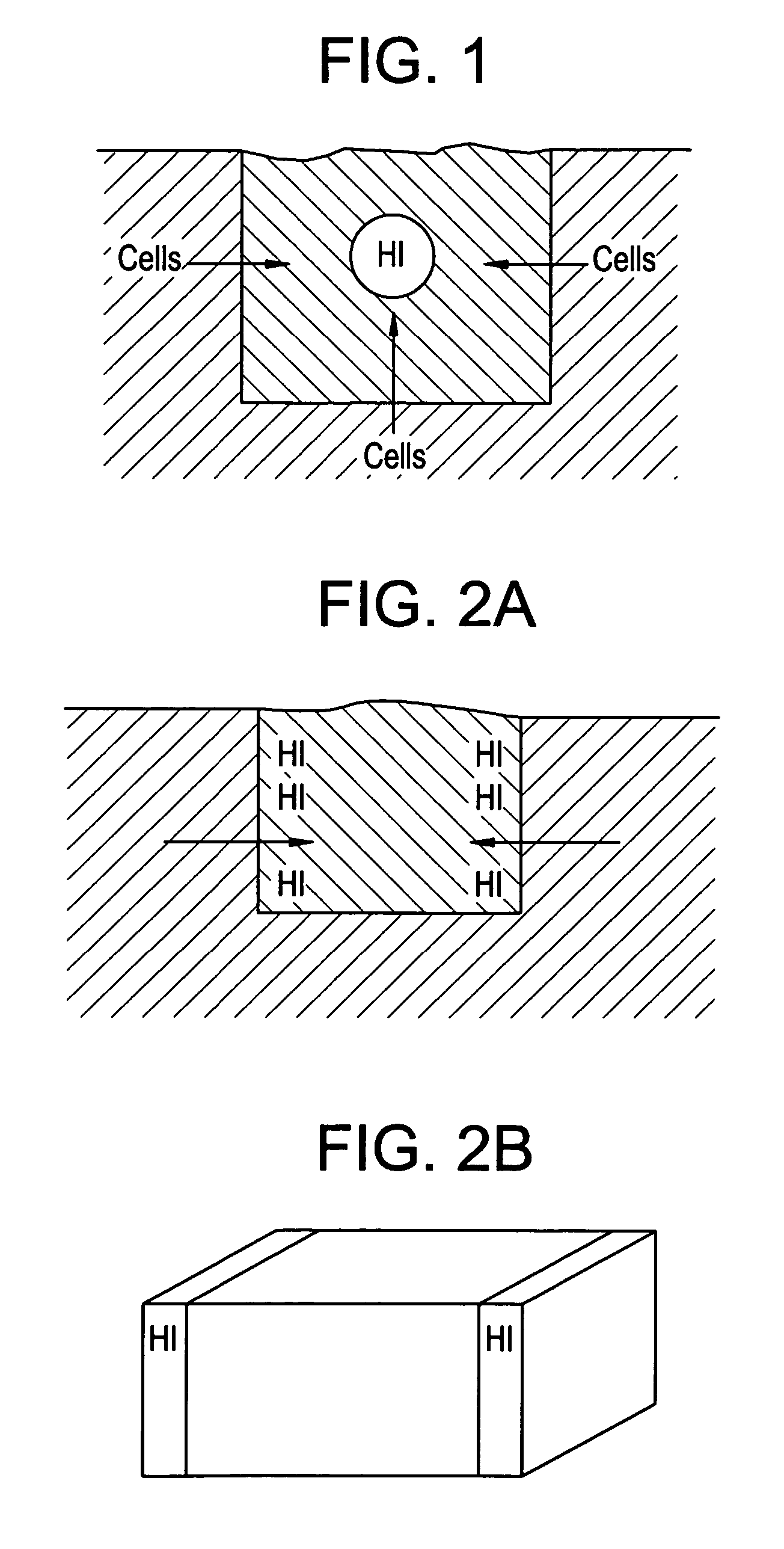
[0009]It is believed that the musculoskeletal repair process may be further enhanced by providing an implant that provides bioactive agents (such as rhGDF-5) to the cellular environment of the implant in a manner that more fully exploits the bioactive agent's characteristics (such as chemotaxis) in a more desirable manner. It is further believed that specifically grading the spatial concentration of a bioactive agent within the implant may provide effects from the bioactive agent that would be superior to implants having a homogeneous agent concentration profile.
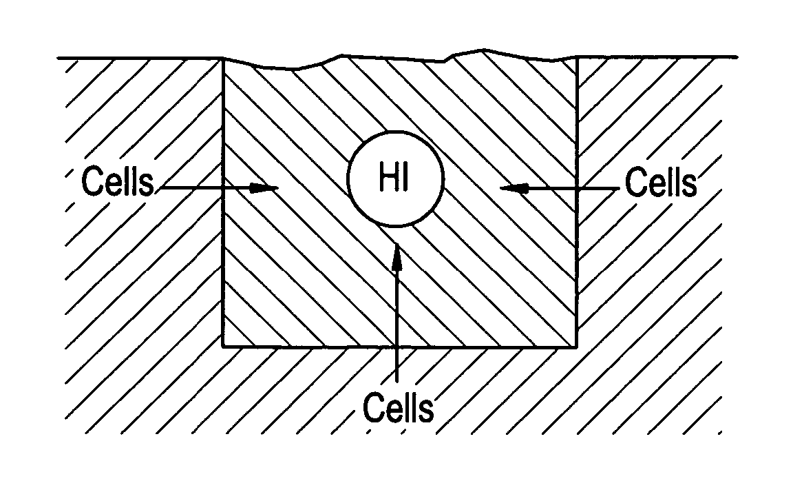
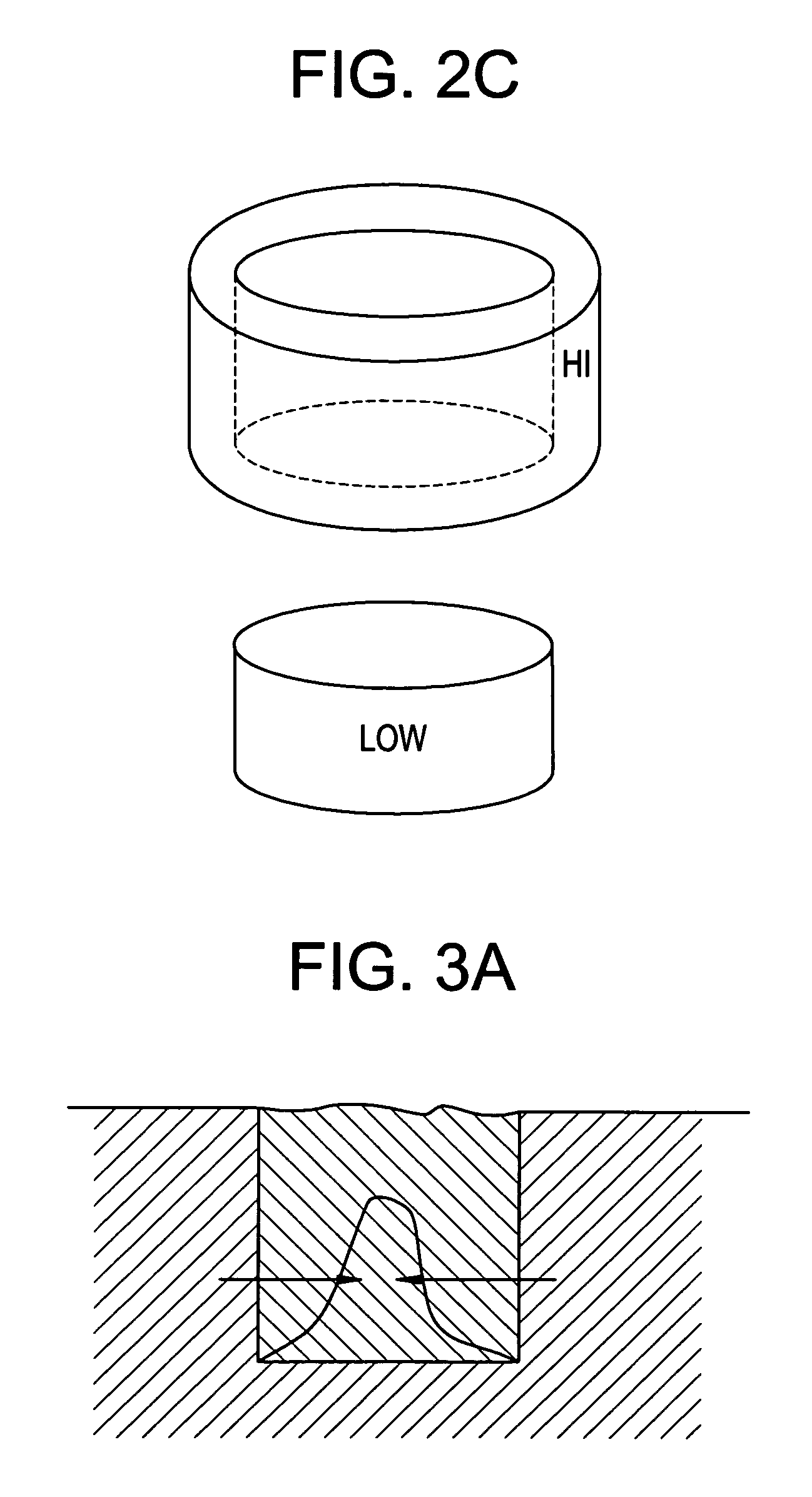
[0010]Accordingly, in some embodiments, the implant includes a bioactive agent presented to the cellular environment in a concentration profile characterized by a spatial gradient. Without wishing to be tied to a theory, it is believed that the musculoskeletal repair process may be further accelerated or enhanced by providing a spatially-graded bioactive agent concentration within the cellular environment of the implant that enhances the chemotactic properties of the bioactive agent.
[0014]In another aspect of the present invention, the implant includes bioactive agents presented to the cellular environment via two different matrix phases of the implant, wherein at least one of the bioactive agents is a member of the BMP superfamily, preferably a GDF, more preferably rhGDF-5. In some embodiments thereof, the first and second phases respectively contain rhGDF-5 and a different bioactive agent. In others, the two phases contain the same rhGDF-5 bioactive agent but at different or similar concentrations. Desirably, the two phases are designed to release the bioactive agents to the cellular environment at different times, thereby allowing the bioactive agents to spur different phases of regeneration or repair. It is believed that specifically tailoring the timing of the release of the bioactive agent(s) within the implant may provide effects from the agent that would be superior to implants having a single, unimodal release profile.
[0019]Preferably, in the dual phase aspect of the novel implant, the bioactive agent associated with the first phase enhances at least one of chemotaxis and proliferation, while the bioactive agent associated with the second phase enhances at least one of differentiation and ECM stimulation. Such an implant would allow the migration and proliferation of native cells into the implant without their premature differentiation. Avoiding premature differentiation of these native cells is advantageous because it allows the cells to migrate, resulting in appropriate cellular distribution through the implant.

Problems solved by technology

Damaged articular cartilage can severely restrict joint function, cause debilitating pain and may result in long term chronic diseases such as osteoarthritis, which gradually destroys the cartilage and underlying bone of the joint.
Injuries to the articular cartilage tissue will not heal spontaneously and require surgical intervention if symptomatic.
Unfortunately, fibrocartilage does not have the same mechanical properties as hyaline cartilage and degrades over time as a consequence of wear.
Patients typically have to undergo repeated surgical procedures, which can lead to the complete deterioration of the cartilage surface.
This manual, labor-intensive process is extremely costly and time consuming.
Although the clinical data suggest long-term benefit for the patient, the prohibitive cost of the operation combined with the traumatic impact of two surgical procedures to the knee, have hampered adoption of this technique.

Method used


Image

  • Musculo-skeletal implant having a bioactive gradient
  • Musculo-skeletal implant having a bioactive gradient
  • Musculo-skeletal implant having a bioactive gradient

Examples


Embodiment Construction

[0045]For the purposes of the present invention, a “matrix” is interchangeable with a “carrier”. “Presented to the cellular environment” means “not sequestered” or “sequestered, then released” or “sequestered”.

[0046]A “bioactive agent” or “agent” is a protein (for example, ECM molecules, growth factors, signaling molecules, etc), gene, small molecule, DNA or RNA fragment, peptide, virus, sugar or mineral.

[0047]The term “chemotaxis” is intended to include hepatotaxic effects and movements.

[0048]It is believed that designing an implant to possess a spatially-graded chemotactic factor concentration profile can be beneficial in two ways. First, the resulting chemotaxis could enhance the uniform distribution of viable cells within the implant. Second, it could enhance tissue integration with the surrounding native tissue.

[0049]It is often the case that an implant contains either no cells or cells present only upon an outer surface of the implant. In such cases, a chemotactic bioactive ag...


PUM


Abstract

This invention relates to an implant for a musculoskeletal defect having a bioactive agent having a concentration gradient.

Description

CONTINUING DATA[0001]This application claims the benefit of U.S. Provisional Patent Application Ser. No. 60 / 606,382, entitled “Musculo-Skeletal Implant Having a Bioactive Gradient”, filed Sept. 1, 2004, the specification of which is incorporated by reference in its entirety.FIELD OF THE INVENTION[0002]The present invention relates to biocompatible tissue implant devices for use in the repair of tissue injuries, organ disease states and / or congenital anomalies as well as methods for making and using such biocompatible tissue implant devices.BACKGROUND OF THE INVENTION[0003]Injuries to tissue, such as cartilage, meniscus, intervertebral disc, skin, muscle, bone, tendon and ligament, where the tissue has been injured or traumatized, frequently require surgical intervention to repair the damage and facilitate healing. Such surgical repairs can include suturing or otherwise repairing the damaged tissue with known medical devices, augmenting the damaged tissue with other tissue, using an ...

Claims


Application Information

Patent Timeline
Patent Type & Authority Patents(United States)
IPC IPC(8): A61F2/00
CPCA61F2/08A61K9/0024A61L27/54A61L27/56A61F2002/0894A61F2250/0035A61F2250/0068A61L2300/414A61L2300/432A61L2300/45A61L2300/602
Inventor KADIYALA, SUDHAKARBINETTE, FRANCOISCOLEMAN, CYNTHIA MARIEKAPUR, THERESA ADAMS
Owner DEPUY SPINE INC (US)
Learn More