

Crystalline VAP-1 and uses thereof
a technology of vascular adhesion protein and vap-1, which is applied in the field of crystalline human vascular adhesion protein1, can solve the problems of increasing level and damage to cells leading to vascular damage, and achieve the effect of inhibiting vap-1 activity
- Summary
- Abstract
- Description
- Claims
- Application Information
AI Technical Summary
Benefits of technology
Problems solved by technology
Method used
Image

Examples
example 1
Production and Purification of Human VAP-1.
[0070]The full-length protein with the N-terminal transmembrane region was expressed in glycosylation-competent CHO cells, as described in Smith et al., 1998. The harvested cells were lysed using a lysis buffer (150 mM NaCl, 10 mM Tris-Base pH 7.2, 1.5 mM MgCl2, 1% NP40). Clarified cell lysate was used for the purification of HVAP-1 based on a monoclonal antibody affinity column and using the ÄKTA™ purifier system (Amersham Biotech). The protein was purified to homogeneity (>95%) using affinity chromatography and after purification the presence of the VAP-1 protein, the 90- and 170-180-kD bands, was confirmed by silver stained SDS-page as described by Smith et al. 1998.
[0071]The purified protein retained its CAO activity as determined using benzylamine as the substrate. Amine oxidase activity was measured using a spectrophotometric method as described (Hoft et al., 1997), 200 μl volume and 1 mM benzylamine as the substrate. The absorbance c...
example 2
Crystallization and Preliminary Analysis
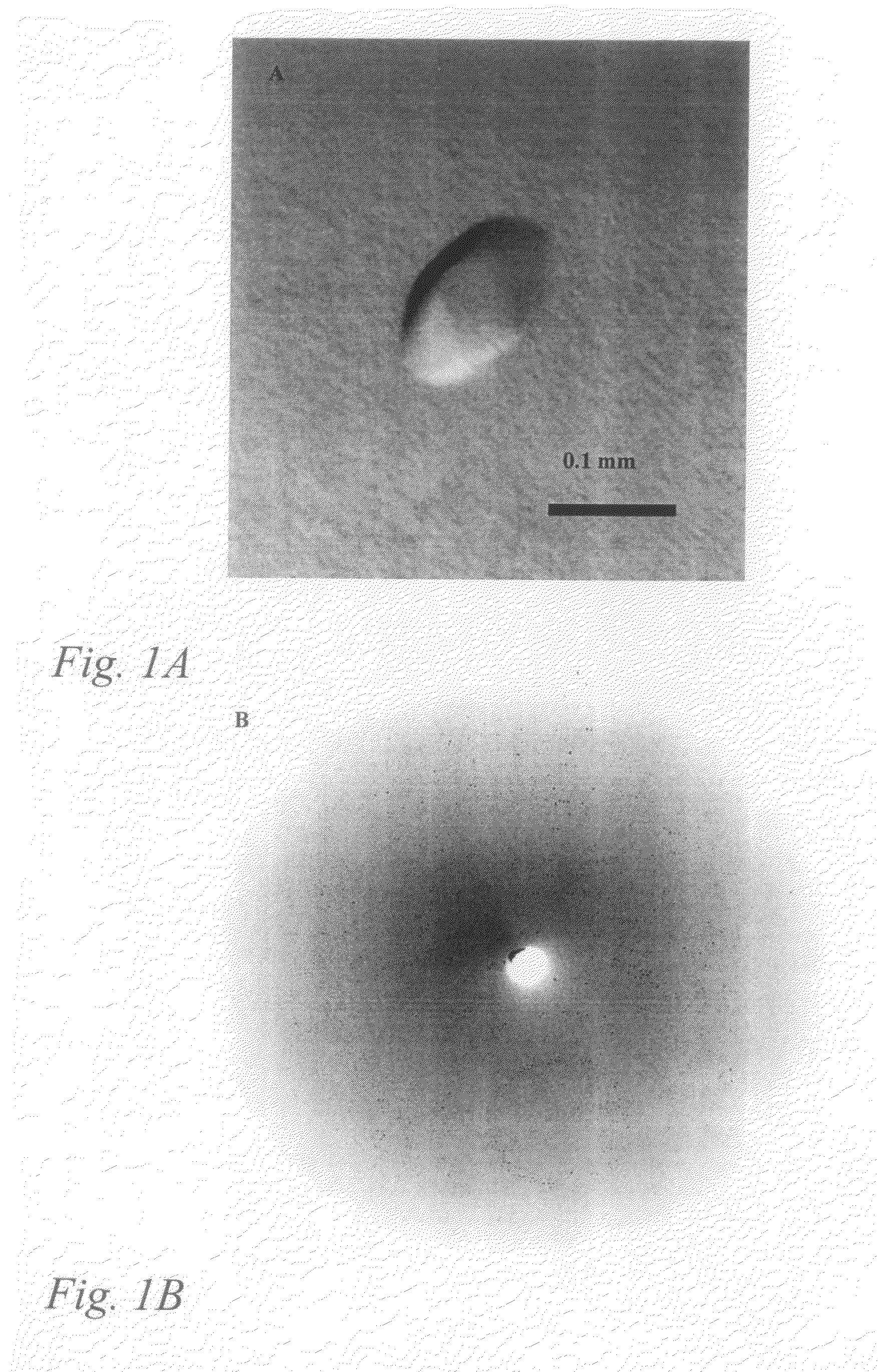
[0073]Initial crystallization conditions for hVAP-1 were screened at room temperature using the Wizard I random sparse matrix crystallization screen (Emerald BioStructures, Inc., USA) and the vapour-diffusion method. Small hexagonal crystals were obtained in a condition containing 1.0 M K / Na tartrate, 100 mM imidazole (pH 8.0) and 200 mM NaCl after several months of incubation. The hanging drops contained 2 μl of protein sample (1.0 mg / ml) in 10 mM potassium phosphate buffer (pH 7.2) and 2 μl of reservoir solution. After optimization the best crystals were obtained using a reservoir solution of 1.0 M K / Na tartrate, 100 mM imidazole .(pH 7.8) and 150-250 mM NaCl as the precipitant. The crystals formed in a few days and grew to a final size of about 0.15×0.15×0.1 mm (FIG. 1).
[0074]One crystal was mounted in a capillary and preliminary X-ray analysis was carried out in-house using a rotating-anode radiation source (Cu Kα radiation, 50 kV, 150 mA)...
example 3
Structure Determination
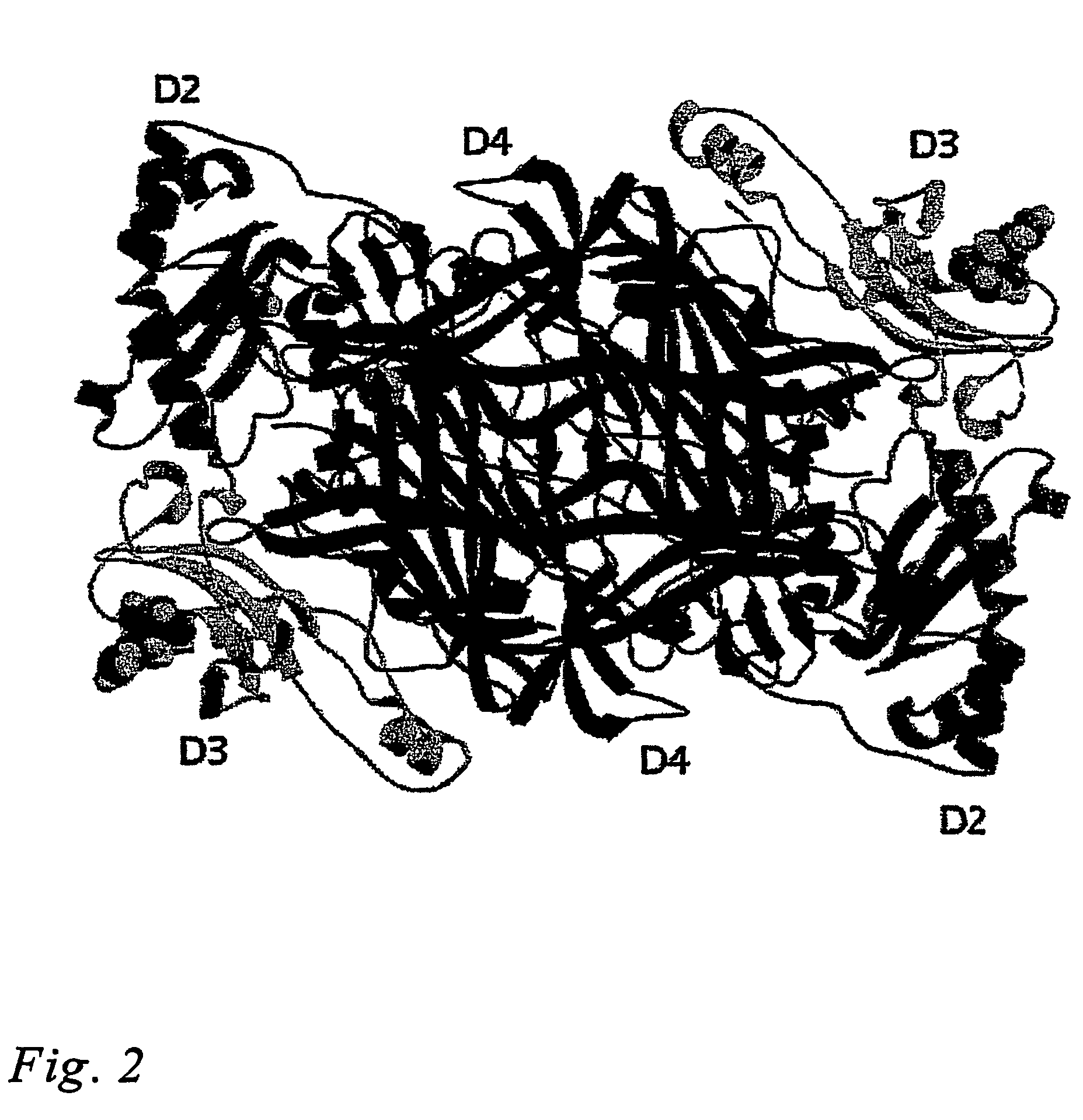
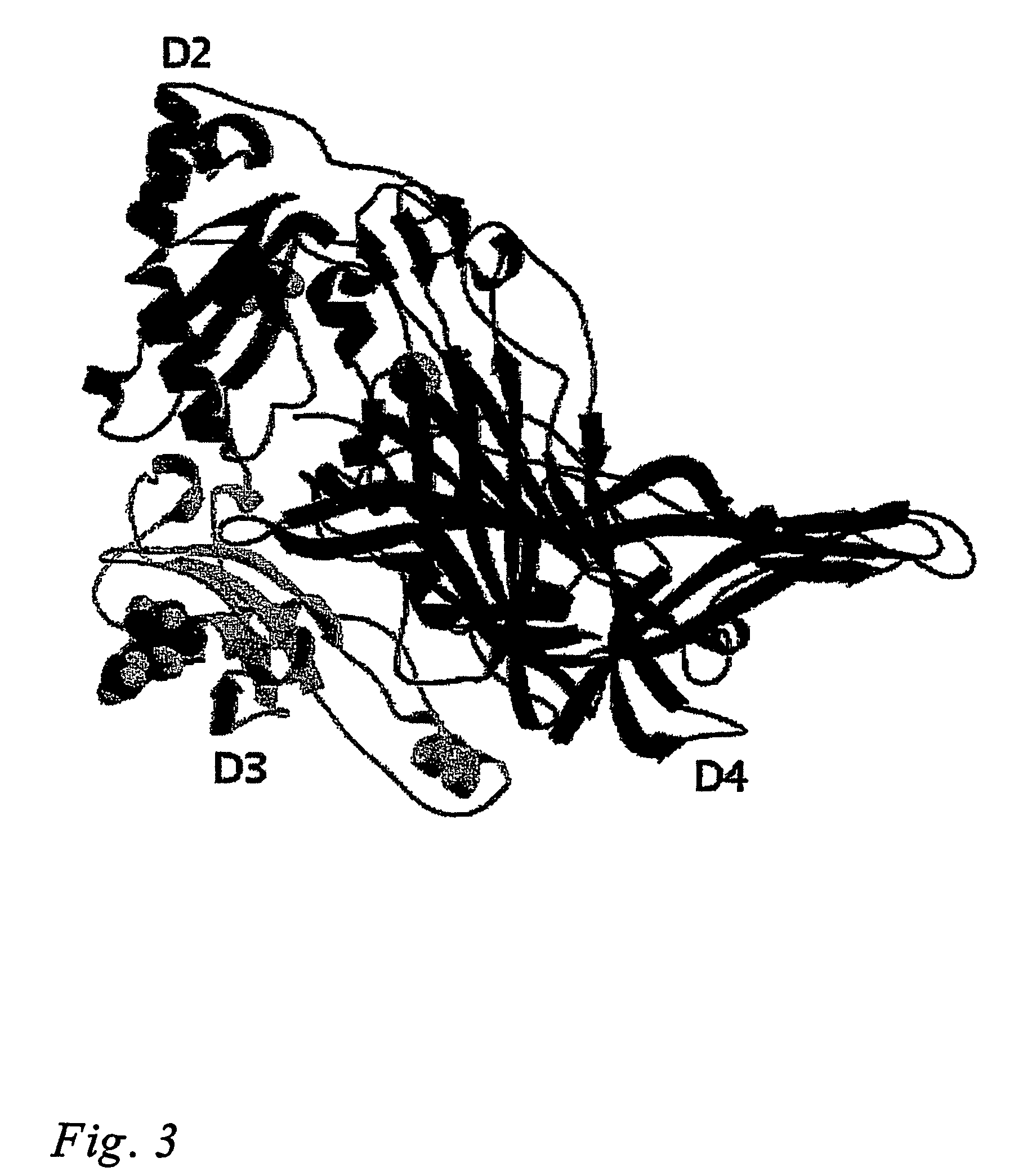
[0077]The structure of VAP-1 was solved by molecular replacement using the program AMORE (Navaza, 1994) of the CCP4i program suite (Collaborative Computational Project, 1994). This method confirmed that the space group of VAP-1 crystals was P6522 with one biological unit, a dimer per asymmetric unit. Out of the dimeric polyalanine backbones of Escherchia coli CAO (residues 93-720; PDB code 1OAC), Pisum sativum CAO (residues 7-634; PDB code 1KSI), Hansenula polymorpha CAO (residues 22-655; PDB code 1A2V) and Arthobacter globiformis CAO (residues 9-623; PDB code 1AV4) tested in molecular replacement, the structure of P. sativum gave the best correlation coefficient (44.1%) and Rfactor (53.4%) and was used a search model.
[0078]Electron density maps were calculated with FFT of CCP4i suite and were predictable enough to trace the VAP-1 polypeptide, even though, at the beginning of building, in several fragments. The model was manually built using the program O and ...
PUM
| Property | Measurement | Unit |
|---|---|---|
| pH | aaaaa | aaaaa |
| pH | aaaaa | aaaaa |
| volume | aaaaa | aaaaa |
Abstract
Description
Claims
Application Information