High sensitivity biomolecule detection with magnetic particles

a biomolecule and magnetic particle technology, applied in the field of biomolecule detection, can solve the problems of limited usefulness of radioactive probes to detect a specific protein in a heterogeneous population, cumbersome and expensive systems, and short shelf life of radioactive probes, so as to improve the binding effect of probes and reduce background nois

Inactive Publication Date: 2009-03-17
FOX JOHN
View PDF2 Cites 22 Cited by
  • Summary
  • Abstract
  • Description
  • Claims
  • Application Information

AI Technical Summary

Benefits of technology

[0011]Recognizing the limitations associated with current techniques for detection, manipulation and separation of biomolecules, the present invention provides methods and systems for the detection and manipulation of biomolecules using magnetic particles. Through its embodiments, the present invention improves specificity and sensitivity while minimizing background, cost and handling issues related to biomolecule detection and manipulation.
[0015]Methods and systems for the detection and separation of nucleic acids, as one example only, using ferrofluids and beads, are also provided. The invention also includes methods and systems to enhance the binding of a probe to a target biomolecule and methods to reduce the background noise in hybridizations and binding assays.

Problems solved by technology

While these techniques may be useful to resolve total protein in a sample, they are limited in their usefulness to detect a specific protein in a heterogeneous population of proteins.
The PhosphorImager instrument, an expensive laser based optical system, and clean image-ready phosphor screens are needed for each sample read, making the system both cumbersome and very expensive.
In addition, radioactive probes have a short shelf life (T2=days to months) and require tight inventory control in a licensed facility.
Although some companies are currently performing genetic screening using this method, the cost is prohibitive for most diagnostic procedures.
One significant problem with this approach is non-specific background.
Several natural occurring molecules either contribute to or quench the fluorescent signal, making this technique prone to a background noise which prevents this system from achieving highly sensitive nucleic acid detection.
While the CCD cameras are robust, CCD based systems do not have the sensitivity of film and the reagents have a one-year shelf life when stored at 4° C.
As with fluorescence detection approaches, this approach is limited by background noise caused by naturally occurring enzymes or compounds contributing to the signal.

Method used


Image

  • High sensitivity biomolecule detection with magnetic particles
  • High sensitivity biomolecule detection with magnetic particles
  • High sensitivity biomolecule detection with magnetic particles

Examples


example 1

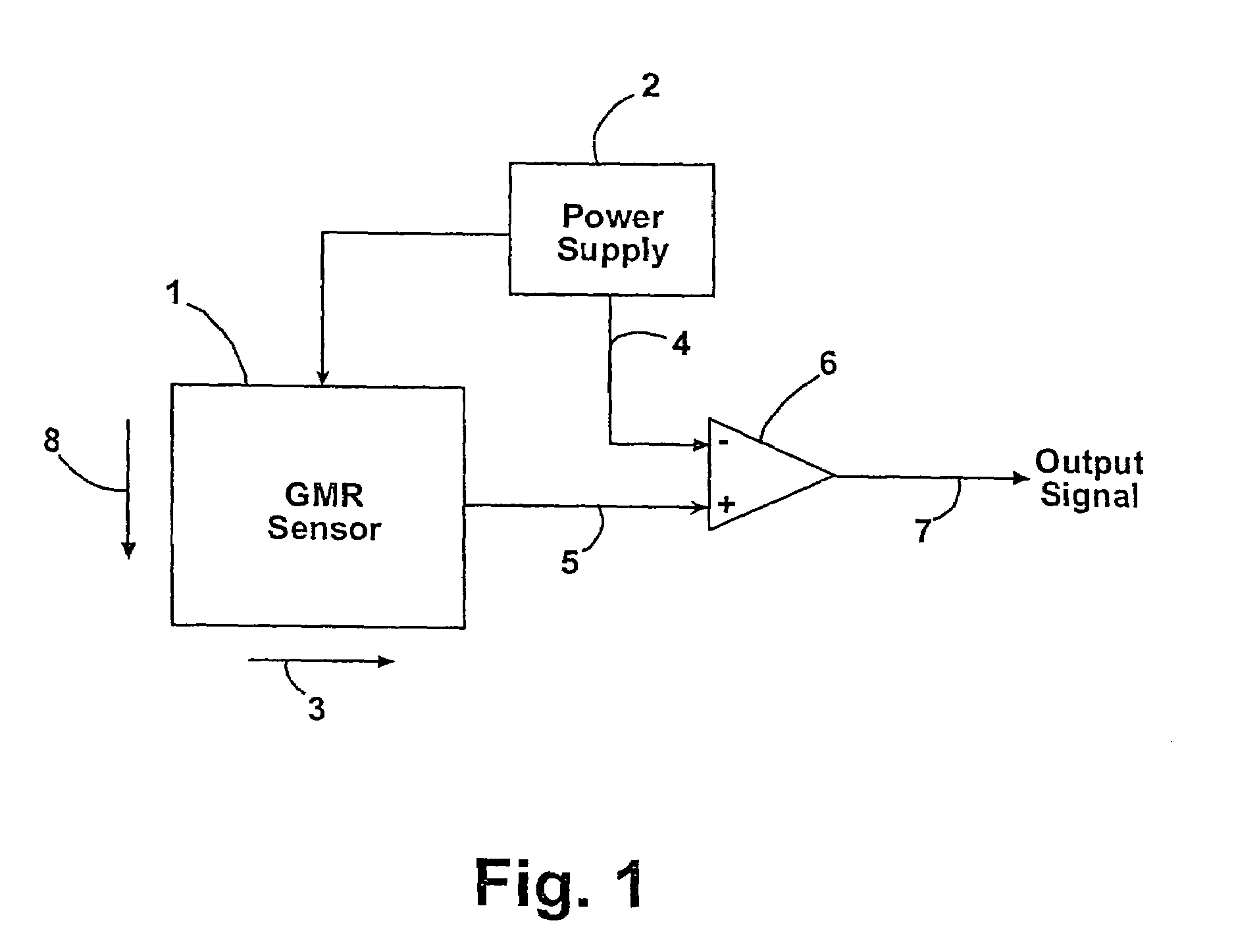
[0079]In this example, the present invention detects the presence and location of a nucleic acid disposed on a support by means of a magnetic label attached to a nucleic acid. As an example, one microliter containing 100 ng plasmid DNA (pHIS, ≈3700 base pairs / molecule) are spotted on a positively charged nylon membrane (Hybond, Amersham) that is subsequently cut into 1 cm×4 cm strips. The strips are air dried for 10 minutes and each strip is dipped in distilled water for 10 seconds. Next, the strips are contacted with a ferrofluid (EMG607; Ferrofluidics, Nashua N.H.) for two minutes, then washed for 2 minutes in distilled water and air dried.

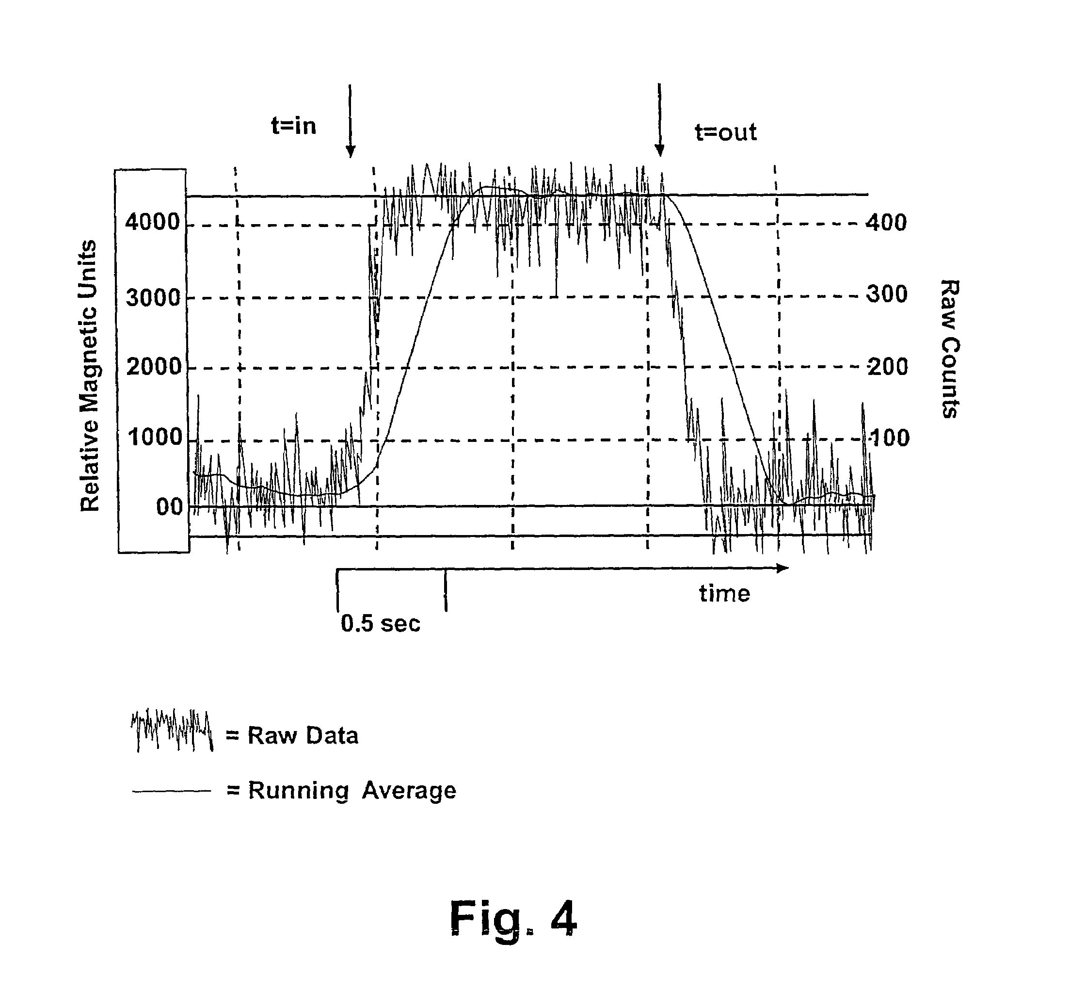
[0080]Samples are measured using a GMR sensor (T15 model; Nonvolatile Electronics Inc., Eden Prairie Minn.) with bias-magnets, and the voltages are recorded by a PC after analog processing and 12 bit A / D conversion. Output from the prototype magnetic detection system unit for the detection of nucleic acids is shown in FIG. 4. The magnetism of th...

example 2

[0082]In this example, the invention detects a protein disposed on a support. In this example, the invention detects the presence and location of a protein disposed on a support by means of a magnetic label attached to a protein. In one example, 1 ul of a solution containing either mouse IgG or goat IgG is spotted on a membrane that is subsequently cut into 1 cm×4 cm strips. The strips are then air dried and each strip is dipped in distilled water. Next, the strips are contacted with 2 ul of ferrofluid (EMG607; Ferrofluidics, Nashua N.H.). The samples are then washed, allowed to air dry, and are measured using a GMR sensor (T15 model; Nonvolatile Electronics Inc., Eden Prairie Minn.) with bias-magnets, and the voltages were recorded by a PC after processing as in Example 1. The results show that ferrofluid efficiently labeled mouse IgG and goat IgG which was disposed on a support and the proteins are effectively detected using the GMR sensor.

example 3

[0083]The invention also comprises hybridization using magnetically labeled nucleic acid probes are provided. In this example, the invention uses a magnetic label attached to a nucleic acid probe to detect a target nucleic acid disposed on a support. A first oligonucleotide of 52 nucleotides (T54) is used as a target nucleic acid and a second complementary oligonucleotide (T55), bound with a ferrofluid, was used as a probe nucleic acid. One microliter of the target nucleic acid T54 (at 0.5 ug / ul) is spotted on a nylon membrane and is crosslinked with UV light (autolink setting; Stratalinker 2500; Stratagene). Subsequently, the membrane is washed briefly with water and air dried. The probe nucleic acid is made by incubating 2 ug of a complementary oligonucleotide, T55, with ferrofluid (1:1 (v:v) in 10:1 total volume). Unbound T55 is separated and removed from the ferrofluid conjugated T55 by washing with water, as described above.

[0084]The hybridization is conducted as follows. The f...


PUM

PropertyMeasurementUnit
atomic numbersaaaaaaaaaa
Tmaaaaaaaaaa

Abstract

The present invention generally relates to the field of biomolecule detection. More specifically, the present invention relates to compositions, methods and systems for the detection and manipulation of biomolecules using magnetic particles.

Description

CROSS-REFERENCE TO RELATED APPLICATION[0001]This application claims priority based on U.S. provisional patent application No. 60 / 150,210 filed Aug. 21, 1999, which is hereby incorporated by reference in its entirety.FIELD OF THE INVENTION[0002]The present invention generally relates to the field of biomolecule detection. More specifically, the present invention relates to compositions, methods, and systems for the detection and manipulation of biomolecules using magnetic particles.BACKGROUND OF THE INVENTION[0003]In basic research, one goal is to understand how genes are distributed within populations and how expression of those genes leads to phenotypic differences. This information has the potential to become a powerful tool for predicting human health trends and has been a driving force behind the search for genetic markers for human disease.[0004]Over the years, many biochemical techniques have been introduced for analyzing the presence and / or amount of a biomolecule in a sample...

Claims


Application Information

Patent Timeline
Patent Type & Authority Patents(United States)
IPC IPC(8): G01N25/18B03C1/01C12N15/10C12Q1/68C12Q1/6834C12Q1/6837G01N33/543
CPCB03C1/01C12N15/1013C12Q1/6834C12Q1/6837G01N27/745G01N33/54326G01N33/54333C12Q2565/513C12Q2563/143
Inventor FOX, JOHN
Owner FOX JOHN
Why Eureka
Try Eureka