Implantable vascular access device with ceramic needle guard insert

a technology of implantable vascular access and ceramic needle guard insert, which is applied in the direction of medical devices, other medical devices, etc., can solve the problems of difficult fabrication, high cost, and all-metal construction, and achieve the effect of better anatomical fit in the subcutaneous area

Inactive Publication Date: 2010-05-11
PRIMO MEDICAL GRP INC
View PDF24 Cites 95 Cited by
  • Summary
  • Abstract
  • Description
  • Claims
  • Application Information

AI Technical Summary

Benefits of technology

[0030]Accordingly, the present invention provides an improved vascular access port comprising a plastic port base with a metallic dish insert molded (or bonded) into the bottom of the reservoir. In one embodiment, a single reservoir is provided. In another embodiment, plural reservoirs are provided. The metallic bottom of the reservoir provides a hard surface that will resist abrasion and puncture by the access needles used to infuse medication or withdraw blood. The features of both the single and dual ports include ‘duck tail’ rears. This feature is intended to better anatomically fit into the subcutaneous areas around muscle tissue. The plastic must be of a bio-compatible and chemically-compatible material such as Nylon, Poly Acetals, or Polyethersulphone. The chemical resistance is necessary so that the injected medications do not react with the port materials. The yield and creep strength and flexure modulus of the material is important so that the plastic will hold and retain the port top retaining the septum.
[0032]The cup may be made of stainless steel, titanium or ceramic (alumina or zirconium). All these materials are biologically and chemically inert so that they will not react with the body or medications. The insert is formed, cast, machined or molded into a ‘dish’ shape with the edges being of a radius greater than 0.035″. This radius is important for the reduction of coagulation and turbulence of the blood and medication within the reservoir.
[0035]The septums for these ports are intended to assist in the location (and in the case of the dual port) differentiation of the reservoir location. The combinations for these may vary for application preferences. The two styles include concave and convex shapes. After the surgical implantation, the location is determined by palpitation of the skin over the port. For the illustration shown, the concave side feels different than the convex side. The convex feature has a nipple shape. Both septums are designed to fit into the port body and be retained by the port top. The septum is intended to be self sealing under the pressures produced by the vascular system and the injection pressures of injections. This is achieved by controlling the rubber hardness and the dimensions relative to the retained assembly. The interference fit or squeezing of the septum rubber creates a residual pressure radially toward the center of the septum. The softness of the rubber allows the punctures to reseal without leakage. It is preferred that non-coring needles are used by clinical staff to avoid excessive reduction of material and coring or skiving material into the reservoir which flow into the blood stream. The rubber material commonly used for septums is silicone, but other biocompatible elastomer materials may be used.
[0036]Advantageously, the present invention provides low cost, yet durable subcutaneous vascular access port(s). The port(s) can be manufactured from bicompatible grades of plastics and metals which meets the safety and clinical parameters of implanted devices. This plastic port features a metallic (titanium, stainless steel or ceramic) dish which will be insert molded pressed, or bonded into the base of the port reservoir to prevent potential penetration of needles through the base of a typical style plastic port. Also advantageously, the port of the present invention features a titanium shield molded into a plastic port top around the rubber septum to prevent needle mislocation, false needle locations and potential medication misdirection. The port septums have incorporate in them indication features to distinguish which port reservoir the surgeon or nurse is accessing and better locates the reservoir center. These features are utilized in other dual lumen ports by an indication on the port itself, but not on the septum.

Problems solved by technology

Additionally, all-metal constructions, such as titanium, or stainless steel provide a port which is both biocompatible and compatible with the injected fluid.
However, all-metal constructions present the disadvantages that they are relatively heavy, difficult to fabricate and relatively expensive.
Additionally, all-metal ports produce large Magnetic Resonance Imaging (MRI) artifacts.
However, ports constructed from plastic have the disadvantage that infused fluids may react with the plastic body of the port.
All-plastic ports contain the disadvantage that they cannot maintain a sealing engagement with the septum after repeated percutaneous injections.
Additionally, all-plastic ports are susceptible to nicks and scratches on the interior surface by the accessing needle.
These nicks and scratches could lead to nidus, blood clots, or precipitation formations.
These shortcomings include quality control problems during manufacturing, and expensive molding processes.
This design has shortcomings associated with it, including defects in the plastic housing which may cause an improperly sealed septum.
Although these materials are nonreactive, access ports constructed utilizing titanium or stainless steel materials produce an interfering or blurred image of the body of the patient in the vicinity of the implanted access port when diagnostic imaging techniques such as magnetic resonance imaging (“MRI”), CAT scans, or computerized tomography are used.
Therefore, the use of metallic access ports limits the diagnostic imaging techniques that may be used relative to those areas of the body in which an access port is implanted.
A further problem relating to the materials for and manufacture of access ports is the deleterious impact of some manufacturing procedures on the fluids which flow through the fluid cavities and related structures located between the fluid cavities and the catheter.
This manufacturing process often leaves sharp edges, seams and corners in the areas where the fluid cavity is to direct the flow of the fluid through an exit passageway.
In addition, the production of the circular fluid cavities often results in the creation of areas within the housing in which fluid flow is retarded.
As the flow of fluids through dead spaces is retarded, stagnation occurs, resulting in some fluid being trapped within these dead spaces.
If the access port is used to withdraw or transfuse blood, blood trapped in these dead spaces may form clots and block the flow of fluid through the fluid cavity.
This results in an undesirable seam being formed where the adjacent parts abut one another.
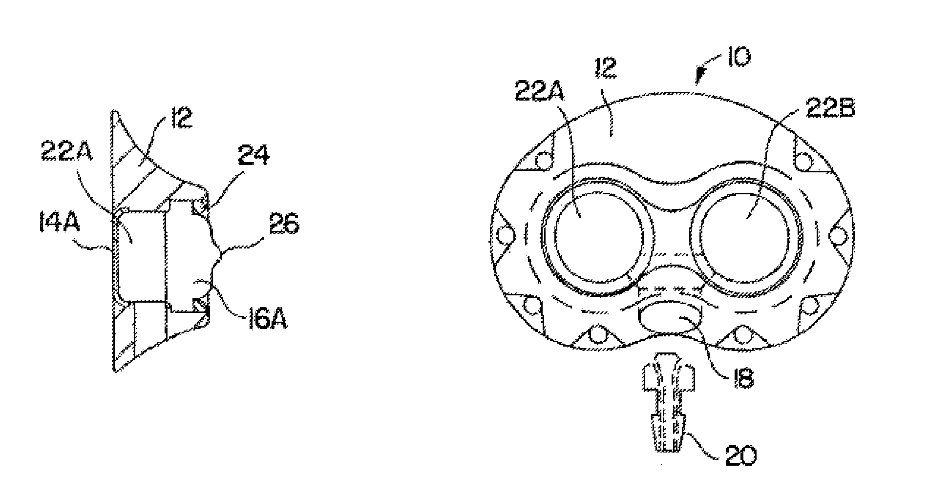
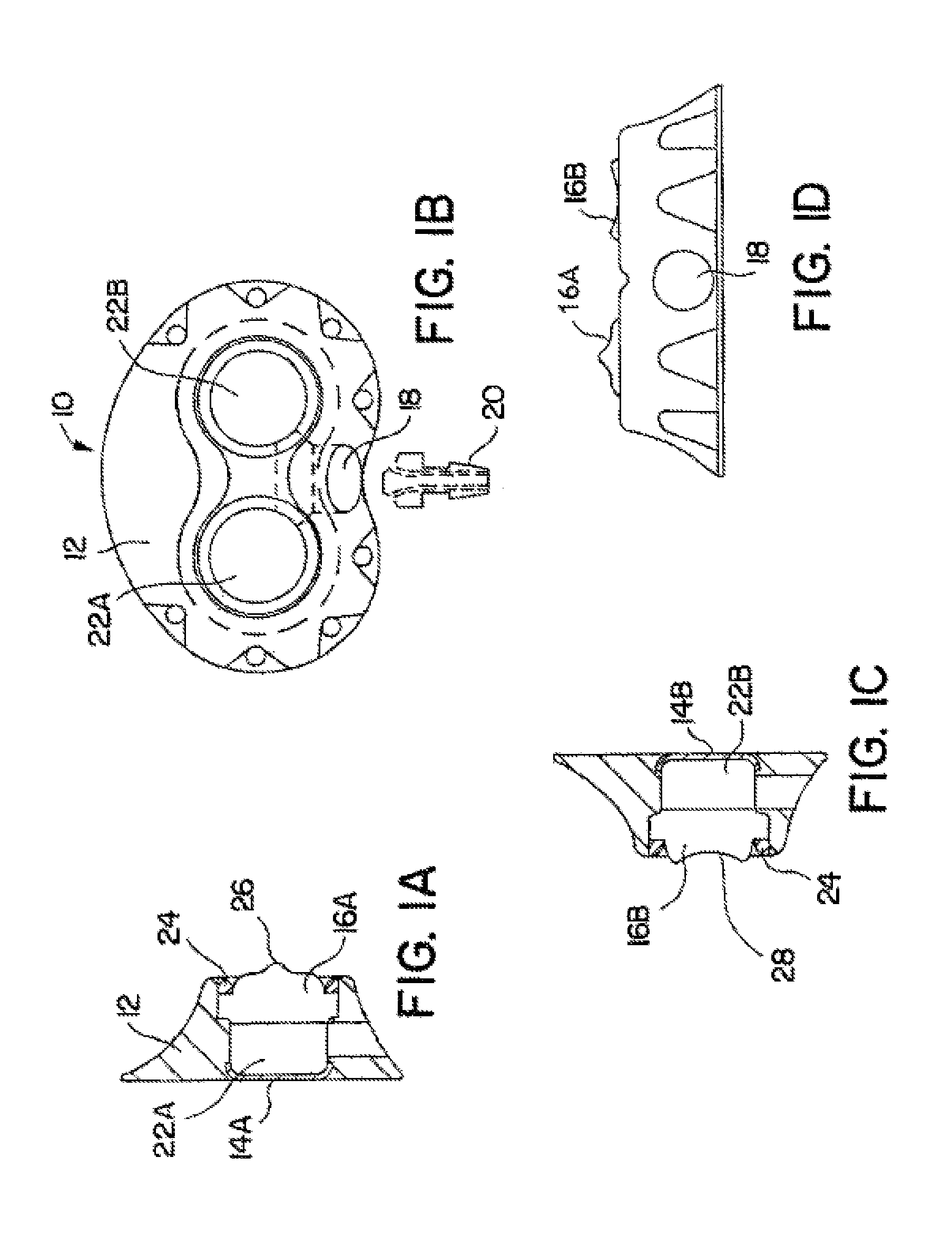
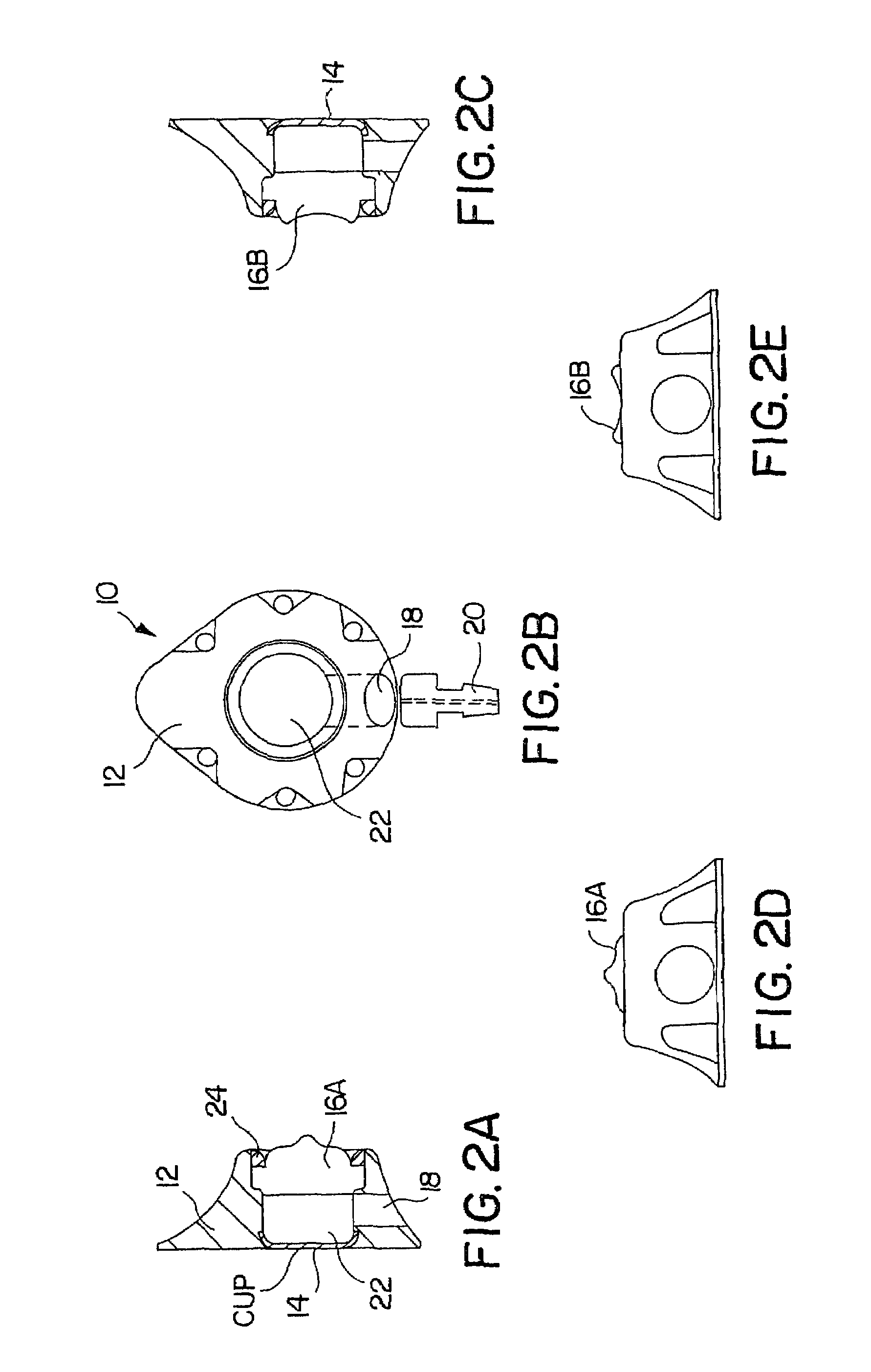
A further problem encountered in the design and construction of access port relates to the positioning of the septums within the housing of the access port.
The distancing of the septums to facilitate their differentiation, however, results in a corresponding distancing of the fluid cavities.
Naturally, this complicates the process of manufacture and increases its cost, as well as the changes of structural failure.
The use of such a metal component will interfere with the production of an access port which is free of limits as to the diagnostic imaging techniques that may be used relative to those areas of the body in which an access port is implanted.
In addition, the integral nature of such metal outlet passageways raises the possibility of leakage of medication through the interstices between the metal tubes and the body of the access port.
This type of arrangement increases the size of the overall access port and its cost of manufacture by adding thereto the necessity of fabricating and assembling of the hub element.
An additional set of problems encountered in the use of access ports relates to the actual connection of the catheter to the access port.
When utilizing this system, however, it is difficult to determine the amount of engagement of the catheter onto the outlet stem.
Some catheter connection systems do not allow visual verification of attachment.
As a result, leakage and failure can occur.
While this practice alleviates many of the problems with leakage and failure due to catheter slippage, this system severely limits the type of the catheter usable with the access port.

Method used


Image

  • Implantable vascular access device with ceramic needle guard insert
  • Implantable vascular access device with ceramic needle guard insert
  • Implantable vascular access device with ceramic needle guard insert

Examples


Embodiment Construction

[0043]FIGS. 1A-1D depict various views of the preferred dual-port implantable access device 10 of the present invention. The port 10 generally comprises a housing member 12 defining fluid chambers 22A and 22B. The chambers are sealed by the housing 12, bottom cup members 14A and 14B, and self-sealing septum members 16A and 16B. In this embodiment, the housing 12 is preferably formed of titanium, stainless steel, ceramic, and / or other biocompatible material. The septum 16A and 16B is preferably formed of silicon or other semi-permeable materials that permit ingress and egress of needles to deliver fluid to the chambers 22A and / or 22B. An exit port 18 is provided in communication with chambers 22A and 22B, which delivers fluid out of the chambers 22A and / or 22B to a predetermined location, via stem 20 and attached catheter (not shown), as is understood in the art.

[0044]The septums 16A and 16B are formed with a generally circular shape, and, as shown in the drawings, may include a nipp...


PUM


Abstract

The present invention provides an improved vascular access port comprising a port base with a metallic dish insert molded (or bonded) into the bottom of the reservoir. In one embodiment, a single reservoir is provided. In another embodiment, plural reservoirs are provided. The metallic bottom of the reservoir provides a hard surface that will resist abrasion and puncture by the access needles used to infuse medication or withdraw blood. Additionally, the single and dual ports can include exit ports that are intended to better anatomically fit into the subcutaneous areas around muscle tissue.

Description

[0001]This application is a continuation application of application Ser. No. 09 / 582,406, filed Jun. 23, 2000, now U.S. Pat. No. 6,527,754, which claims priority from PCT Application Serial No. PCT / US99 / 28695, filed Dec. 3, 1999, which claims priority from U.S. Provisional Application Ser. No. 60 / 111,257, filed Dec. 7, 1998, all of which are incorporated herein by reference.BACKGROUND OF THE INVENTION[0002]1. Field of the Invention[0003]The present invention relates generally to a subcutaneously implantable vascular access port. More specifically, the present invention relates to an access port having a single needle-penetrable, self-sealing septum which affords repeated access to a plurality of distinct fluid cavities having staggered outlet ducts in communication with a plural lumen catheter.[0004]2. Description of Related Art[0005]Access portals, or ports, provide a convenient method to repeatedly deliver medicants to remote areas of the body without utilizing surgical procedures....

Claims


Application Information

Patent Timeline
Patent Type & Authority Patents(United States)
IPC IPC(8): A61M31/00A61M39/02
CPCA61M39/0208A61M2039/0226A61M2039/0211
Inventor TALLARIDA, STEVEN J.ETTLINGER, MARK
Owner PRIMO MEDICAL GRP INC
Why Eureka
Try Eureka