Detection of micro metastasis of melanoma and breast cancer in paraffin-embedded tumor draining lymph nodes by multimarker quantitative RT-PCR

a technology of paraffin-embedded tumors and micro metastases, applied in the field of micro metastasis detection of melanoma and breast cancer, can solve the problems of malignant neoplasms' tendency to disseminate cells, high labor intensity of approaches, and often underestimate the presence of micrometastatic disease in procedures. , to achieve the effect of accurate determination, high sensitivity and specificity

Inactive Publication Date: 2011-03-22
JOHN WAYNE CANCER INST
View PDF16 Cites 17 Cited by
  • Summary
  • Abstract
  • Description
  • Claims
  • Application Information

AI Technical Summary

Benefits of technology

[0021]The present invention provides a number of unexpected advantages. First, it allows to determine stages of the cancers more accurately as compared to the existing methods. The methods and kit of the present invention allow accurate determination of a need for further lymph node removal surgery and / or adjuvant treatment. Second, the present invention demonstrates the better reliability of molecular marker panels of the present invention in prediction of the disease outcome and recurrence as compared to conventional prognostic factors such as age, gender, Breslow thickness, Clark level, site, ulceration. The panels of the present invention provide an accurate prediction of disease recurrence in histopathology negative SLN (p<0.0001) and survival (p<0.0001). The methods of the present invention diagnose metastasis in SLN with high sensitivity and specificity.

Problems solved by technology

One of the most devastating aspects of cancer is the propensity of cells from malignant neoplasms to disseminate from their primary site to distant organs and develop into metastases.
However, such approach is labor-intensive.
Also, about 80% of patients who undergo elective lymph node dissection have no evidence of metastases and do not benefit from this procedure.
But this procedure often underestimates the presence of micrometastatic disease.
Despite initially encouraging results, the clinical and survival significance of these occult metastases detected by RT-PCR assays has not been demonstrated conclusively.
Furthermore, conventional single mRNA marker RT-PCR assays are limited in their ability to discriminate cancer cells from normal cells that also carry the marker and, thus, these methods suffer from reduced specificity and reliability.
In addition, tumor heterogeneity has caused sensitivity problems where a single mRNA marker was employed—although all tumor cells within a primary tumor or metastasis may express the same marker gene, the level of specific mRNA expression can vary considerably.
Thus, despite the identification of melanoma and breast cancer markers, these markers cannot individually detect tumor cells in a highly specific and sensitive manner (U.S. Pat. No. 6,057,105).
Although this study reported a great improvement of detection sensitivity compared to H&E staining and IHC, the study did not determine clinicopathologic importance of the detection.
Also, since the study limited the follow-up period to 12 months and recurrence of melanoma, typically, takes 3-8 years, it failed to establish an ability of the selected markers to predict disease recurrence and patient's overall survival.
But the study did not establish the prognostic significance of the method (Bilchik, A. J., 2000).
Although the methods of the U.S. Pat. No. 6,057,105 are more sensitive than RT-PCR with a single marker, they fail to predict a long-term (at least 3 years) disease recurrence and survival.
However, similarly to the U.S. Pat. No. 6,057,105, the U.S. Pat. No. 6,037,129 does not conduct a retrospective long-term (greater than 5 years) analysis of the samples and, thus, does not evaluate prognostic significance of the selected markers.
Also, the selected set of the markers has a limited utility as it produced a large number (40%) of false positive results.
In summary, none of the currently available methods provides a reliable prediction of disease recurrence, patient's prognosis and survival, particularly in AJCC stage I, II and III patients.

Method used


Image

  • Detection of micro metastasis of melanoma and breast cancer in paraffin-embedded tumor draining lymph nodes by multimarker quantitative RT-PCR
  • Detection of micro metastasis of melanoma and breast cancer in paraffin-embedded tumor draining lymph nodes by multimarker quantitative RT-PCR
  • Detection of micro metastasis of melanoma and breast cancer in paraffin-embedded tumor draining lymph nodes by multimarker quantitative RT-PCR

Examples


example 1

Patients and Tumors

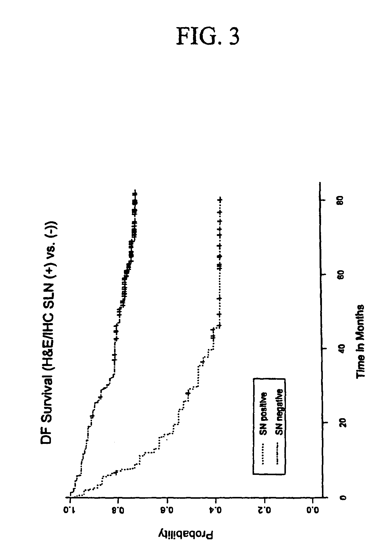
[0060]SLN specimens were obtained in consultation with the surgeon and pathologist at the John Wayne Cancer Institute (JWCI). Informed human subject IRB consent was obtained from patients for the use of all specimens. All patients had AJCC stage I or II malignant melanoma (staged by current AJCC staging system) (Balch, C. M., 2001) which are defined as those patients with no clinical evidence of regional or metastatic disease. SLND was performed in the patients as previously described (Morton, D. L., 1992). Total 308 SNs obtained from 215 patients were assessed for histopathologic examination and qRT-PCR assay. The patients ranged in age from 18.7 to 91.3 years old (mean age 50.9±15.9 SD), consisting of 127 males and 88 females. The patients who had histopathologically (H&E / IHC) proven metastatic melanoma in SLNs at SLND subsequently received complete lymph node dissection of the lymphatic basin. Patients were followed up by clinical diagnostic examinations in the...

example 2

Histopathologic Examination and RNA Isolation

[0063]Histopathologic examination by the pathologists was performed on each collected SN as previously described (Bostick, P. J., 1999, which is incorporated herin by the reference). Each SN was bisected, and an 8-μm imprint slide was then prepared from the tissue surface and stained with Diff-Quik I & II (Dade International, Miami, Fla.) for the pathologist's intraoperative diagnosis. If melanoma cells were identified in the frozen section, a complete lymph node dissection was then performed (Bostick, P. J., 1999). Six immediately adjacent frozen sections were cut on the cryostat to a thickness of 12 μm each and stored at −80° C. until processed at a later date (Bostick, P. J., 1999; Miyashiro, I., 2001). The remainder of the bisected node, which was not frozen, was then placed in 10% formalin and embedded in paraffin, and 4-μm-thick sections were examined with H&E staining. Adjacent 4-μm-thick sections werre evaluated by IHC using antib...

example 3

Dilution Study

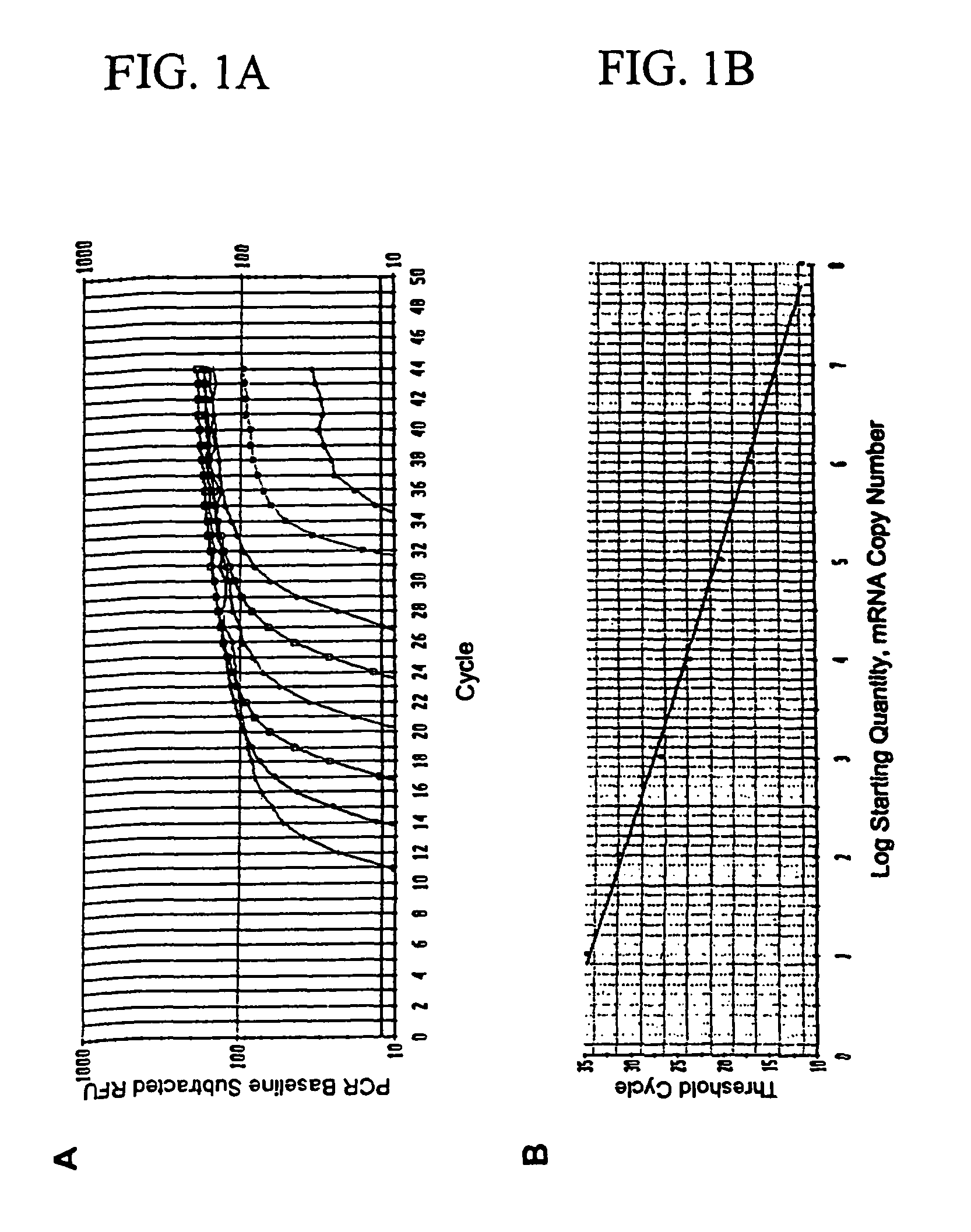
[0065]To verify the specificity of detecting occult metastasis, total RNA was extracted from 1000 melanoma cells of obviously metastatic melanoma tissue (n=8) in PE sections using laser capture microdissection (LCM). Five-μm thick sections were cut from each tissue specimen embedded in paraffin using a microtomb and mounted on a slide. Then, tissue sections were deparaffinized with xylene and subsequently stained with H&E. After drying a slide, 1000 metastatic melanoma cells were microdissected using the PixCell II LCM System (Arcturus Engineering, Mountain View, Calif.) as manufacture's procedure. Microdissected tissues were digested with proteinase K and total RNA was extracted using the Paraffin Block RNA Isolation Kit (Ambion). RNA isolated from 1000 melanoma cells was dissolved in molecular grade water, and was serially diluted by ½, 1 / 10, 1 / 20, 1 / 50, 1 / 100, 1 / 500, and 1 / 1000. Reverse transcriptase reaction and qRT-PCR assays were performed for each marker on the ...


PUM

PropertyMeasurementUnit
thicknessaaaaaaaaaa
volumeaaaaaaaaaa
thickaaaaaaaaaa

Abstract

The invention provides a quantitative realtime RT-PCR assay for detection of metastatic breast, gastric, pancreas or colon cancer cells or metastatic melanoma. The assay allows to predict disease recurrence and survival in patients with AJCC stage I and II, and III disease using multimarker panels. The method for detecting metastatic melanoma cells utilizes panels of markers selected from a group consisting of MAGE-A3, GalNAcT, MART-1, PAX3, Mitf, TRP-2, and Tyrosinase. The method for detecting metastatic breast, gastric, pancreas or colon cancer cells in paraffin-embedded samples utilizes panels of markers selected from a group consisting of C-Met, MAGE-A3, Stanniocalcin-1, mammoglobin, HSP27, GalNAcT, CK20, and β-HCG.

Description

[0001]This invention claims priority to the U.S. Provisional Patent Application No. 60 / 426,216 filed Nov. 14, 2002.BACKGROUND OF THE INVENTION[0002]The present invention relates to methods of detection of micro metastasis in lymph nodes and methods of predicting the recurrence of the disease and survival. More specifically, the invention is directed to detection of micro metastasis in cancerous tissue and lymph nodes by using multimarker real-time reverse transcriptase polymerase chain reaction assay.DESCRIPTION OF THE PRIOR ART[0003]The incidence of malignant melanoma has been increasing in the United States over the past decade (Balch, C. M., 2001; Jemal, A. J., 2002; and Rigel, D. S., 1996). Breast cancer is another common form of malignant disease whose incidence is increasing. One of the most devastating aspects of cancer is the propensity of cells from malignant neoplasms to disseminate from their primary site to distant organs and develop into metastases. Despite advances in ...

Claims


Application Information

Patent Timeline
Patent Type & Authority Patents(United States)
IPC IPC(8): C12Q1/68C12P19/34G01N33/53
CPCC12Q1/6886C12Q2600/118C12Q2600/112
Inventor HOON, DAVE S. B.TAKEUCHI, HIROYA
Owner JOHN WAYNE CANCER INST
Why Eureka
Try Eureka