Tissue engineered osteochondral implant

a technology implant, which is applied in the field of tissue engineered osteochondral implants, can solve the problems of limited repair response, articular cartilage, and lack of durability or mechanical properties of normal articular cartilage, and achieve the effect of small pore size and enhanced gravity

Active Publication Date: 2011-04-26
RUSH UNIV MEDICAL CENT +1
View PDF16 Cites 28 Cited by
  • Summary
  • Abstract
  • Description
  • Claims
  • Application Information

AI Technical Summary

Benefits of technology

In some embodiments, the method further includes, prior to contacting the scaffold, filling pores of the scaffold with a biocompatible filler agent. Filling the pores prevents growth of the overlying cells into the scaffold material beyond a limited depth. Filling the pores comprises using positive pressure from above to urge the filler agent into the pores; or using negative pressure (suction) from below to urge the filler agent into the pores. Alternatively filling the pores comprises adding the filler agent into the pores by the force of gravity, or by several fold enhanced gravity means such as by centrifugation. The same purpose, which is exclusion of growth of cells into at least a portion of the plurality of the pores for at least a portion of the time of culture, can be achieved by using a scaffold material with small pore sizes.

Problems solved by technology

Damage to the articular cartilage, subchondral bone, or both can result from traumatic injury or a disease state.
Injuries that do not penetrate subchondral bone are associated with a limited repair response.
Fibrocartilage lacks the durability or mechanical properties of normal articular cartilage, and eventually degrades from normal joint use.
Secondary degeneration at the site of the injury can ultimately result in poor joint function, pain, and disability.
The potential for donor site morbidity, produced by harvest of the autologous graft, however, presents a major remaining disadvantage.
Further, potential donor site morbidity limits effective treatment of a defect to a greater extent as the size of the defect increases.

Method used


Image

  • Tissue engineered osteochondral implant
  • Tissue engineered osteochondral implant
  • Tissue engineered osteochondral implant

Examples


Embodiment Construction

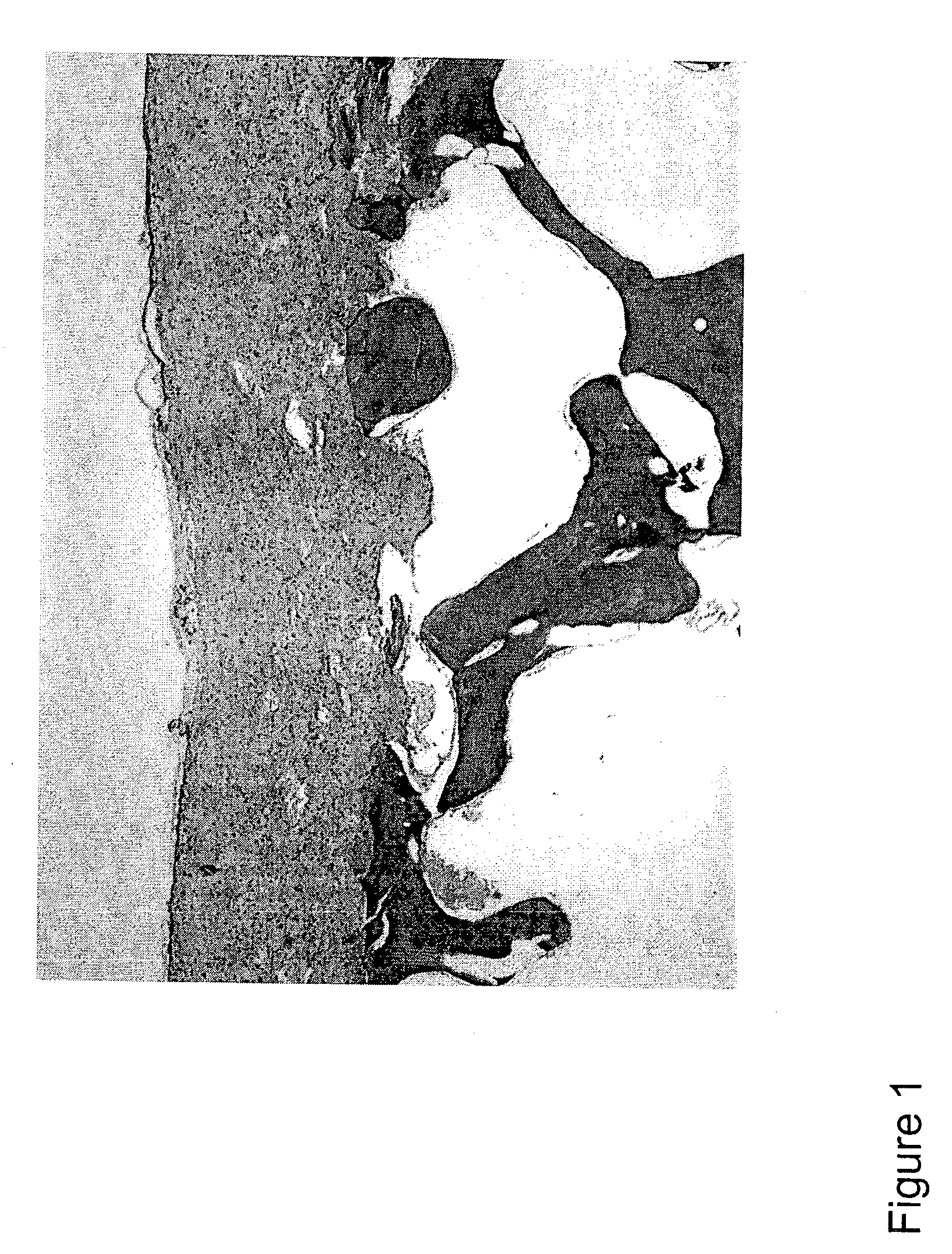
The present invention relates to a transplantable osteochondral implant and a method for its production. The implant includes engineered cartilage tissue attached to a biocompatible support scaffold. In the present methods, an osteochondral implant is produced in vitro by culturing isolated chondrogenic cells for an amount of time effective for the formation of a chondrogenic cell-associated matrix. The chondrogenic cells with cell-associated matrix are previously cultured on a porous biocompatible support scaffold in the presence of a growth factor, for a time effective for allowing both the formation of an engineered cartilage tissue and attachment of the engineered cartilage tissue to the biocompatible support scaffold.

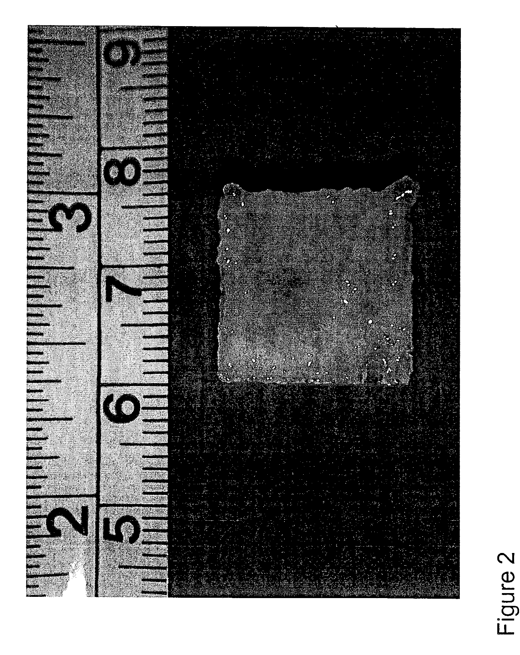
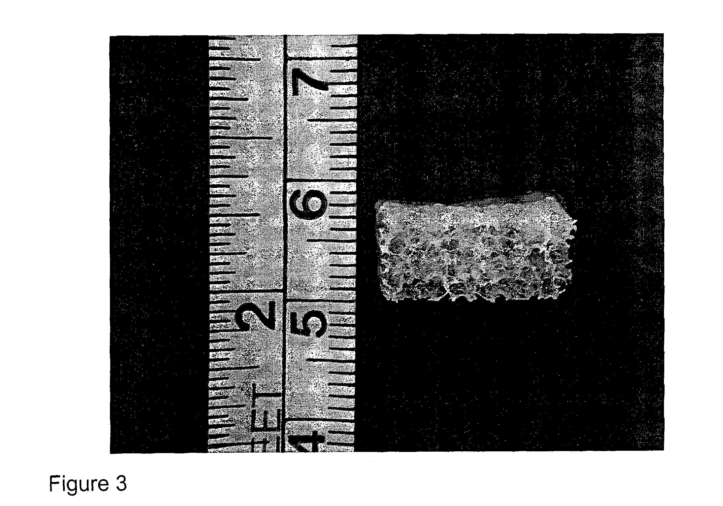
The porous biocompatible support scaffold can be composed of natural cancellous bone, demineralized natural cancellous bone, or bone substitute material such as calcium phosphate, or collagen, or hydroxyapatite or a combination of these materials and is of a thickn...


PUM

PropertyMeasurementUnit
thicknessaaaaaaaaaa
thicknessaaaaaaaaaa
thicknessaaaaaaaaaa

Abstract

Compositions, methods of production and use, and kits for an osteochondral graft involving both articular cartilage and underlying bone are provided.

Description

FIELD OF INVENTIONThe present invention relates to compositions for and methods of production of an osteochondral tissue graft comprising both articular cartilage and underlying bone, and methods for surgical implantation of the compositions into human joints for the purpose of filling defects, or replacing damaged or degraded cartilage and bone, and kits having such a graft.BACKGROUND OF THE INVENTIONHealthy human joint surfaces are covered by articular cartilage, which provides a resilient, durable surface with low friction, for distribution of mechanical forces and protection of the underlying bone. Cartilage is an avascular tissue, having a small number of chondrocytes encapsulated within an extensive extracellular matrix. Damage to the articular cartilage, subchondral bone, or both can result from traumatic injury or a disease state.There are significant differences in the ability of natural tissues to respond in vivo to different types of injury or disease. Injuries that do no...

Claims


Application Information

Patent Timeline
Patent Type & Authority Patents(United States)
IPC IPC(8): A61F2/02A01N63/00A61BA61B1/00A61F2/00A61F2/28A61F2/30A61F15/00A61L27/24A61L27/38A61L27/48A61L27/56G09B17/04G09B23/30G09B25/00
CPCA61L27/3608A61L27/3612A61L27/3654A61L27/3683A61L27/3852A61L27/56G09B23/30A61F2/30756A61F2/28Y10S623/901A61F2002/2817A61F2002/2835A61F2002/30762A61F2002/30764A61F2002/30766A61F2310/00293A61F2310/00365A61F2310/00359A61L27/3817
Inventor MASUDA, KOICHIHEJNA, MICHAEL J.PFISTER, BRIAN E.
Owner RUSH UNIV MEDICAL CENT
Why Eureka
Try Eureka