Method of preparing a hydrogel network encapsulating cells
a technology of hydrogel network and encapsulation cell, which is applied in the field of preparing a hydrogel network encapsulating cell, can solve the problems of limited performance and often inadequate mechanical strength, and achieve the effect of high mechanical strength
- Summary
- Abstract
- Description
- Claims
- Application Information
AI Technical Summary
Benefits of technology
Problems solved by technology
Method used
Image
Examples
example 1
Agarose / PHEMA Hydrogel Synthesis and Mechanical Testing
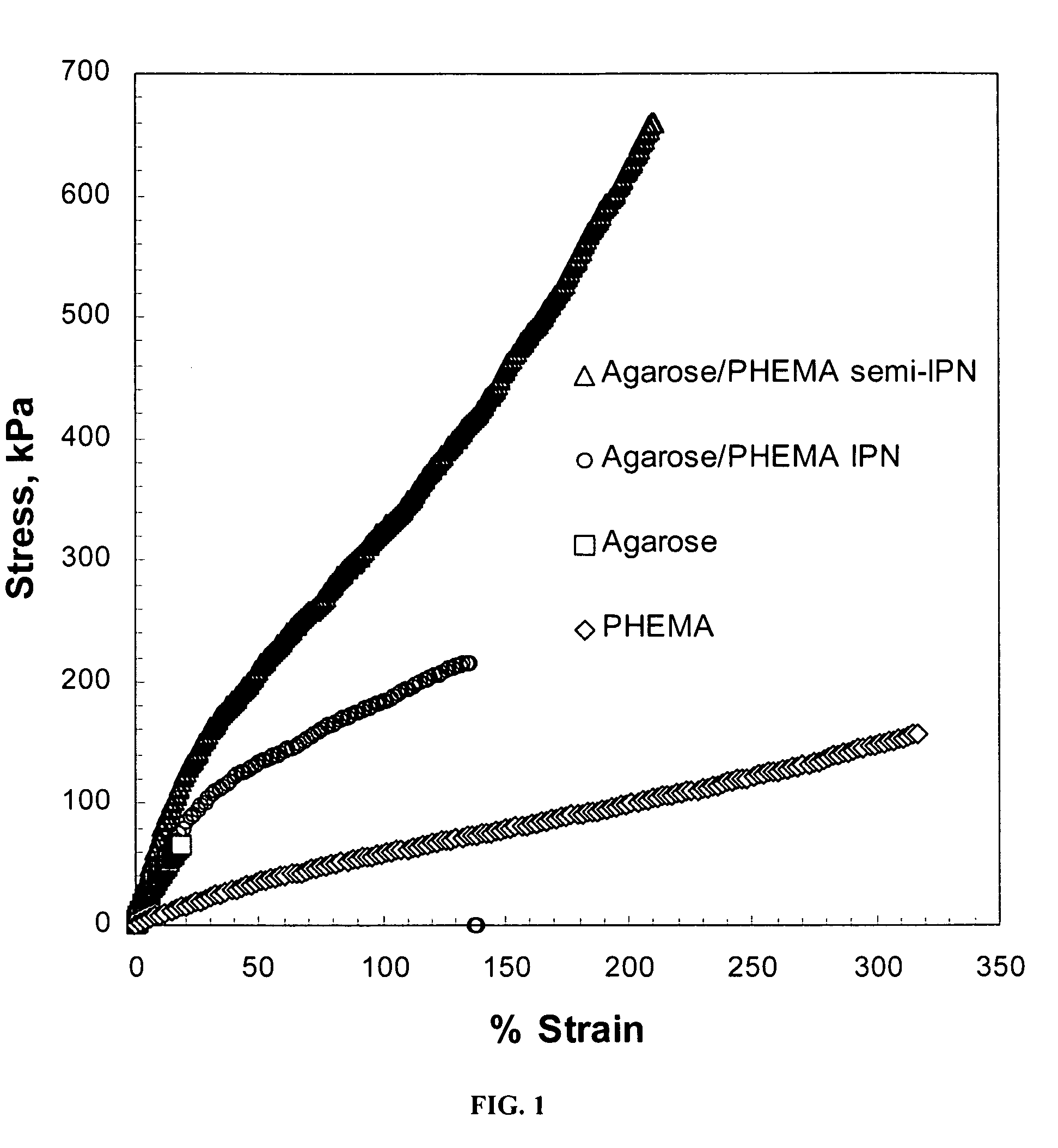
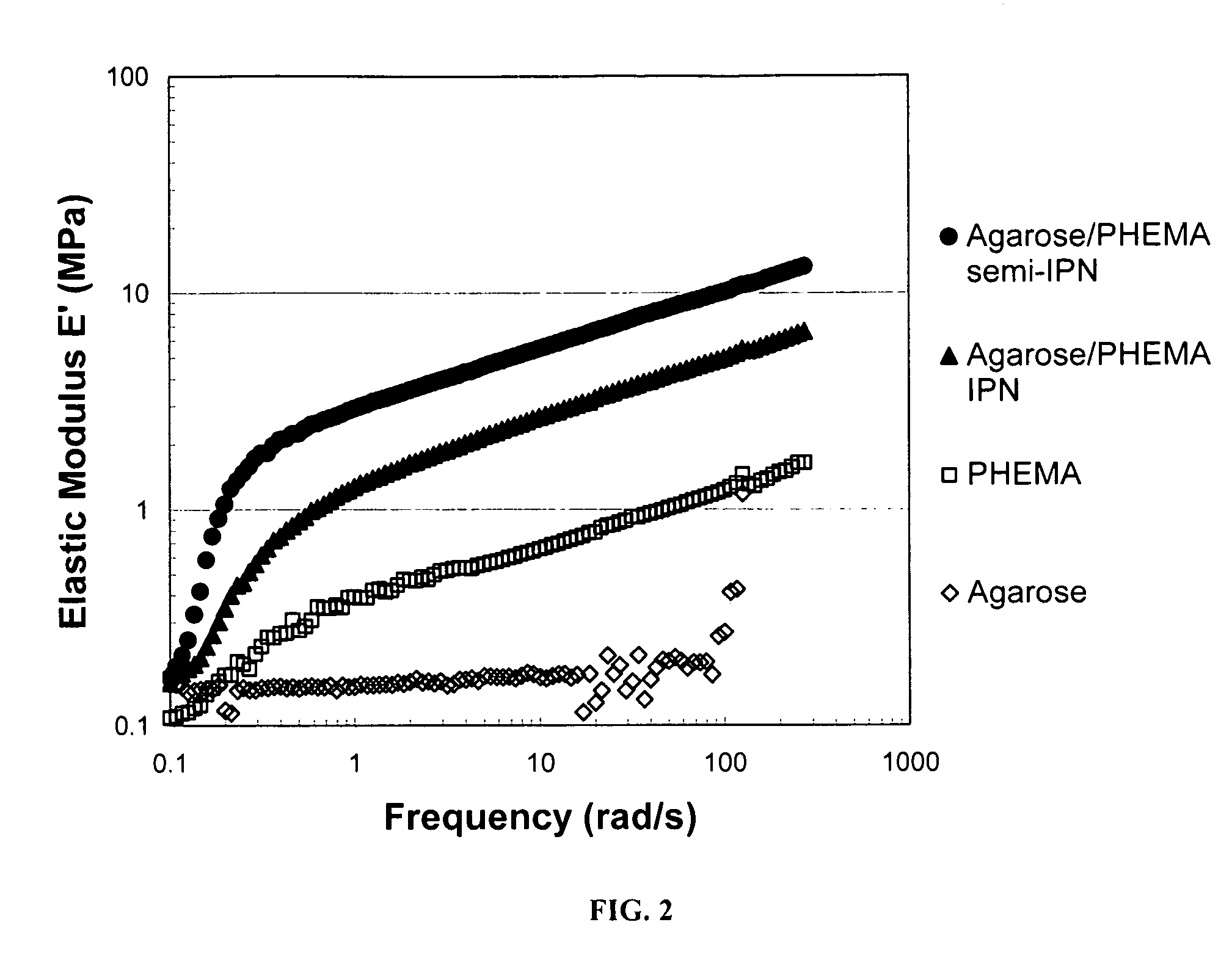
[0043]In this example, a hydrogel network formed from agarose as the rigid polysaccharide component and poly(2-hydroxymethacrylate) (“PHEMA”) as the ductile polymer phase was prepared, and compared to agarose and PHEMA gels alone. In general, agarose formed a physical gel upon cooling from an aqueous solution. The agarose gel was then soaked a solution comprising a HEMA monomer with a photoinitiator, with and without the crosslinker N,N′-methylene-bis-acrylamide. After equilibration, the HEMA monomer was photopolymerized with UV light. The hydrogel network was then subjected to both transient (ultimate) and dynamic tensile tests and compared the single polymer gels with the semi-IPN (uncrosslinked PHEMA) and IPN (cross-linked PHEMA).
[0044]More specifically, agarose as a dry powder (>99% pure with a gelling temperature of 40-43° C. lot number 456755 / 1), the monomer 2-hydroxyethyl methacrylate (HEMA) (>99% pure), the initiator α-k...
example 2
Agarose / PEG-DA Hydrogel Synthesis and Mechanical Testing
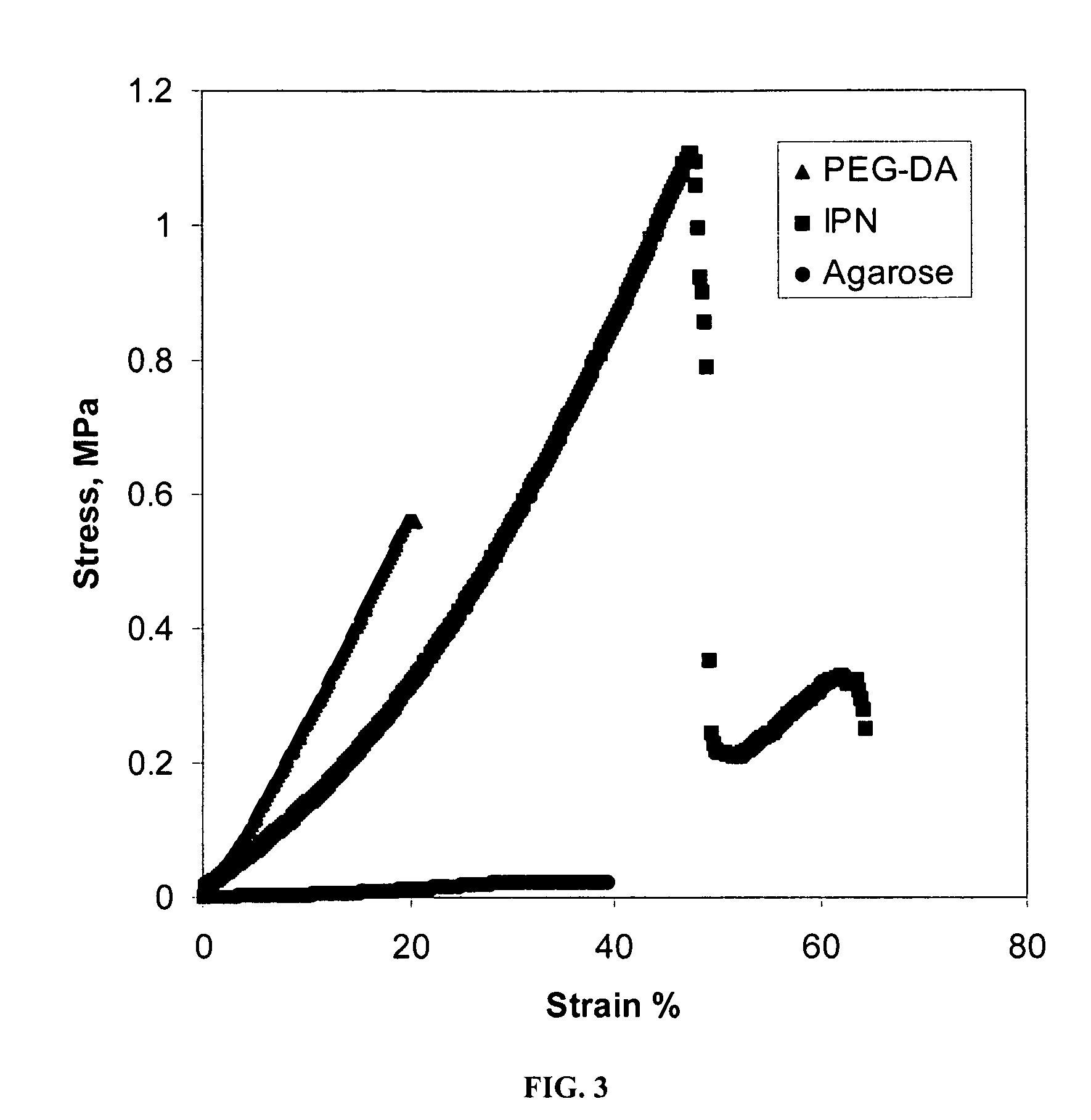
[0048]In this example, a hydrogel network formed from agarose as the rigid polysaccharide component and PEG-DA as the chemically cross-linked component was prepared, and compared to agarose and PEG-DA gels alone.
[0049]More specifically, agarose powder (Type VII [gelling temperature60° C.] cell culture-tested, Sigma Aldrich, St. Louis, Mo.) was dissolved in hot water to form a clear, 2 wt % solution and about 0.4 mL aliquots were added to cylindrical molds of silicon rubber to form disks upon gelation. They were refrigerated at about 4° C. for 24 hours (without cells). A solution of 23 wt % PEG-DA (nominal molecular weight 750, Sigma Aldrich, St. Louis, Mo.) and 0.1 wt % Irgacure 2959 photoinitiator (Ciba Specialty Chemicals, Tarrytown, N.Y.) was then prepared. Agarose gel disks were then placed in excess PEG-DA solution and permitted to soak for about 24 hours to absorb PEG-DA. After about 24 hours, the gel was removed from the...
example 3
Cell Encapsulation in Agarose-PEG-DA IPN
[0052]In this example, chondrocytes were encapsulated in a hydrogel comprising agarose and a chemically crosslinked polymer or copolymer synthesized from PEG-DA. To isolate the chondrocytes, articular cartilage was obtained under aseptic conditions from the ankle of six-week old female pigs. Cells were harvested within six hours of slaughter. The cartilage was digested in collagenase type II (Worthington, Biochemical Corp., Lakewood, N.J.) in culture medium. The medium was Dulbecco's Modified Eagle Medium (DMEM) with 4.5 g / L D-glucose 10% fetal bovine serum, 1% non-essential amino acids, ascorbic acid (50 μg / mL), and 1% penicillin-streptomycin-fungicide (Fungizone) (Cambrex). After suspension in phosphate buffered saline (PBS), the cells were encapsulated in agarose at a concentration of approximately 1×10⁶ cells / mL as described below.
[0053]To synthesize the hydrogel network agarose / PEG-DA having living cells encapsulated therein, the chondroc...
PUM
| Property | Measurement | Unit |
|---|---|---|
| molecular weight | aaaaa | aaaaa |
| molecular weight | aaaaa | aaaaa |
| water content | aaaaa | aaaaa |
Abstract
Description
Claims
Application Information
 - R&D
- Intellectual Property
- Life Sciences
- Materials
- Tech Scout
- Unparalleled Data Quality
- Higher Quality Content
- 60% Fewer Hallucinations
Browse by: Latest US Patents, China's latest patents, Technical Efficacy Thesaurus, Application Domain, Technology Topic, Popular Technical Reports.
© 2025 PatSnap. All rights reserved.Legal|Privacy policy|Modern Slavery Act Transparency Statement|Sitemap|About US| Contact US: help@patsnap.com


