

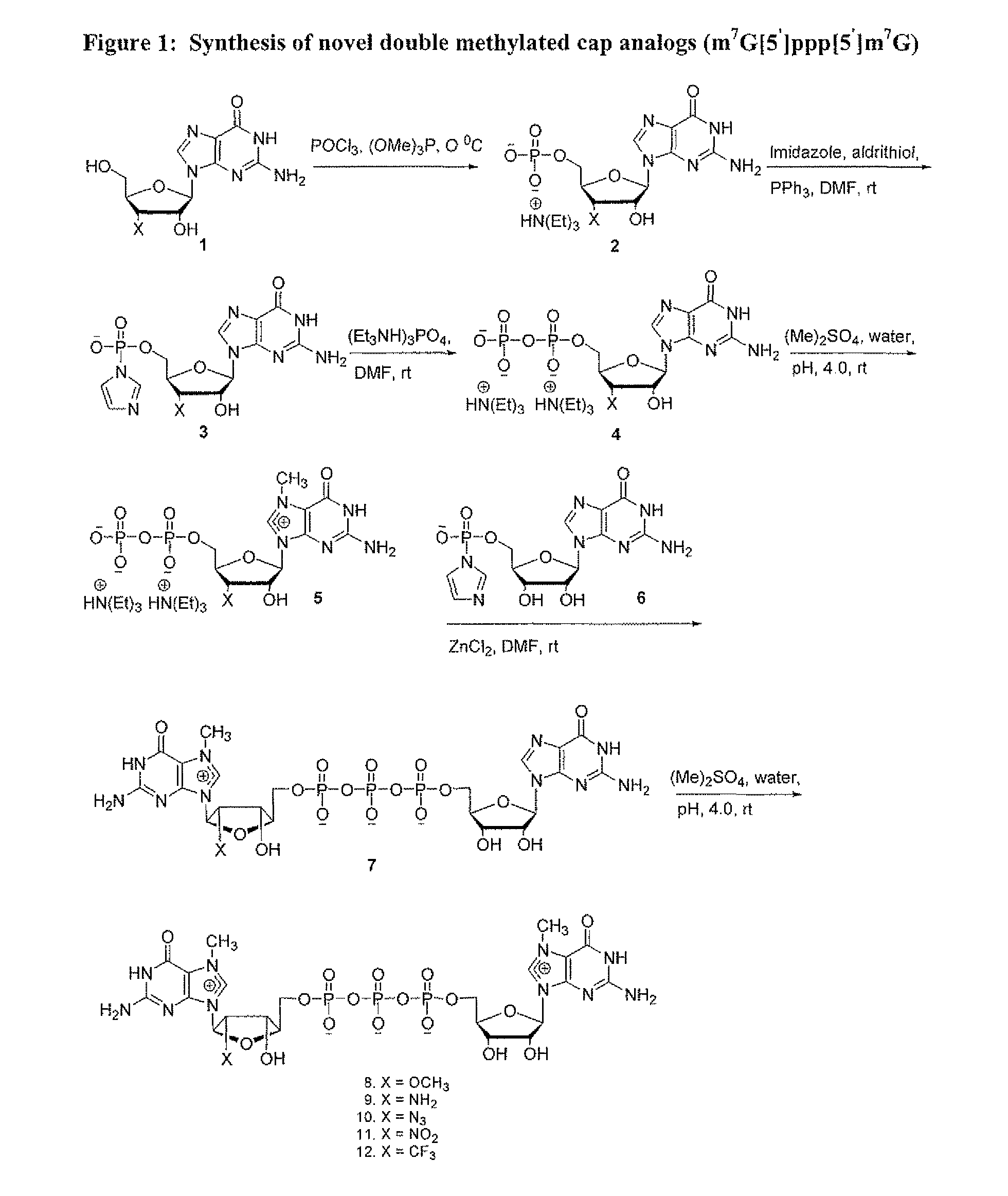
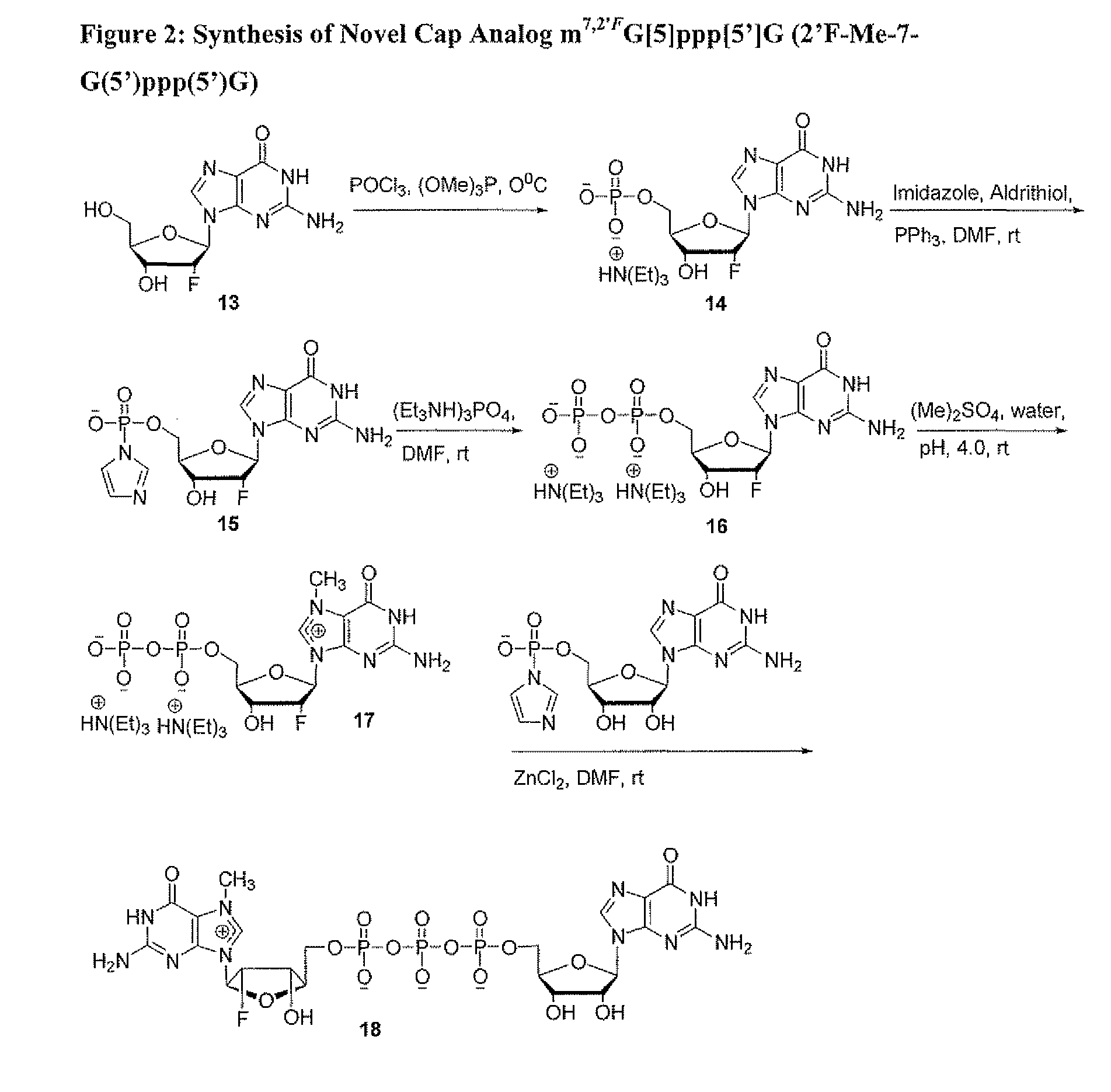
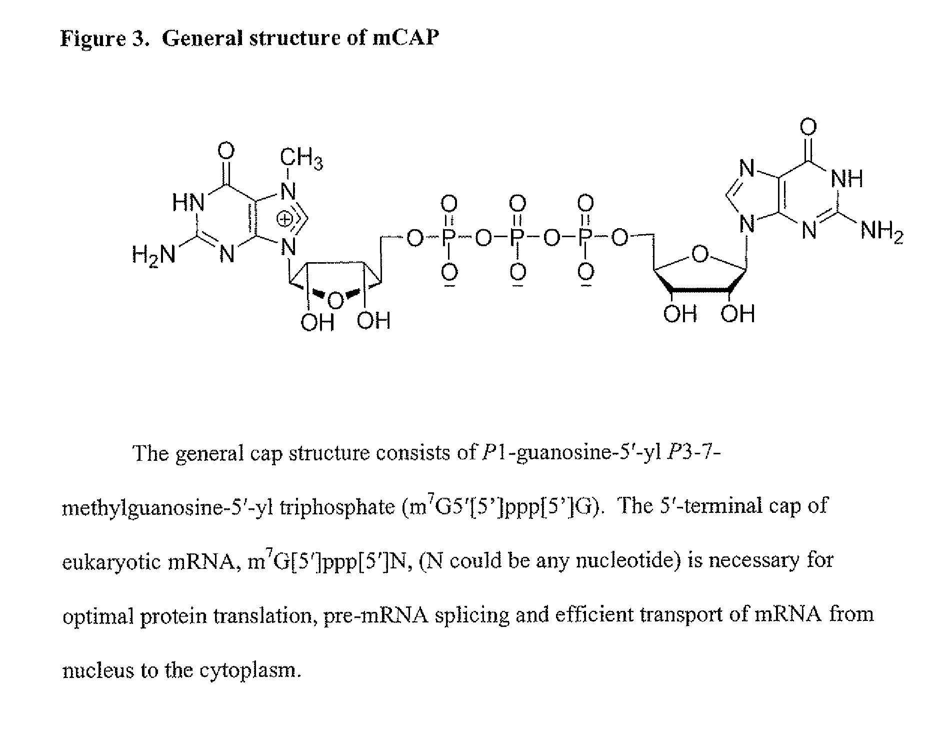
Dinucleotide MRNA cap analogs
a technology of mrna cap and mrna cap, which is applied in the field of mrna cap analogs of mrna caps, can solve the problems of pseudosymmetric dinucleotides and the propensity to the 3 being almost impossible to d
- Summary
- Abstract
- Description
- Claims
- Application Information
AI Technical Summary
Benefits of technology
Problems solved by technology
Method used
Image

Examples
example 1
[0082]Synthesis of 7-Methyl Guanosine 5′-diphosphate In a clean, dry 2000 mL round bottom flask equipped with a stirring bar and under a stream of argon slowly dissolve dry and finely powdered guanosine 5′-diphosphate (1), (10.0 g, 20.5 mmol), either in free acid or sodium as a counter ion form, in 200 mL water, adjusting pH to 4.0 with glacial acetic acid. Dimethyl sulfate (20 mL, 119.04 mmol) was then added over a period of one hour with constant stirring at room temperature and the reaction continued for an addition hour during which time a decrease in pH was observed but pH was kept between pH 3.8 to 4.0 by drop-wise addition of 10 mM NaOH and methylations was monitored by analytical HPLC for progress. Methylation was determined to be 98% complete within 2 h. After 2 h, the reaction mixture was extracted with CHCl3(3×200 mL) to remove unreacted excess dimethyl sulfate.
[0083]The resulting aqueous layer was further evaporated on a rotary evaporator to remove any chloroform traces,...
example 2
Synthesis of Nucleoside-5′-diphosphates
[0084]Although the following procedure illustrates synthesis of guanosine-5′-diphosphate, one of skill in the art would be able to use the procedure for the synthesis of adenosine-5′-diphosphate, uridine-5′-diphosphate, and cytidine-5′-diphosphate and analogs thereof. In a clean, dry 500 mL round bottom flask equipped with a stirring bar the triethylammonium salt of guanosine 5′-monophosphate (10.0 g, 21.5 mmol) in anhydrous dimethylformamide (200 mL) was stirred together, triethylamine was added (2.4 mL, 142.8 mmol) and allowed to stir for 5 min, followed by the addition of Imidazole (5.86 g, 86.1 mmol), 2,2′-dithiodipyridine (7.4 g, 33.58 mmol), and triphenylphosphine (8.9 g, 33.9 mmol). Stirring was continued for 2 h at room temperature. The reaction was allowed to go to completion as determined by HPLC and then poured slowly into a mixture of sodium perchlorate (7 g) in acetone (1500 mL), and then cooled for 30 min. at 4° C. The reaction mi...
example 3
Synthesis of 3′-O-Methyl Guanosine Monophosphate (3′-O-Me-GMP) TEA Salt (Compound 2)
[0086]In a clean, dry 500 mL round bottom flask equipped with a stirring bar and under a stream of argon slowly add dry and finely powdered 3′-O-Me-Guanosine (1), (6 g, 20 mmol) to a mixture of trimethylphosphate ((OMe)3P) (50 mL) and phosphorous oxychloride (POCl3) (6 mL, 60 mmol) at 0° C. in small portions with continuous stirring under argon. The mixture was kept at 0-4° C. and allowed to stir at least 19 hrs. Diethyl ether (200 mL) was added to extract the excess phosphorous oxychloride and to simultaneously precipitate the 3′-O-methylguanosine-5′-phosphodichloridate, which was then pelleted by centrifugation and dissolved in 100 mL ice-cold 5% NaHCO3 in water. The resulting aqueous solution was adjusted to pH ˜1.5 using 1 N NaOH. After stirring at 0-4° C. for an additional 20 h, the pH was adjusted to 7.0 and the resulting mixture was applied to a column of DEAE Sephadex A25. The column was wash...
PUM
| Property | Measurement | Unit |
|---|---|---|
| pH | aaaaa | aaaaa |
| height | aaaaa | aaaaa |
| diameter | aaaaa | aaaaa |
Abstract
Description
Claims
Application Information