Method of determining sperm capacitation

a sperm capacitation and method technology, applied in the field of male fertility, can solve the problems of no sensitive and simple markers for capacitation, inability to immediately fertilize an egg, and inability to achieve sperm capacitation

Active Publication Date: 2013-02-05
CORNELL UNIVERSITY +1
View PDF5 Cites 14 Cited by
  • Summary
  • Abstract
  • Description
  • Claims
  • Application Information

AI Technical Summary

Problems solved by technology

Ejaculated sperm are not immediately able to fertilize an egg.
Currently there are no sensitive and simple markers for capacitation that can be used in a clinical setting.
However, visualization of these events using polyacrylamide gel electrophoresis and immunoblotting can take upwards of 48 hours to perform, making it ill-suited for clinical purposes.
However, it is unclear how other lipids are organized in the sperm plasma membrane and if the organization of these lipids might reflect changes associated with the process of capacitation.

Method used


Image

  • Method of determining sperm capacitation
  • Method of determining sperm capacitation
  • Method of determining sperm capacitation

Examples


example 1

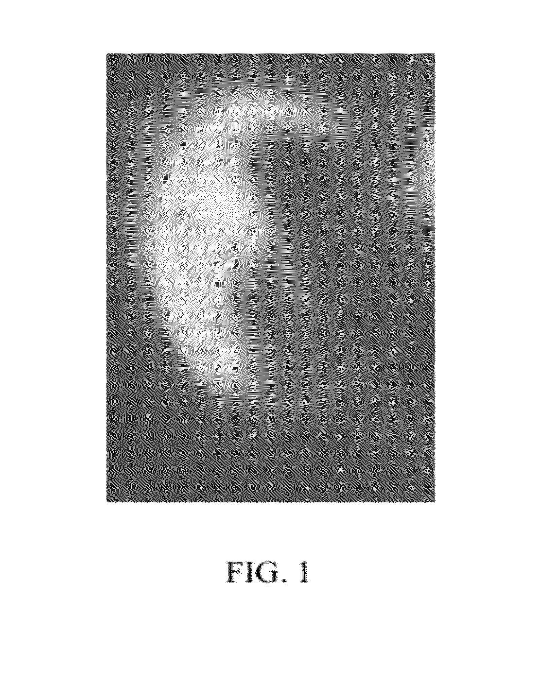
[0053]To localize the ganglioside, GM1, in living, motile sperm, 5×10⁶ murine sperm were incubated with 10 μg / ml Cholera toxin b subunit with an Alexa-Fluor fluorescent tag (“CTb,” Molecular Probes, Eugene, Oreg.) in 750 μl of modified Whitten's medium (22 mM HEPES, 1.2 mM MgCl2, 100 mM NaCl, 4.7 mM KCl, 1 mM pyruvic acid, 4.8 mM lactic acid hemi-calcium salt, pH 7.35). Glucose (5.5 mM), NaHCO3 (10 mM), and (2-hydroxypropyl)-β-cyclodextrin (2-OHCD; 3 mM) were supplemented as needed for 10 minutes at 37° C. in the dark. After washing at 37° C., CTb was seen specifically in the plasma membrane overlying the acrosome (FIG. 1). The fluorescence was detected with a Nikon TE2000 microscope equipped with an OpenLab / Volocity imaging system (Improvision, Lexington, Mass.). The same pattern of fluorescence was seen when sperm were fixed with 4% paraformaldehyde and 0.1% glutaraldehyde.

example 2

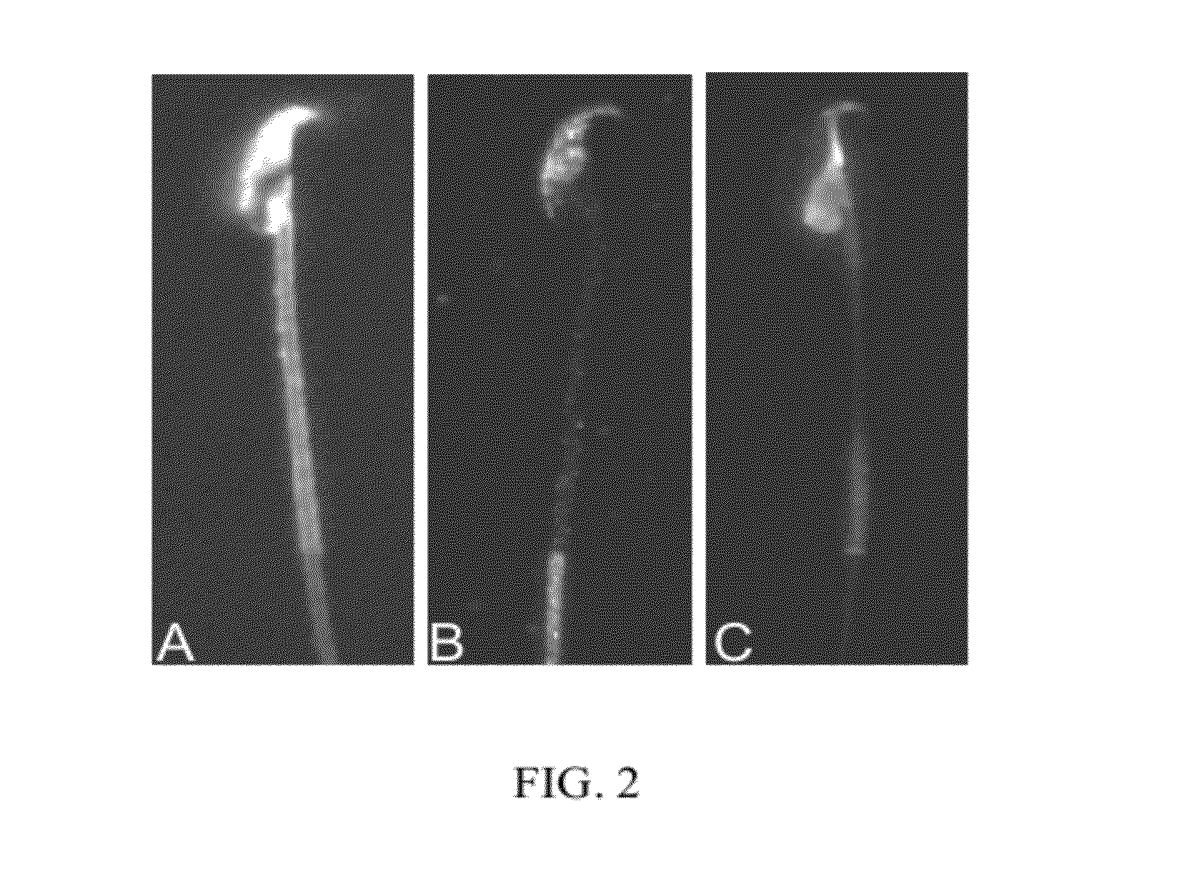
[0054]To investigate the existence of lipid rafts in murine sperm, sterols, caveolin-1, and the ganglioside, GM1, were localized by fluorescence methods. Caveolin-1 is often used as a marker for lipid rafts in that it requires interaction with a sterol to attach to a membrane, and often fractionates with lipid raft membrane sub-domains. It should be noted that caveolin cannot be detected in living cells by indirect immunofluorescence because the epitopes recognized by the caveolin antibody are intracellular. Similarly, filipin must be visualized in fixed cells as the autofluorescence is weak and easily quenched, hence movement of the cells during a long exposure time would cause a loss of resolution.

[0055]To localize sterols in murine sperm, we incubated 1×10⁶ sperm with 0.005% filipin (w / v, in dimethyl formamide) (Sigma, St. Louis, Mo.) in 1 ml of modified Whitten's medium (i.e. non-capacitating conditions). Sperm were washed twice by centrifugation and resuspended in medium contai...

example 3

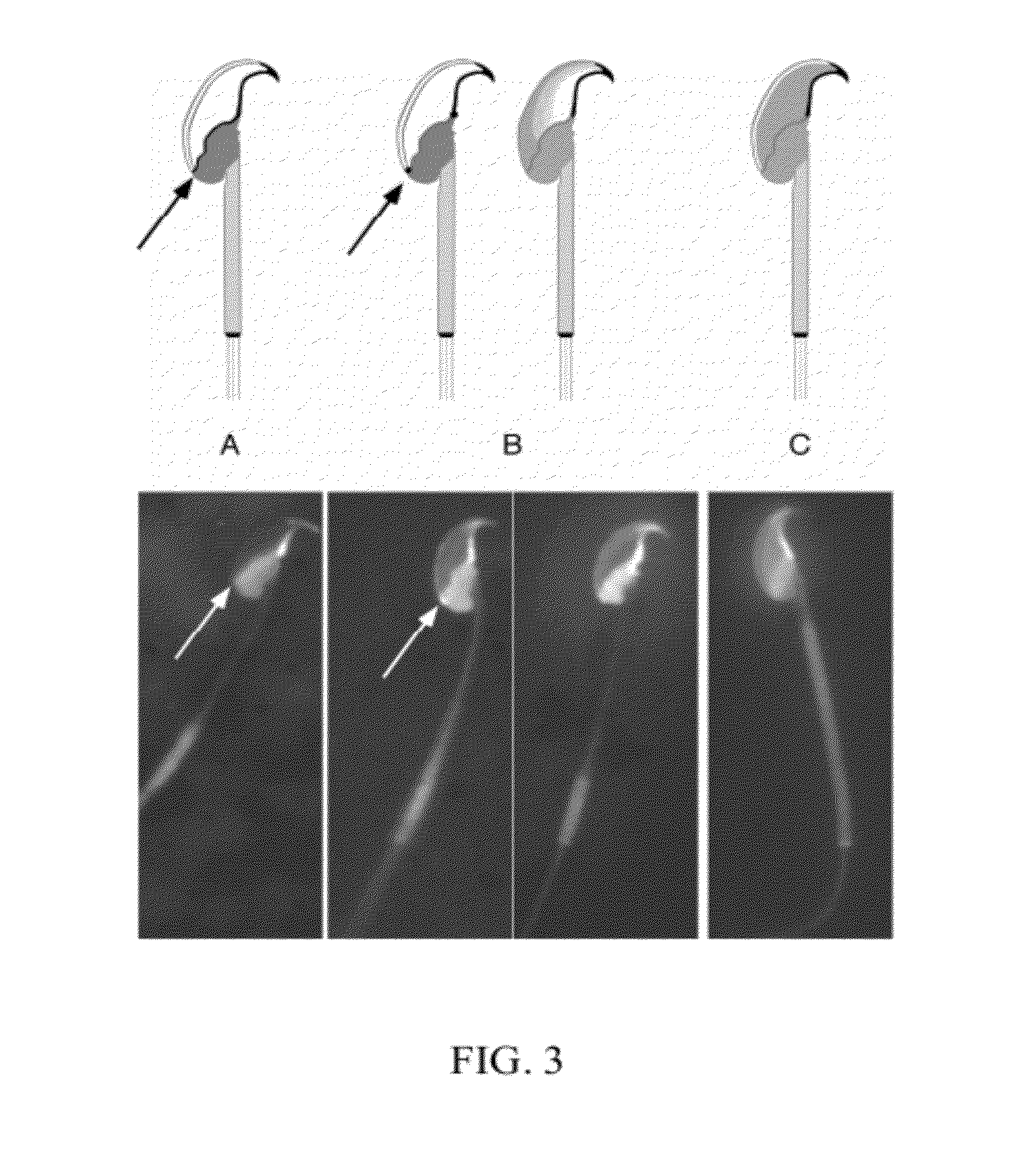
[0056]This example demonstrates the changes in the pattern of GM1 distribution (redistribution) that are observed under conditions which are known to induce or which accompany the process of capacitation. In Example 2, it was demonstrated that the plasma membrane overlying the acrosome was enriched in sterols, whereas the post-acrosomal plasma membrane was enriched in GM1, when the sperm were incubated under non-capacitating conditions and then fixed in 0.004% paraformaldehyde. This segregation was remarkably consistent in non-capacitated sperm. It has been documented that exposure of sperm to sterol acceptors such as 2-hydroxy-propyl cyclodextrin (2-OHCD) or bovine serum albumin (BSA) causes the loss of most FSC from the plasma membrane overlying the acrosome, and allows some FSC to diffuse laterally into the post-acrosomal plasma membrane. This finding is consistent with the loss of sterols causing an increase in membrane fluidity, and promoting lateral diffusion. To investigate t...


PUM


Abstract

This invention describes unique patterns of distribution of ganglioside GM1 in non-capacitated sperm and demonstrates that the pattern of distribution of GM1 undergoes changes that can be correlated with the process of capacitation and / or with acrosomal exocytosis. Accordingly, the present invention discloses a method for determining the ability of sperm to respond to capacitation and / or acrosomal exocytosis stimuli. The method comprises determination of distribution pattern for GM1. The method can be used for both diagnostic and predictive purposes when assessing male reproductive fitness, and can also be used to assess the effects of any agent or environment on sperm including cryoprotective agents and protocols, and contraceptive agents.

Description

[0001]This application is a Continuation-in-part of U.S. Non-provisional application Ser. No. 11 / 591,061, filed on Nov. 1, 2006, which is a divisional application of U.S. Non-provisional application Ser. No. 10 / 898,041, filed on Jul. 23, 2004, which in turn claims priority to U.S. provisional application No. 60 / 489,443, filed on Jul. 23, 2003, the disclosures of all of which are incorporated herein by reference.[0002]This work was supported by grant numbers KO1-RR00188, P01-HD-06274 and RO1-HD-045664 from the National Institutes of Health. The government has certain rights in the invention.FIELD OF THE INVENTION[0003]This invention relates generally to the field of male fertility and more specifically provides a method for determining sperm quality, particularly with regard to assessing the ability of sperm to undergo capacitation by following the pattern of distribution of GM1 ganglioside.DESCRIPTION OF RELATED ART[0004]Ejaculated sperm are not immediately able to fertilize an egg....

Claims


Application Information

Patent Timeline
Patent Type & Authority Patents(United States)
IPC IPC(8): A01N1/02
CPCC12N5/0612A01N1/02G01N33/5091G01N33/5035G01N1/30G01N2405/10
Inventor TRAVIS, ALEXANDER J.KOPF, GREGORY S.
Owner CORNELL UNIVERSITY
Why Eureka
Try Eureka