
Guidance and implantation of catheters

a catheter and implantation technology, applied in the field of neoplasia treatment, can solve the problems of severe and irreparable damage to the tissues in and around the intended treatment field, chemotherapy may not be able to eradicate tumor cells, and all cancerous cells are likely to maim or destroy the organ in which the cancer originated

Inactive Publication Date: 2013-12-03
PEAK BIOSCI
View PDF54 Cites 5 Cited by
  • Summary
  • Abstract
  • Description
  • Claims
  • Application Information

AI Technical Summary

Benefits of technology

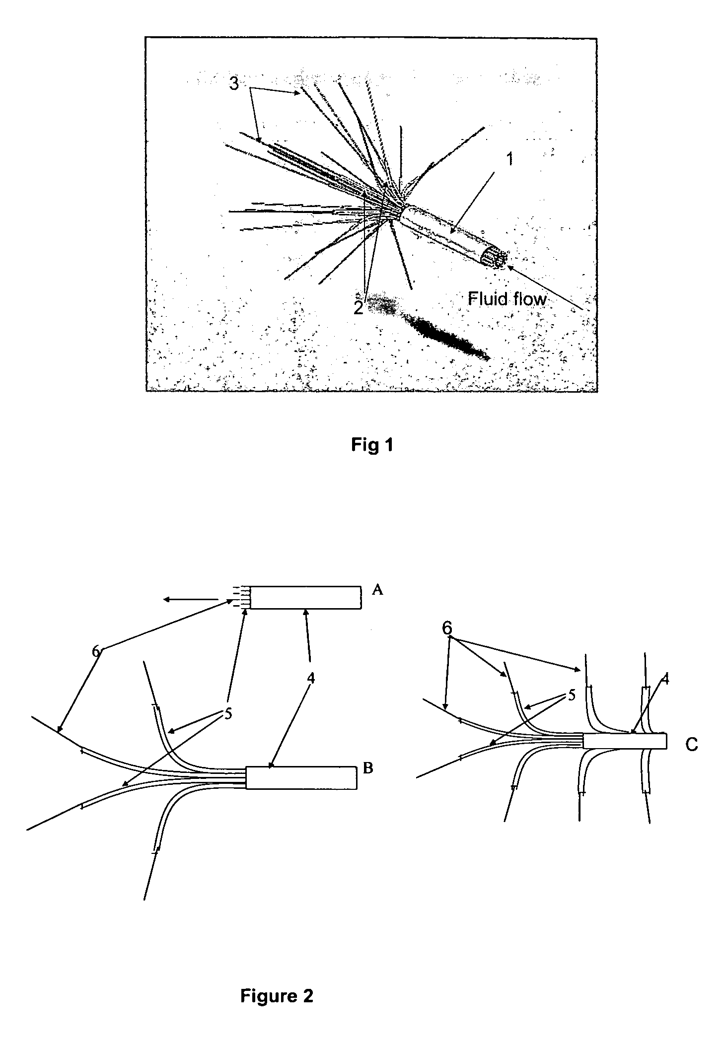
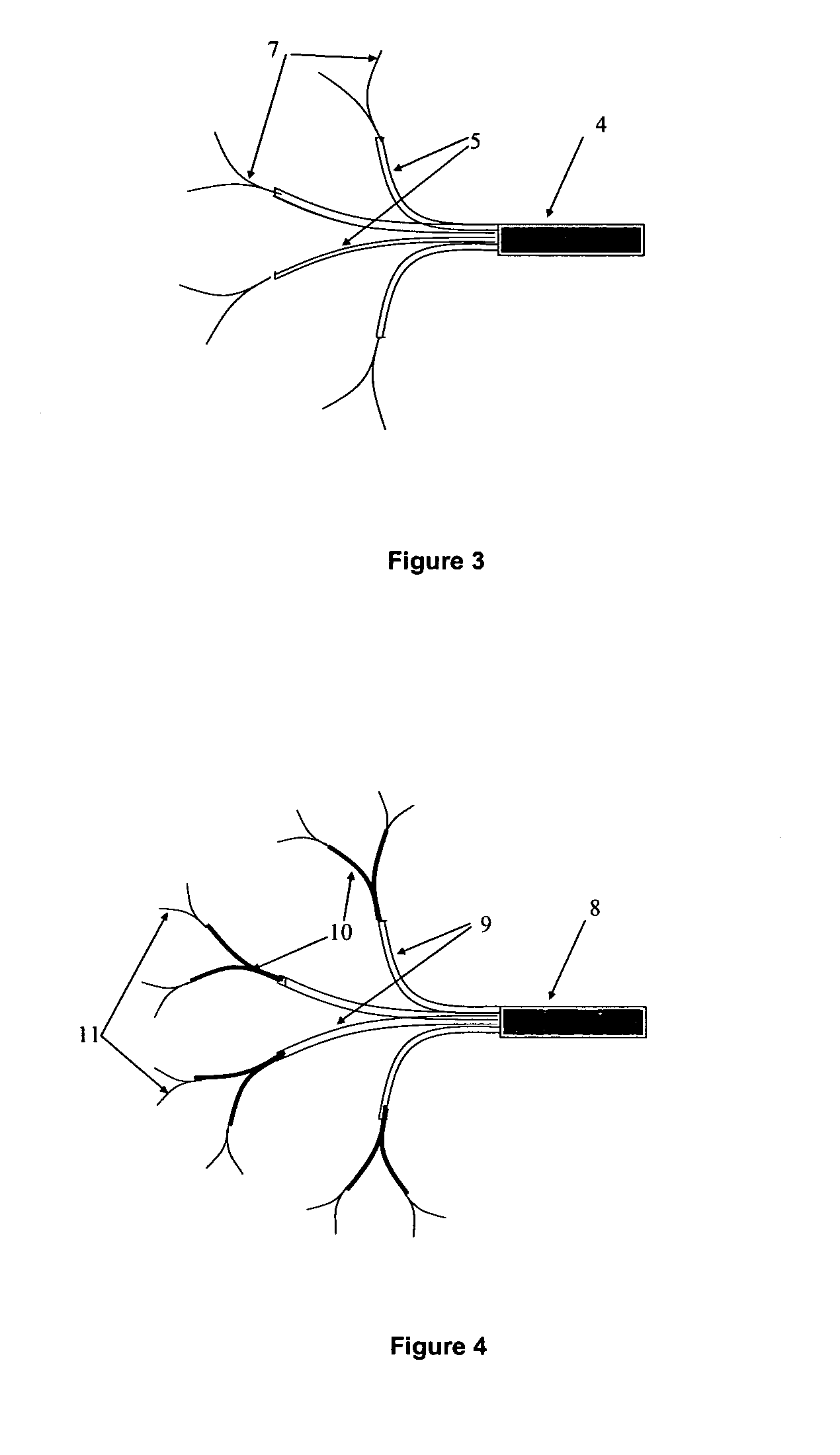
[0028]In various embodiments, the plurality of catheters and the catheter guide device can be adapted to remain connected after the catheters have been emplaced within the tissue to provide an anchoring effect. For example, one subset of catheters can be emplaced within the tissue at an angle other than orthogonal to the guide structure and a second subset can be emplaced at another angle other than orthogonal to the guide structure, for example in a splayed fashion, to provide an anchoring effect.

Problems solved by technology

However, this is not the case for many solid tumors that have advanced to later stages.
Locally advanced tumors may arise in tissues throughout the body, but unlike early stage tumors may not be amenable to complete surgical excision or complete ablation using radiation treatments.
Due to the invasion of the surrounding tissues by tumor processes, any surgical procedure that would serve to remove all the cancerous cells would also be likely to maim or destroy the organ in which the cancer originated.
Similarly, radiation treatments intended to eradicate the cancerous cells left behind following surgery frequently lead to severe and irreparable damage to the tissues in and around the intended treatment field.
However, when a tumor has infiltrated into otherwise healthy tissues surrounding the site where the tumor originated, even combination treatments including surgery plus radiation therapy, or surgery plus radiation therapy plus chemotherapy may not be capable of eradicating the tumor cells without causing severe damage to the tissues in the treatment field.
Neither surgeons nor radiotherapists have the tools to eliminate individual tumor cells, microscopic tumor processes, or tumor-associated vasculature from the otherwise normal tissue surrounding locally advanced solid tumors.
Despite the above mentioned benefits, and its widespread use, conventional radiation therapy cannot cure locally advanced solid tumors, because, in order to gain access to a tumor mass, x-ray beams usually must pass completely through the body; therefore, exposure to normal tissues is inevitable.
In addition, x-ray beams lack the microscopic accuracy needed to eliminate individual cancer cells from the treatment field.
X-rays cannot eradicate or cure most types of locally advanced solid tumors, because they lack the specificity needed to kill cancer cells while sparing the normal cells in the treatment field.
Unfortunately, this approach is of limited value even in instances when the x-ray beam can be focused precisely on the treatment field.
The main problem is that x-rays cannot discriminate between the cancer cells and normal cells within the treatment field.
Because of this conventional radiation is associated with side effects, often severe, including mucositis, alopecia, dermatitis, proctitis, enteritis, and tissue necrosis.
Regardless of how precisely one defines the treatment field, and regardless of how precisely the x-ray beam is projected through the treatment field, x-rays will damage normal cells in the treatment field, and radiotherapy beams do not have the microscopic accuracy needed to eliminate individual cancer cells from the treatment field.
Thus, even the most precise digital pre-treatment planning cannot overcome the inherent deficiencies of ionizing radiation.
Most types of chemotherapy also suffer from a lack of tumor specificity and also cause collateral damage to normal tissues, since chemotherapeutic agents are distributed throughout the body and exert their effects on normal cells as well as malignant cells.
The deficiencies of current treatment modalities are especially glaring with respect to specific types of cancer, for example glioblastoma multiforme (GBM), a highly aggressive type of cancer that constitutes the most common form of brain malignancy.
Indeed, after nearly 35 years of investigations involving hundreds of experimental treatments and thousands of GBM patients participating in clinical trials, the prognosis of patients with newly diagnosed GBM is dismal.
However, the challenge is great, because the majority of chemical entities do not diffuse far into brain tissue or other types of solid tissues.
Currently available methods of convection enhanced delivery have several limitations and drawbacks.
One of the biggest problems is to determine the optimal position of the catheter tips.
Optimal catheter placement is especially challenging given the highly variable size and shape of surgical resection cavities.
Regional differences in anatomy and fluid dynamics increase the challenge of accurate catheter placement.
Another problem that aggravates the optimal positioning of catheter tips is tissue swelling.
This makes it quite difficult to accurately position the tips of catheters into the perimeter of the surgical resection cavity.
Insertion of such thick catheters from inside of the surgical resection cavity is challenging because of the minimum depth requirement, by their limited pliability, and by their sheer bulk.
The use of 2.5 mm OD catheters may increase the risk of hemorrhage and / or trauma to nervous tissues as compared with thinner catheters.
Given the above constraints it is very difficult to consistently arrange the tips of thick catheters into an orderly distribution around many surgical resection cavities.
Another limiting factor is that each catheter supplies a large proportion of the intended treatment field, e.g. 33% of the treatment field for 3 catheters, and 50% of the treatment field for 2 catheters.
A high proportional flow per catheter is an unavoidable consequence of using only a few catheters, and has the effect of reducing the accuracy of convection enhanced delivery.
In addition, the use of a high fractional flow per catheter, and the fact that the catheters must be inserted from the surface of the brain, limits the surgeon's available options for catheter insertion.
Based upon clinical experience from many convection enhanced delivery studies involving patients with brain tumors, 2-3 catheters appear to be insufficient to provide optimal biodistribution of drugs around many surgical resection cavities.
In this regard, a major issue revealed by studies of gene expression profiling, is that tumors are genetically and metabolically much more heterogeneous than previously anticipated.
Despite the recognition that 125IUDR has a unique cell killing capability, and despite many years of research aimed at exploiting this mechanism of action, including the concept of directly introducing 125IUDR into tumors (for example, see Kassis et. al., “Treatment of tumors with 5-radioiodo-2′-deoxyuridine,” U.S. Pat. No. 5,077,034), these agents have not been successfully applied to the treatment of cancer.
The delivery of 125IUDR and related agents to solid tumors, using systemic or local administration, has proven to be extremely challenging.

Method used


Image

  • Guidance and implantation of catheters
  • Guidance and implantation of catheters
  • Guidance and implantation of catheters

Examples


Embodiment Construction

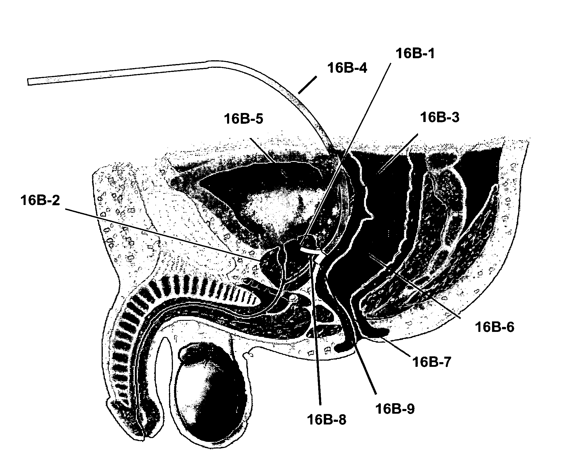
[0070]The inventive catheter system enables the stereotactic emplacement of a plurality of catheters into a target tissue or field of treatment. The emplacement can be guided by an electronic, radiofrequency, ultrasound, or video-assisted computerized digital navigation and stereotactic system. The individual sources of bioactive agent or drug emanating from each catheter provides an overlapping field of treatment in the target tissue to expose the entire treatment field to an antineoplastic agent, radiopharmaceutical agent, or other pharmaceutical agent. The inventive catheter guidance and guidance system may be used to create essentially any 3-dimensional spatial array of catheters within the target tissue using a catheter guide template, comprising catheter guide tubes or passageways, or both, to guide the catheters into the spatially defined implantation in the target tissue or field of treatment.

[0071]Various embodiments of the present invention involve a catheter guide device ...


PUM

PropertyMeasurementUnit
angleaaaaaaaaaa
outer diameteraaaaaaaaaa
biocompatibleaaaaaaaaaa

Abstract

A catheter system adapted for navigating, guiding and implanting a catheter or a plurality of catheters in a spatially-defined implantation within the tissue of a patient is provided. The system can include a tissue navigation system and a probe to inform the navigation system to guide emplacement of the catheters within a target tissue. The probe can provide images, such as fiberoptic visual images, or ultrasound images, or can provide radiolocation data, to guide the catheter emplacement. The catheters supply a pressurized liquid including a bioactive agent, such as can be used in the treatment of cancer, for example 123I- or 125I-IUDR. The system and methods provided can be used in the treatment of locally advanced tumors, such as cancers of the brain, head or neck, esophagus, prostate, ovary, liver, pancreas, bladder or rectum.

Description

CLAIM OF PRIORITY TO RELATED APPLICATIONS[0001]This application is a U.S. National Stage Filing under 35 U.S.C. 371 from International Patent Application Serial No. PCT / US2008 / 003711, filed on Mar. 20, 2008, and published on Sep. 25, 2008 as WO 2008 / 115566, which claims the priority of U.S. Provisional Patent Application Ser. No. 60 / 895,916, filed on Mar. 20, 2007, and U.S. Provisional Patent Application Ser. No. 60 / 917,226, filed on May 10, 2007, which applications and publication are incorporated herein by reference in their entirety.BACKGROUND OF INVENTION[0002]In the treatment of neoplasia, such as solid tumors in the early stages, surgical excision or ablation with radiation often provides a successful form of therapy. However, this is not the case for many solid tumors that have advanced to later stages. Locally advanced or locally invasive solid tumors are primary cancers that have extensively invaded or infiltrated into the otherwise healthy tissues surrounding the site wher...

Claims


Application Information

Patent Timeline
Patent Type & Authority Patents(United States)
IPC IPC(8): A61B5/05
CPCA61K51/0491Y10T137/0402A61P35/00
Inventor DALKE, WILLIAM D.LILLEHEI, KEVINJELLISON, THOMAS A.GERBER, MICHAEL J.MATSUURA, JAMES E.WARREN, STEPHEN L
Owner PEAK BIOSCI
Learn More