Oligonucleotide microarray for identification of pathogens

a technology of oligonucleotide microarray and pathogen detection, which is applied in the field of test kits and can solve the problems of difficult to provide accurate diagnosis, increase the “noise” of electrophoresis gel, and procedures that require highly trained staff and are very time-consuming, so as to achieve easy and rapid determination

Active Publication Date: 2014-06-03
HONEYWELL INT INC
View PDF16 Cites 5 Cited by
  • Summary
  • Abstract
  • Description
  • Claims
  • Application Information

AI Technical Summary

Benefits of technology

[0008]In one aspect, the present invention relates to a method of easily and rapidly determining whether a polynucleotide containing a target nucleic acid is present in a biological sample or test sample.

Problems solved by technology

Different pathogenic infections commonly have similar symptoms resulting in difficulties in providing an accurate diagnosis of the cause of the infection or disease.
These procedures require highly trained staff and are very time consuming.
However, as the number of PCR primers contained in a primer reagent is increased, “noise” on an electrophoresis gel is increased.
For this reason, when a PCR reaction is used, it is difficult to determine the presence of more than only a few genes.
Additionally, it is necessary to design the primers so that the PCR products amplified by the PCR reaction are distinguishable on electrophoresis gels, which often have limited resolution.
Designing PCR primers that produce suitably distinguishable PCR products with low noise is difficult and time consuming.
Thus, a strictly PCR / electrophoresis-based approach is inefficient for screening for expression of a large number of genes.

Method used


Image

  • Oligonucleotide microarray for identification of pathogens
  • Oligonucleotide microarray for identification of pathogens
  • Oligonucleotide microarray for identification of pathogens

Examples


example 1

[0061]DNA Microarray Fabrication Process

1. Glass Surface Washing.

[0062]Clear slide immersed in H2SO4 / H2O2, 2:1 for 30 min. Wash with H2O three times, and then with methanol for twice. Dry under a stream of pressed air, then baked at 80° C. for 10 min.

2. Glass Silanization.[0063]Sonicated in 2% N-(2-aminoethyl)-3-aminopropyltrimethoxysilane (EDA) in 1 mM HAc for 30 min at room temperature (RT). Wash with H2O three times, and then with methanol for twice. Dry under a stream of N2, then baked at 110° C. for 15 min.

3. Modify Silanized Slide with Heterobifunctional Crosslinker

[0064]Immersed in the crosslinker solution (384 mg PDITC (2 mmol) in 80 ml 10% anhydrous pyridine in DMF) for 2 hr at RT. Rinsed with the DMF three times and then dichloroethane twice. Dry under N2.

4. Microarray Print and DNA Immobilization

[0065]Spot 40 uM NH2-oligodeoxynucleotide (ODN) in 0.1M carbonate buffer (pH 9.0) on the active glass slide at 60% humidity. React over 1 hr at 37° C. under saturated humidity. Ri...

example 2

DNA Microarray Hybridization Process

1. PCR Amplification and Labeling

[0068]Add 1 ul DNA sample to 100 ul PCR reaction buffer, which contains 10 mM Tris-HCl (pH 9.0), 50 mM KCl, 3.75 mM MgCl2, 0.1% Triton X-100, 0.1 mM dNTP (each), and 0.2 uM Cy5 labeled primers (each). Then add 5 u Tag DNA Polymerse and PCR amplified at the following temperature condition:[0069]i). 96° C. 2 min[0070]ii). 95° C. 20 s[0071]iii). 60° C. 20 s[0072]iv). 72° C. 30 s[0073]Go to step ii for 28 cycles.[0074]v). 72° C. 2 min[0075]The reaction takes about 1 hr and 20 min total.

2. DNA Microarray Hybridization[0076]Dilute the PCR solution with 5 volumes hybridization buffer which contain 0.75M NaCl, 75 mM sodium citrate, and 0.1% SDS pH 7.2. Hybridization at 61° C. for 1 hr.

3. DNA Microarray Washing[0077]Wash the hybridized slide with 0.3M NaCl, 30 mM sodium citrate, and 0.1% SDS for 15 min for 3 times at 50° C., then wash with 0.3M NaCl, 30 mM sodium citrate, for 15 min at 50° C. Rinse with H2O then dry under p...

example 3

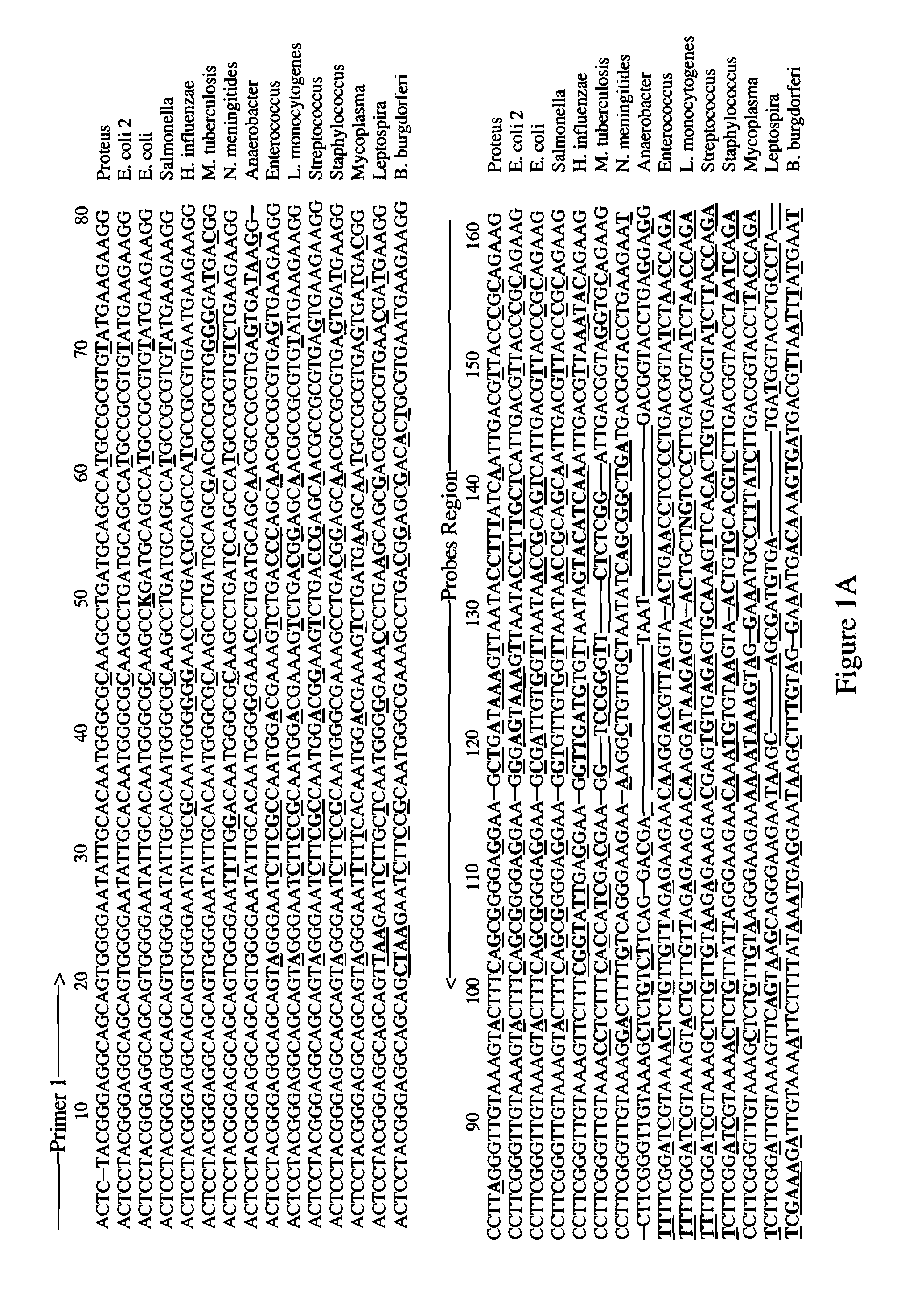
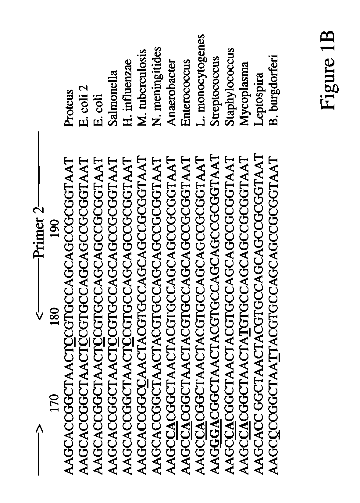
[0079]Cells are harvested directly from sections of tissue isolated from a patient. DNA is extracted from these cells using any known protocol. About 2-10 ng of DNA is amplified via PCR using two primer pairs: SEQ ID NOS: 1-2 and SEQ ID NOS: 7-8. [32P]-dCTP is included in the PCR reaction in order to label the amplified DNA, thus generating target nucleic acid detecting reagents with marker molecules.

[0080]The target nucleic acid detecting reagents with marker molecules are purified via spin column in order to remove excess [32P]-dCTP and denatured by incubation at 95° C. followed by immediate cooling on ice. The denatured target nucleic acid detecting reagents with marker molecules are then incubated under highly stringent conditions in the presence of an oligonucleotide microarray that comprises probes comprising SEQ ID NOS: 13-52. The hybridization is for 2 hours in 0.5 M NaHPO4, 7% sodium dodecyl sulfate (SDS), 1 mM EDTA at 65° C., followed by 3 washes in 0.1×SSC / 0.1% SDS at 68°...


PUM

PropertyMeasurementUnit
diameteraaaaaaaaaa
temperaturesaaaaaaaaaa

Abstract

A method for detecting a target nucleic acid of a pathogen in a test sample, the method comprising preparing a target nucleic acid detecting reagent and contacting the target nucleic acid detecting reagent with an oligonucleotide microarray. A kit for detecting a target nucleic acid of a pathogen in a test sample is also described. The kit comprises at least one primer pair and an oligonucleotide microarray comprising at least one probe.

Description

[0001]This application claims the benefit of U.S. Provisional Patent Application 60 / 755,504 filed Dec. 30, 2005.BACKGROUND OF THE INVENTION[0002]The present invention relates to test kits and methods for detecting the presence of a pathogen in a test sample. The test sample may be obtained from a patient or alternatively from a source of food or drink that may contain pathogens.[0003]Diseases such as encephalitis, cephalomeningitis, meningitis, pneumonia, enterogastritis, endocarditis, urinary infectiosus, are caused by infection by a pathogen. The pathogens include different bacteria, viruses, fungi and protozoa. Different pathogenic infections commonly have similar symptoms resulting in difficulties in providing an accurate diagnosis of the cause of the infection or disease. For example, encephalitis and meningitis patients generally manifest fever, headache, and convulsion. Identifying the pathogens responsible for these symptoms quickly and accurately is important to determining...

Claims


Application Information

Patent Timeline
Patent Type & Authority Patents(United States)
IPC IPC(8): C12Q1/68C07H21/04C07H21/02
CPCC12Q1/689C12Q1/701C12Q1/6837C12Q2531/113C12Q2565/501Y02A50/30
Inventor GU, YUANDONGXU, LEON
Owner HONEYWELL INT INC
Why Eureka
Try Eureka