
Combined rapid susceptibility assay and microorganism identification system

a technology of rapid susceptibility assay and microorganism identification, which is applied in the field of combined antimicrobial susceptibility testing and microorganism identification platform, can solve the problems of life-threatening and debilitating systemic and localized microbial infections, add significantly to patient suffering, and mortality resulting from infectious agents remains particularly high, so as to improve the sensitivity and accuracy of the assay.

Inactive Publication Date: 2014-09-09
BECKMAN COULTER INC
View PDF92 Cites 14 Cited by
  • Summary
  • Abstract
  • Description
  • Claims
  • Application Information

AI Technical Summary

Benefits of technology

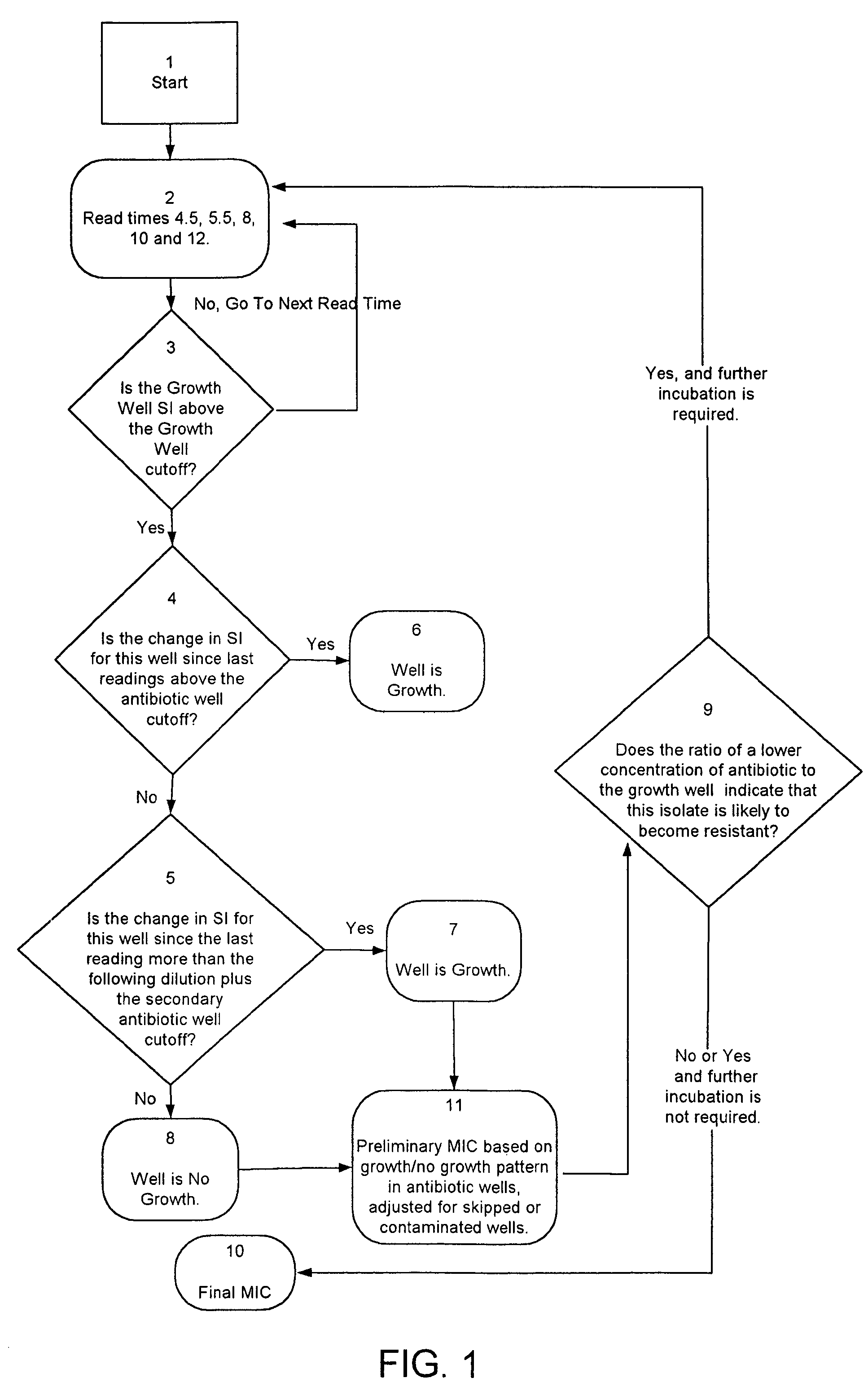
The present invention provides a combined system that can quickly and accurately identify microorganisms and determine their resistance to antimicrobial agents. The system is fully automated and hands-free, and uses a hybrid panel with multiple wavelength optics and novel methods for determining incubation time and susceptibility results. The system can provide standardized results in a rapid and reliable format.

Problems solved by technology

However, life threatening and debilitating systemic and localized microbial infections remains a major healthcare problem.
Mortality resulting from infectious agents remains particularly high among infants, the elderly, the immunosuppressed, patients in long-term care facilities, and skilled nursing homes, Moreover, the emergence of multi-drug resistance organisms such as vancomycin resistant enterococci (VRE) and methacillin resistant Staphylococcus aureaus (MRSA) have increased the challenges of caring for hospitalized patients.
Hospital acquired infections (nosocomial infections) caused by organisms such as VRE, MRSA and pseudomonas add significantly to patient suffering, increased hospital stays, iatrogenic mortality, and increased healthcare costs.
Drug resistance, specifically antibiotic resistance, often occurs when the antibiotic used to treat an infection is either improperly selected, prescribed in a fashion that does not effectively eradicate the infectious agent, or as a result of poor patient compliance.
Furthermore, when ineffective or unnecessary antibiotics are prescribed any infecting bacteria present continues to multiply unabated often resulting in life threatening complications necessitating expensive, aggressive treatments including otherwise needless hospitalization.
However, stains are largely non-specific and seldom definitive.
However, most clinical samples taken directly from infected patients are contaminated and contain PCR inhibitors.
Furthermore, PCR assays are expensive, highly specialized, and require multiple probes in order to identify an unknown organism.
Consequently, PCR is seldom, if ever used to replace standard microbial culture and identification techniques as the front line test in the clinical microbiology laboratory.
Moreover, the labor and material costs associated with such methods are high, consequently only a limited number of biochemical substrates are used in the initial screen.
If the results obtained after the first 18-24 hours of incubation are inconclusive, additional tests must be conducted which further delay definitive results in reaching the physician.
Both the Kirby-Bauer and broth dilution methods are laborious to set up, require skilled personnel to interpret and require a minimum of 18-24 hours before results are available.
The miniaturization of the biochemical assays and drug-containing tubes from 15 mL to 200 μL resulted in difficulty reading plates because visible growth is often barely detectable.
Consequently, much of the time and cost savings associated with the inoculation and set-up was lost reading and reporting results.
This is especially problematic when fluorogenic substrates are used.
In certain cases the growth rate may lag sufficiently so that when a pre-determined reading interval is reached there will be insufficient growth, and hence fluorescence to be detectable.
This in turn could be reported to the clinician that would initiate antimicrobial therapy based on an erroneous result.

Method used


Image

  • Combined rapid susceptibility assay and microorganism identification system
  • Combined rapid susceptibility assay and microorganism identification system
  • Combined rapid susceptibility assay and microorganism identification system

Examples


example 1

Standard Turbidimetric Assay Protocol 1

Gram Positive Assays

[0148]Although the following procedure relates to the use OF MICROSCAN® Dried Gram Positive MIC / Combo and Dried Gram Positive Breakpoint Combo panels, it is intended to be exemplary and not limiting. The standard turbidimetric assay protocol described herein is summarized below for the purpose of example but may be viewed in greater detail in the “DADE® MICROSCAN® Dried Gram Positive Procedural Manual,” Dade International Inc., West Sacramento, Calif. 95691 (1997), the disclosures of which are incorporated by reference herein.

[0149]The antimicrobial susceptibility tests are dehydrated miniaturizations of the broth dilution susceptibility test. Various antimicrobial agents are diluted in Mueller-Hinton broth with calcium and magnesium or Mueller-Hinton broth with additional supplementation to concentrations bridging the range of clinical interest.

[0150]For other specific tests, the use of different broths may be appropriate. ...

example 2

Standard Turbidimetric Assay Protocol 2

Gram Negative Assays

[0188]Although the following procedure relates to the use of MICROSCAN® Dried Gram Negative MIC / Combo and Dried Gram Negative Breakpoint Combo panels, it is intended to be exemplary and not limiting. The standard turbidimetric assay protocol described herein is summarized below for the purpose of example but may be viewed in greater detail in the “DADE® MICROSCAN® Dried Gram Negative Procedural Manual,” Dade International, Inc., West Sacramento, Calif. (1996), the disclosures of which are incorporated by reference herein.

[0189]The antimicrobial susceptibility tests are dehydrated miniaturizations of the broth dilution susceptibility test. Various antimicrobial agents are diluted in Mueller-Hinton broth with calcium and magnesium or Mueller-Hinton broth with additional supplementation to concentrations bridging the range of clinical interest. Breakpoint Combo panels use concentrations equivalent to the categorical breakpoints...

example 3

Fluorescent ID Procedure

[0225]Although the following procedure relates to the use of MICROSCAN® Rapid Panels, it is intended to be exemplary and not limiting. The standard fluorescent ID protocol described herein is summarized below for the purpose of example but may be viewed in greater detail in the “Dade MICROSCAN® Rapid Gram Positive Procedural Manual,” Dade Behring Inc., West Sacramento, Calif. (1998), the disclosures of which are incorporated by reference herein.

[0226]The antimicrobial susceptibility tests are dehydrated miniaturizations of the broth dilution susceptibility test. Various antimicrobial agents are serially diluted in autoclaved distilled water with fluorogenic compounds to concentrations bridging the range of clinical interest. Trimethoprim / Sulfamethoxazole contains thymidine phosphorylase to reduce thymidine levels in the medium.

[0227]Fluorogenic substrates or fluorometric indicators are used for the identification of Micrococcaceae, Streptococcaceae, Listeria ...


PUM


Abstract

In response to the need for highly-sensitive antibiotic susceptibility assays and identification assays that do not require extensive incubation times, the present invention provides automated assay methods and systems that permit the determination of antibiotic susceptibilities and / or microorganism identification in a timeframe that is substantially shorter than has previously been attainable using a hybrid system that combines turbimetric and fluorescence determinations using a single, clear-plastic assay platform. Related devices, kits, and components thereof are also disclosed.

Description

RELATED APPLICATIONS[0001]This application is a divisional of U.S. patent application Ser. No. 10 / 967,062, filed Oct. 15, 2004, which is a continuation of U.S. patent application Ser. No. 09 / 553,223, filed Apr. 20, 2000, the contents of which are hereby incorporated by reference in its entirety, which claims priority of provisional application Ser. No. 60 / 131,829 filed Apr. 29, 1999, entitled “Antibiotic Susceptibility Testing Methods and Devices” and provisional application Ser. No. 60 / 137,819 filed on May 27, 1999, entitled “Antibiotic Susceptibility and Microbial Identification Testing Methods and Systems”.FIELD OF THE INVENTION[0002]A combined anti-microbial susceptibility testing and microorganism identification platform is disclosed. Specifically, the combined anti-microbial susceptibility testing and microorganism identification platform disclosed herein is a hands-free walk-away system ideally suited for clinical microbiology laboratories. More specifically, the automated, c...

Claims


Application Information

Patent Timeline
Patent Type & Authority Patents(United States)
IPC IPC(8): C12Q1/02C12Q1/18C12Q1/04G01N21/64C12Q1/08G01N21/27G01N35/00G01N35/02
CPCG01N35/028G01N35/0092G01N2035/0097G01N2035/0093
Inventor WILLIAMS, GREGORY B.NOTHAFT, DANIEL M.ENSCOE, GLENN F.BURTNER, KATHLEEN N.KANGAS, MONTE E.
Owner BECKMAN COULTER INC
Learn More