Method and device for the treatment of hypertrophic cardiomyopathy

a cardiomyopathy and hypertrophic technology, applied in the field of hypertrophic cardiomyopathy, can solve the problems of increased pressure in the left ventricle, increased obstruction of the ventricle, so as to reduce the risk of fully sedating the patient, prevent unwanted movement of the tubular blade, and facilitate the effect of performing

Inactive Publication Date: 2014-12-09
UNIV OF SOUTH FLORIDA
View PDF14 Cites 8 Cited by
  • Summary
  • Abstract
  • Description
  • Claims
  • Application Information

AI Technical Summary

Benefits of technology

[0018]The proposed technique is non surgical, allowing use of mild, local anesthetics, as opposed to the general anesthetic used in traditional approaches. This reduces risks of fully sedating the patient. Moreover, this technique is easier to perform than the current available standard methods, making it the preferred procedure by both patients and operators.
[0019]The claimed invention includes a catheter with a cutting device at its distal end. The cutting device has an outer shell with a void and a plurality of holes disposed at its distal end. A tubular blade is slideably disposed within the outer shell and has

Problems solved by technology

Problems occur when the septum between the heart's lower chambers, or ventricles, is thickened.
The narrowing of the left ventricular outflow tract disrupts the proper function of the mitral valve, resulting in outflow obstruction and increased pressure in the left ventricle.
When this occurs, the mitral valve frequently leaks, causing the blood to go back into the left atrium.
HCM is the most common cause of sudden cardiac death in people under the age of 30.
Excessive resection of muscle can cause a ventricular septal defect, a serious complication that may occur in up to 2% of patients.
Other complications include inadequate intraoperative protection of the hypertrophied muscle against ischemia, causin

Method used


Image

  • Method and device for the treatment of hypertrophic cardiomyopathy
  • Method and device for the treatment of hypertrophic cardiomyopathy
  • Method and device for the treatment of hypertrophic cardiomyopathy

Examples


example 1

Method of Performing a Myectomy Using the Device

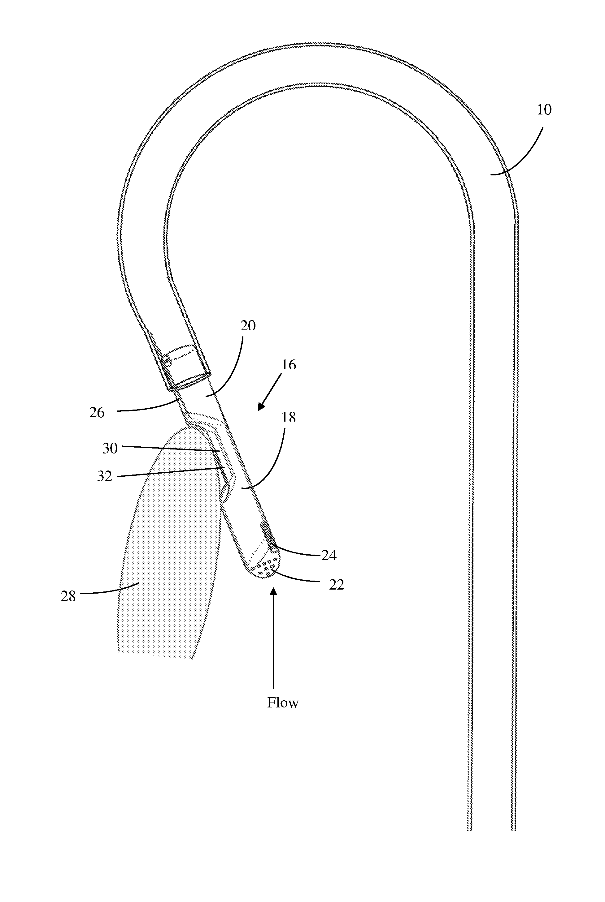
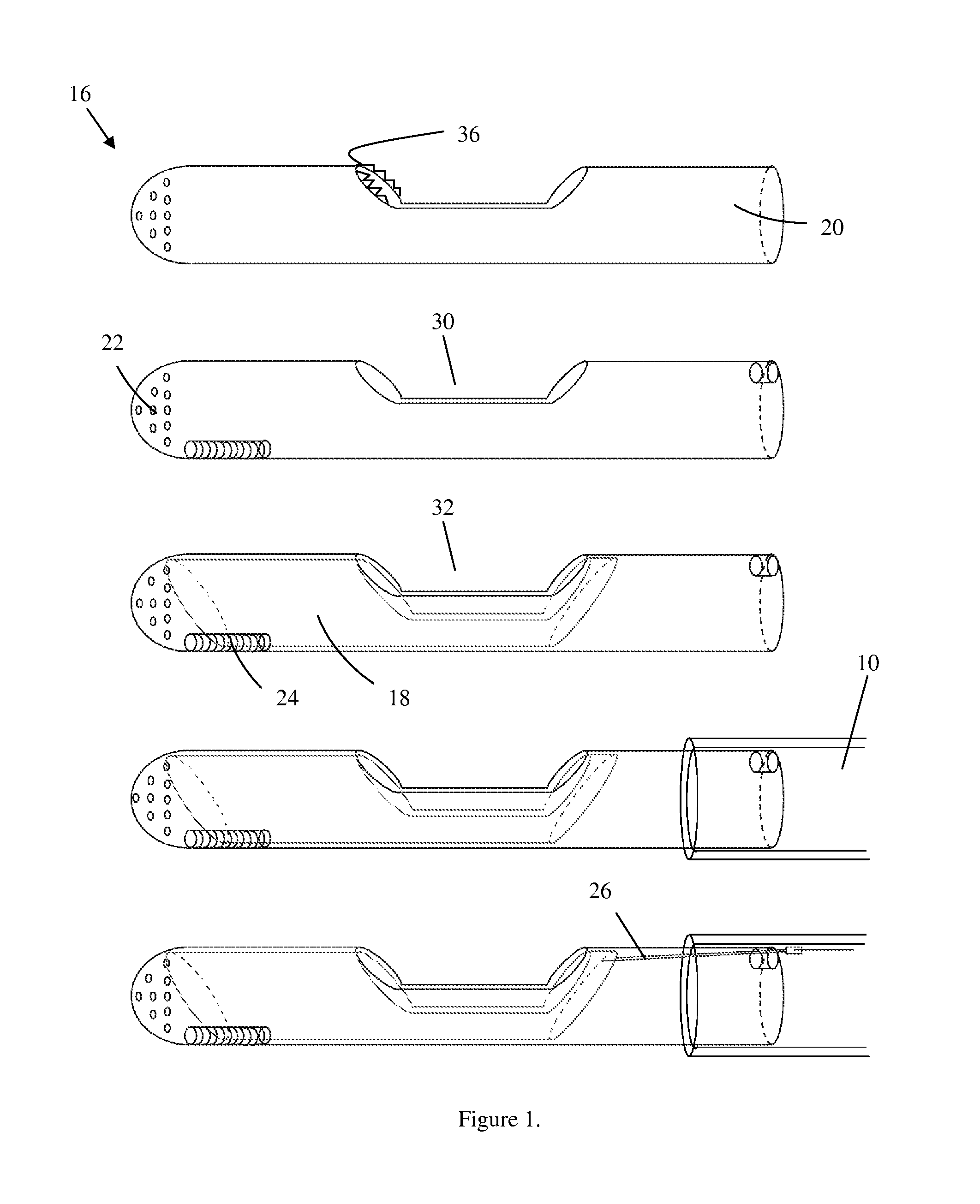
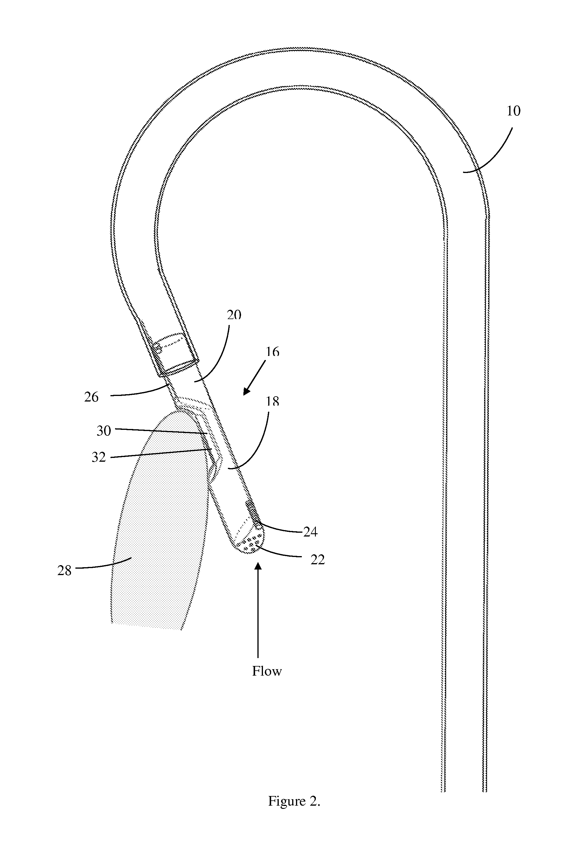
[0030]The femoral or brachial artery is punctured, providing access to the aorta (blood vessel). The sheath is inserted for secure hemostasis and a guide wire is advanced into the left ventricle via trans-aortic route. The device is then filled with normal saline to prevent an air embolism, and the device is then threaded via a hole at the distal end and advanced up to the left ventricle. The cutting tip 16 is positioned adjacent to the myocardium 28 that is to be excised, as seen in FIG. 2, under the guidance of transesophageal, transthoracic, or intracardiac echocardiography, and / or fluoroscopic guidance, then the guide wire is removed. After confirming the position of the cutting tip 16, the operator slides the tubular blade 18 within the outer shell 20 covering to resect the septum. The tubular blade 18 action will be controlled via the proximal end which is outside the patient's body. During this procedure, the tubular blade 18 is...

example 2

Method of Using the Device to Excise Tissue or Plaques

[0035]The device is also useful in removal of tissues in organs, such as where tissue needs to be removed from an organ, tumor removal, and circulatory plaque and thrombosis removal, such as deep vein thrombosis clot removal. In such instances, the device is advanced to the excision site, as described above. Once the cutting tip is adjacent to the target tissue or clot the blade is used to cut the target tissue or clot. The excised tissue is then removed from the patient via the catheter using negative pressure.

example 3

Method of Proximal Insertion and Implementation of the Device

[0036]Under more severe conditions, the device may also be inserted more closely to the organ of targeted implementation and not threaded through distal vessels. For instance, in HCM patients who are severely obstructed, the device may be inserted between the ribs in a location near the heart (mini thoracotomy), in a manner that punctures the heart cavity. In such insertion techniques, the tubular construct connecting the device to the suction apparatus may be comprised of non-flexible polyethylene or other solid plastic material. This embodiment may also utilize a proportionally larger device fabricated in the same manner as above. Once positioned adjacent to the target tissue, the method of utilizing the device may be performed by a surgeon's hand or by a robotic device.


PUM


Abstract

A method and device for the treatment of hypertrophic cardiomyopathy. The device includes a cutting device for resection of a thickened myocardium. The operator positions the cutting device adjacent to the myocardium that is to be resected and then slides a tubular blade within an outer shell of the cutting device to resect the septum.

Description

CROSS-REFERENCE TO RELATED APPLICATIONS[0001]This application claims priority to U.S. Provisional Patent Application No. 61 / 176,738, entitled “PATEL'S SEPTAL MYECTOMY DEVICE”, filed on May 8, 2009, the contents of which are hereby incorporated by reference.BACKGROUND OF THE INVENTION[0002]1. Field of the Invention[0003]This invention relates to a method and device for the treatment of hypertrophic cardiomyopathy. More specifically, this invention relates to a method and device that retracts the thickened myocardium.[0004]2. Description of the Prior Art[0005]Hypertrophic cardiomyopathy (HCM) is a complex type of heart disease that affects the heart muscle. It causes thickening of the heart muscle (especially the ventricles, or lower heart chambers), left ventricular stiffness, mitral valve changes, and cellular changes.[0006]Thickening of the heart muscle (myocardium) occurs most commonly at the septum. The septum is the muscular wall that separates the left and right side of the hea...

Claims


Application Information

Patent Timeline
IPC IPC(8): A61B17/32A61B19/00A61B17/22
CPCA61B17/320783A61B17/32A61B17/320016A61B2017/320064A61B18/1492A61B17/22A61B2017/00243A61B19/00A61B17/32002A61B18/1445A61B2017/320032A61B2018/00351A61B2018/00589
Inventor PATEL, VINOD
Owner UNIV OF SOUTH FLORIDA
Why Eureka
Try Eureka