Trans-catheter ventricular reconstruction structures, methods, and systems for treatment of congestive heart failure and other conditions

a ventricular reconstruction and transcatheter technology, applied in the field of transcatheter ventricular reconstruction structures, methods, systems for treating congestive heart failure and other conditions, can solve the problems of affecting the normal function of the heart muscle, affecting the effective reshaping of the ventricular chamber, and affecting so as to reduce the distance between two locations, reduce the effect of ventricular chamber reshaping, and less or minimally invasiv

Active Publication Date: 2015-03-17
BIOVENTRIX A CHF TECH
View PDF101 Cites 47 Cited by
  • Summary
  • Abstract
  • Description
  • Claims
  • Application Information

AI Technical Summary

Benefits of technology

The present invention provides improved medical devices, systems, and methods for reducing the distance between two locations in tissue, optionally in a less or minimally invasive manner. The invention can be used to treat congestive heart failure and other progressive heart diseases by reconfiguring abnormal heart geometry that may be contributing to heart dysfunction. The invention involves advancing an implant into the heart through a minimally invasive epicardial access tool, with the implant system being secured in place using tension between two anchors. The invention also provides a method for treating congestive heart failure by advancing a first anchor and a tension member into the heart, with the tension member being pulled from the first anchor through the septum and the exterior wall of the left ventricle. The invention allows for controlled pressure application between the anchors, resulting in the effective reshaping of the ventricular chamber and mitigation of congestive heart failure.

Problems solved by technology

However, in many cases congestive heart failure may be triggered by a heart attack or myocardial infarction.
Heart attacks can cause scar tissue that interferes with the heart muscle's healthy function, and that scar tissue can progressively replace more and more of the contractile heart tissue.
More specifically, the presence of the scar may lead to a compensatory neuro-hormonal response by the remaining, non-infarcted myocardium leading to progressive dysfunction and worsening failure.
People with heart failure may have difficulty exerting themselves, often becoming short of breath, tired, and the like.
The increased overall volume and higher intracardiac pressures result in congestion in the tissues.
Fluid may also collect in the lungs, interfering with breathing (especially when lying down).
Congestive heart failure may also be associated with a decrease in the ability of the kidneys to remove sodium and water, and the fluid buildup may be sufficient to cause substantial weight gain.
With progression of the disease, this destructive sequence of events can cause the progressive deterioration and eventual failure of the remaining functional heart muscle.
If the heart becomes sufficiently damaged, even more drastic surgery may be considered.
These surgical therapies can be at least partially effective, but typically involve substantial patient risk.
With congestive heart failure, the left ventricle often dilates or increases in size.
This can result in a significant increase in wall tension and stress.

Method used


Image

  • Trans-catheter ventricular reconstruction structures, methods, and systems for treatment of congestive heart failure and other conditions
  • Trans-catheter ventricular reconstruction structures, methods, and systems for treatment of congestive heart failure and other conditions
  • Trans-catheter ventricular reconstruction structures, methods, and systems for treatment of congestive heart failure and other conditions

Examples


experiment 1

Implantation in a Pig Cadaver Heart #1

[0071]A frozen heart was thawed and placed into expanding foam in a position representative of that in the body seen via a median sternotomy. A variety of baskets were provided for grasping a guide wire passed into the right ventricle across the septum from the left ventricle. The shape of the catheters and baskets varied with target locations along the right ventricular septum that the guide wire would be was to enter.

[0072]As shown in FIGS. 25a-c, basket configured to retrieve a wire from the apex of the right ventricular septum, from the mid-portion of the right ventricular septum, and from the infundibulum (pulmonary outflow tract) of the right ventricular septum were provided, respectively.

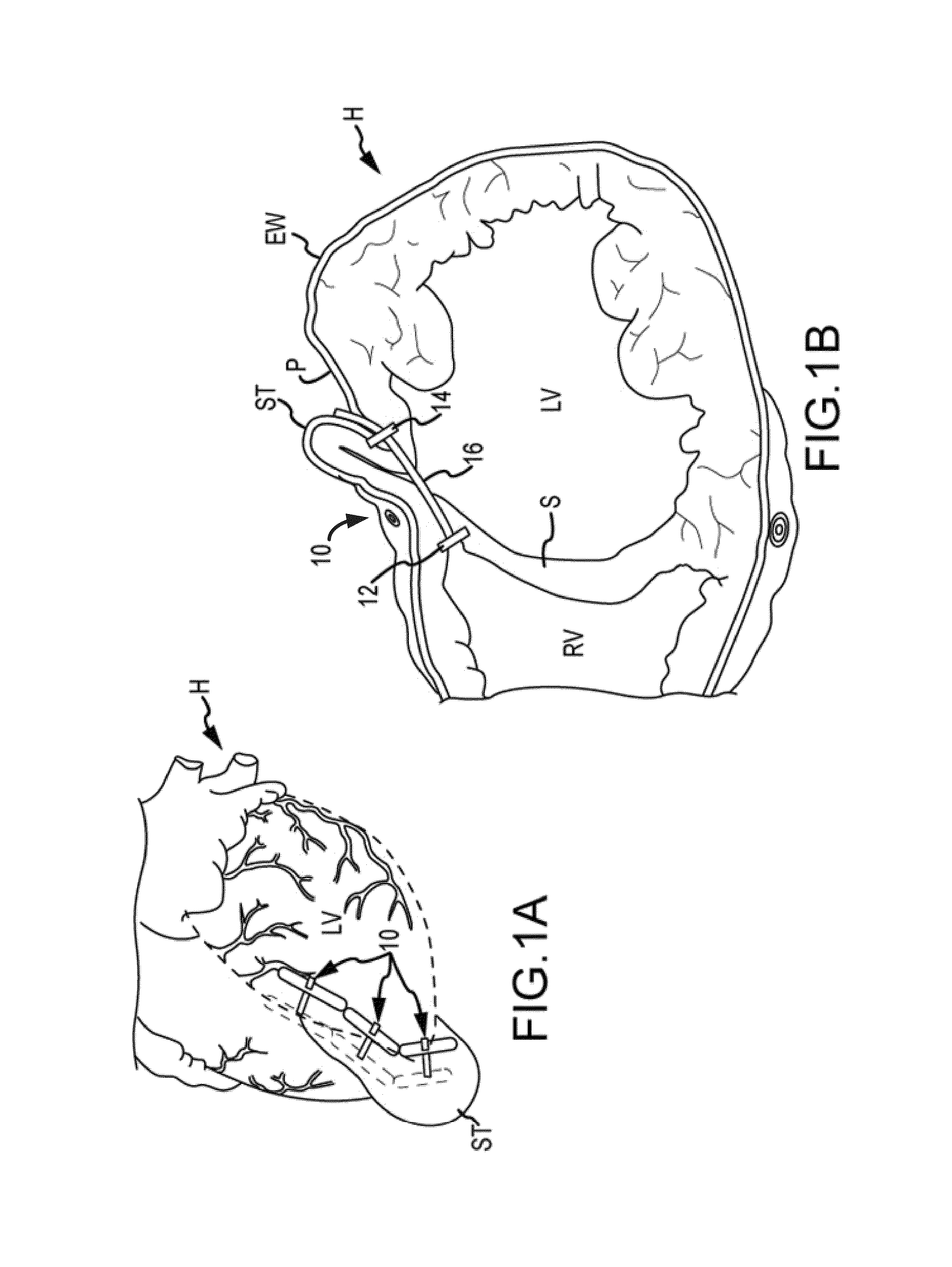
[0073]The apex basket was placed into the right ventricle through the opened right atrium and imaged via fluoroscopy. A curved needle was passed through the eipcardial surface of the left ventricle (LV) lateral to the LAD, through the LV cavity, across th...

experiment 2

Implantation in a Human Cadaver Heart

[0086]67-year-old male. Cause of death: Heart failure

[0087]Serology: NEG

[0088]Height: 71 inches; Weight: 237 lbs

[0089]A frozen heart was thawed, placed into expanding foam in a position representative of that in the body seen via a median sternotomy. A variety of baskets were provided for grasping a guide wire passed into the right ventricle across the septum from the left ventricle, as described above.

[0090]The surgical approach was from the right atrium (RA). The basket was placed into the right ventricle through the opened right atrium and imaged via fluoroscopy. A curved needle was then passed through the epicardial surface of the left ventricle (LV) lateral to the LAD, through the LV cavity, across the ventricular septum and into the right ventricle (RV) in the vicinity of the basket. Following positioning of the needle, a guide wire is passed through the needle and into the basket via fluoroscopic control. The wire position is confirmed in ...

experiment 3

Implantation in a Live Sheep Heart

Weight: 84.7 kg

Age: adult

Sex: male

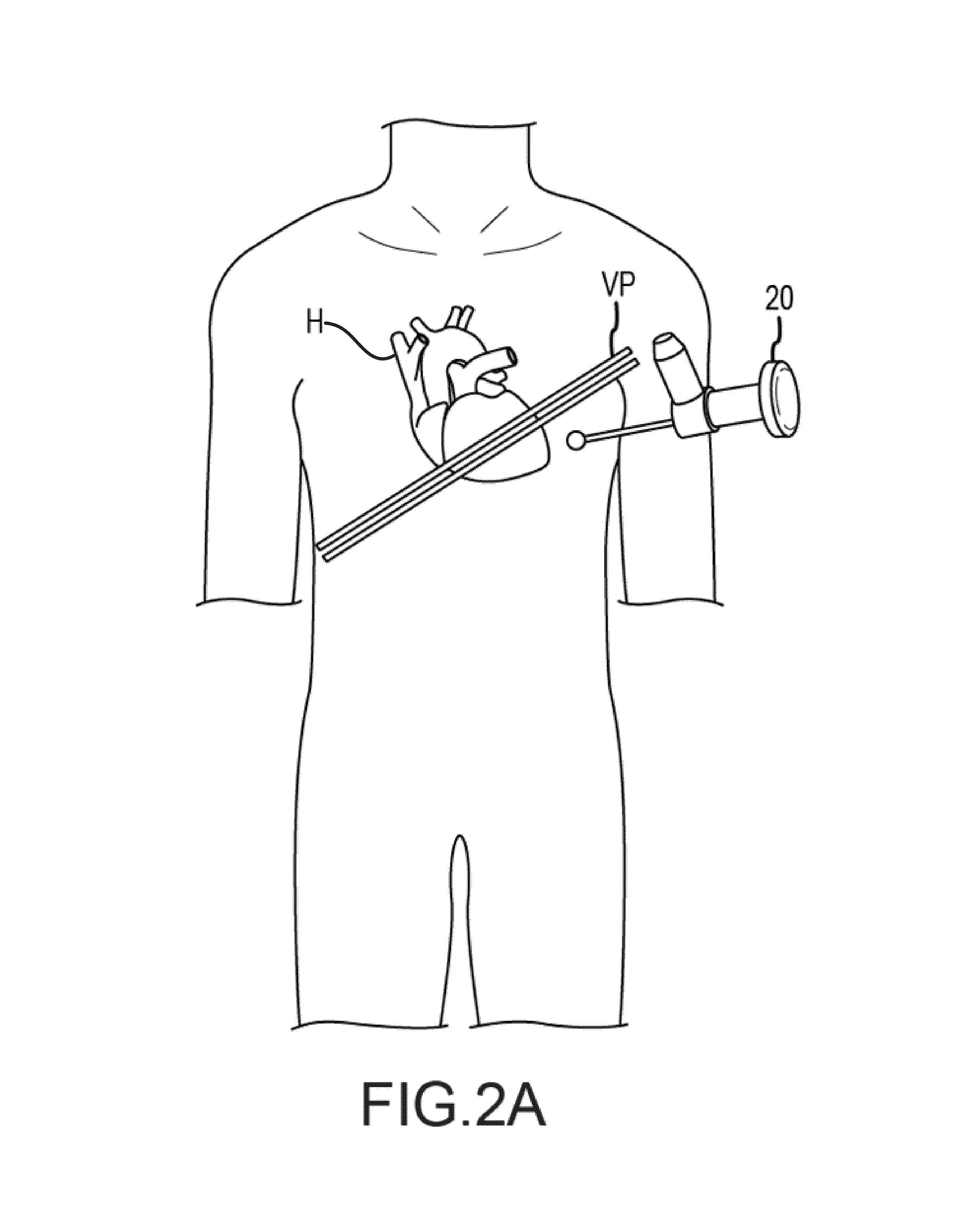
[0101]With the animal under general anesthesia, arterial and venous lines were placed. The chest was opened through a median sternotomy and the pericardium was opened in the midline. A pericardial cradle was created and the right atrial (RA) appendage was exposed. A purse-string suture (4-0 Prolene) was placed on the RA and a 14 Fr. sheath was passed into the right ventricle (RV) through the RA under fluoroscopic guidance.

[0102]In FIG. 33a, the heart is exposed through a mid-sternotomy and pericardiotomy. The LAD and apex are labeled, and (see FIG. 33b) a tourniquet is place on the RA. A sheath (blue) is passed and under fluoroscopy and (see FIG. 33c) positioned in the RV (in circle).

[0103]An apical basket was passed through the sheath in the RA into the RV, and a needle was passed through the LV epicardium near the apex and aimed toward the basket under fluoroscopic control. A guide wire was passed through the LV a...


PUM


Abstract

Embodiments described herein include devices, systems, and methods for reducing the distance between two locations in tissue. In one embodiment, an anchor may reside within the right ventricle in engagement with the septum. A tension member may extend from that anchor through the septum and an exterior wall of the left ventricle to a second anchor disposed along a surface of the heart. Perforating the exterior wall and the septum from an epicardial approach can provide control over the reshaping of the ventricular chamber. Guiding deployment of the implant from along the epicardial access path and another access path into and through the right ventricle provides control over the movement of the anchor within the ventricle. The joined epicardial pathway and right atrial pathway allows the tension member to be advanced into the heart through the right atrium and pulled into engagement along the epicardial access path.

Description

CROSS-REFERENCES TO RELATED APPLICATIONS[0001]This application is related to and claims the benefit of U.S. Provisional Patent Application No. 61 / 541,624 entitled “Trans-Catheter Ventricular Reconstruction Structures, Methods, and Systems for Treatment of Congestive Heart Failure and Other Conditions,” filed Sep. 30, 2011. This application is also related to and claims the benefit of U.S. Provisional Patent Application No. 61 / 541,975 entitled “Remote Pericardial Hemostasis for Ventricular Access and Reconstruction or Other Organ Therapies,” filed Sep. 30, 2011; U.S. Provisional Patent Application No. 61 / 541,980 entitled “Over-The-Wire Cardiac Implant Delivery System for Treatment of CHF and Other Conditions,” filed Sep. 30, 2011; and U.S. Provisional Patent Application No. 61 / 541,978 entitled “Cardiac Implant Migration Inhibiting Systems,” filed Sep. 30, 2011; the full disclosures of which are incorporated herein by reference in their entirety.[0002]The subject matter of this applic...

Claims


Application Information

Patent Timeline
Patent Type & Authority Patents(United States)
IPC IPC(8): A61B17/00A61B17/04A61B17/22A61B17/30A61B17/34A61B18/14A61B19/00A61F2/24A61M29/02
CPCA61B19/22A61B17/0057A61M29/02A61B17/0401A61F2/2487A61B17/00A61B2018/1425A61B17/0485A61B2017/00243A61B2017/0409A61B2017/0417A61B2017/22038A61B2017/22065A61B2017/306A61B2017/3488A61B17/3417A61B17/3478A61B2017/3458A61B2017/2215A61B34/70A61B17/00234A61B17/12013A61B2017/00323A61B2017/00561A61B2017/0406A61B2017/0414A61B2017/042A61B2017/0422A61B2017/0464A61B2017/0496A61B5/0215
Inventor VAN BLADEL, KEVINANNEST, LONSHELDON, MURRAYHEFLIN, ERNIEWECHSLER, ANDREWSTIGGELBOUT, JOHNARCIA, ROVILBOWER, JOHN
Owner BIOVENTRIX A CHF TECH
Why Eureka
Try Eureka