

Nanowired three dimensional tissue scaffolds
a three-dimensional, tissue scaffold technology, applied in the field of tissue engineering, can solve the problems of weak patch contracting potential, lack of electrical conductivity within the construct, and failure to achieve the effect of enhancing tissue growth and improving electrical communication
- Summary
- Abstract
- Description
- Claims
- Application Information
AI Technical Summary
Benefits of technology
Problems solved by technology
Method used
Image

Examples
example 1
Analysis of Cardiac Cell Organization within a 3D Alginate Scaffold with or without Gold Nanowires
[0034]Materials and Methods
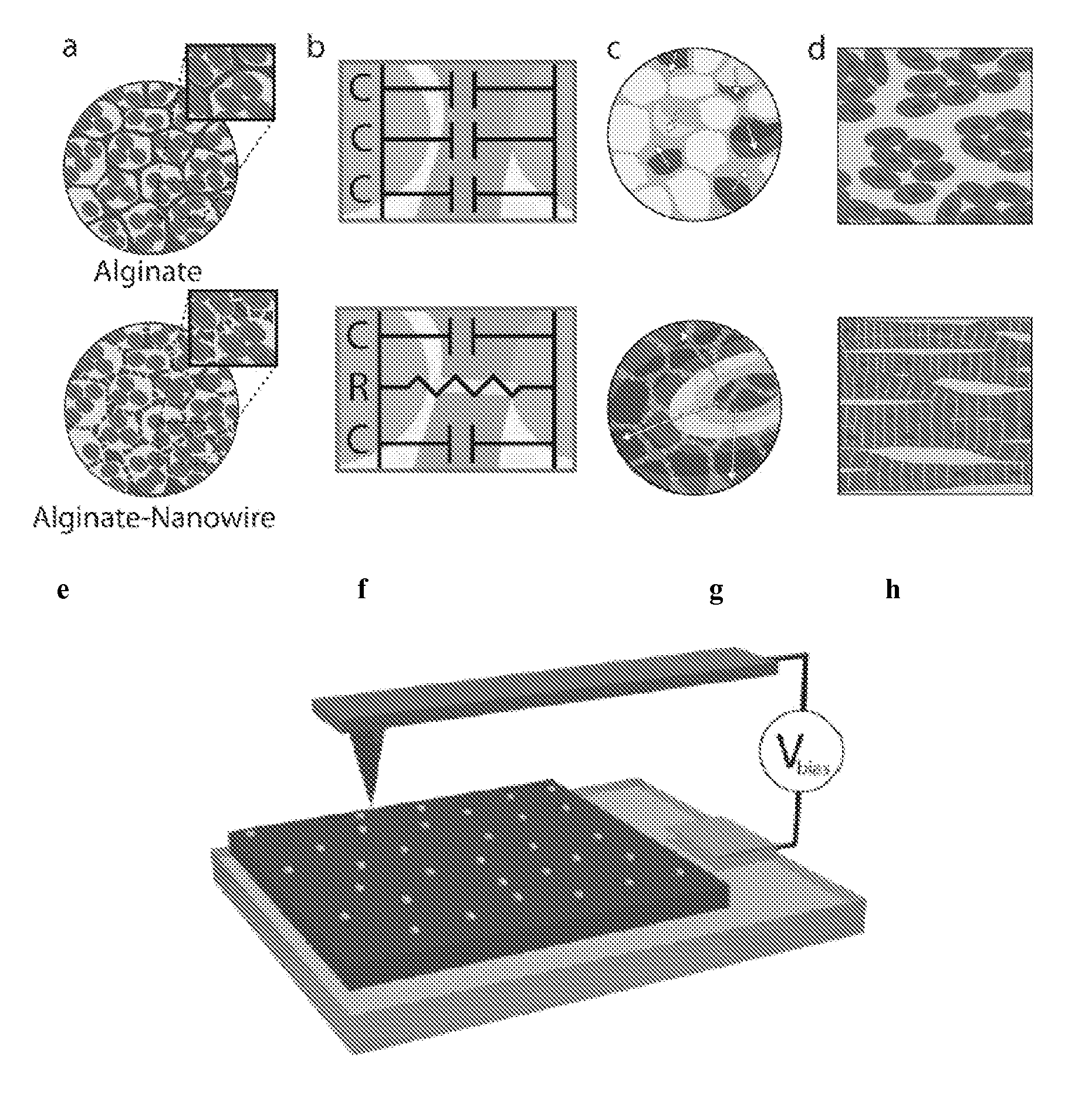
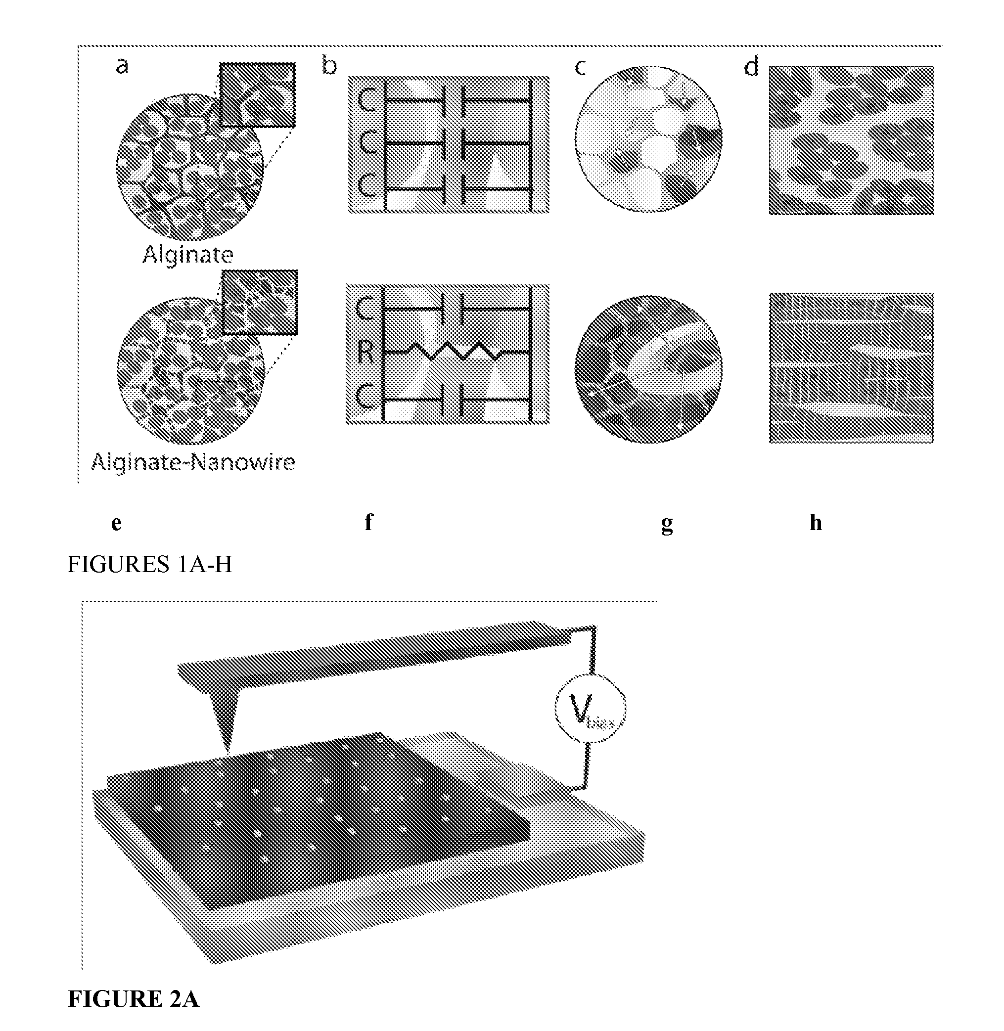
[0035]A nanocomposite scaffold of alginate and NWs (Alg-NW) was created by mixing NWs with sodium alginate, followed by cross-linking with calcium gluconate and lyophilization. The scaffolds were 5 mm in diameter and 2 mm height.
[0036]Preparation of Gold NWs
[0037]First, citrate-capped Au seeds were prepared. A 20 mL aqueous solution of HAuCl4 (0.25 mM) and sodium citrate (0.25 mM) was prepared. Under vigorous stirring, 0.6 mL ice-cold aqueous NaBH4 solution (0.1 M) was added all at once. The solution immediately turned deep red, consistent with the formation of colloidal gold. A typical product contained spherical gold particles about 4 nm in diameter at an estimated density of ca. 7.6×10¹³ particles / ml. The suspension was allowed to stand at room temperature (about 25° C., RT) for several hours before subsequent use to ensure complete degradation of the NaBH4...
example 2
Analysis of Calcium Transient Propagation within a 3D Alginate Scaffold with or without Gold Nanowires Seeded with Cardiac Cells
[0047]Materials and Methods
[0048]Calcium transient propagation within the engineered tissues was assessed at specified points by monitoring calcium dye fluorescence. Calcium transient was assessed at specified points by monitoring calcium dye fluorescence.
[0049]Neonatal rat ventricular myocytes were incubated with 10 μM fluo-4 AM (Invitrogen) and 0.1% Pluronic F-127 for 45 min at 37° C. Cardiac cell constructs (at least 9 samples from each group, from 3 separate experiments) were subsequently washed 3 times in modified Tyrode solution to allow de-esterification. Cell aggregates were electrically paced at 1-2 Hz using a bipolar platinum electrode placed in close contact with the cells using micromanipulator. The calcium transients were imaged using a confocal imaging system (LSM 510, Zeiss). The images were acquired with a EC Plan-Neofluar 10× / 0.30M 27 objec...
example 3
Analysis of Mechanical Properties within a 3D Alginate Scaffold with or without Gold Nanowires Seeded with Cardiac Cells
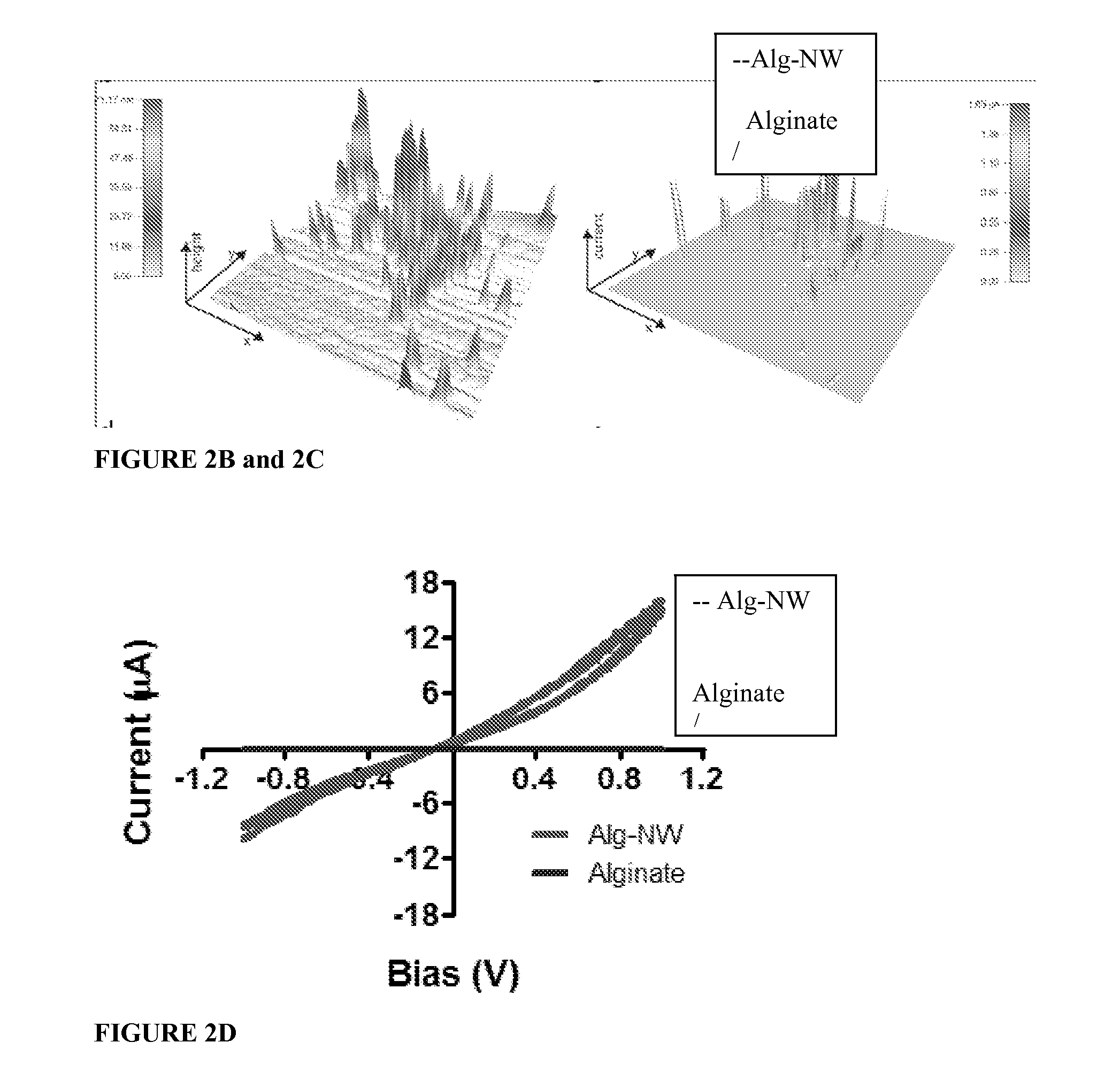
[0053]Rheological testing of the alginate gel prior to lyophilization (which hardens it to its final firmness) showed that viscosity and elastic shear modulus increased with NW concentration, which suggests interactions between the polymer chains and / or the ionic cross-linker (in this case, calcium) and the NWs. The increased viscosity may also be caused by NWs acting as space fillers, increasing the strength of the hybrid material. These results are demonstrated by FIGS. 4A and 4B.
[0054]As shown by FIG. 4A, at higher concentrations of NWs the biomaterial became more viscous. As shown by FIG. 4B, increasing NW concentration increased the elastic shear modulus of the biomaterial. Elemental analysis of the features within the pore wall indicated they are made of gold.
[0055]Scanning electron microscopy (SEM) revealed NWs (0.5 mg / mL) either parallel to or penetrating t...
PUM
| Property | Measurement | Unit |
|---|---|---|
| diameter | aaaaa | aaaaa |
| pore size | aaaaa | aaaaa |
| length | aaaaa | aaaaa |
Abstract
Description
Claims
Application Information