Medical device having a surface comprising nanoparticles
a technology of nanoparticles and nanoparticles, which is applied in the field of medical devices, can solve the problems of limited porousness of nanoparticle coatings and the risk of microbe penetration into the surface, and achieve the effect of reducing the risk of detrimental infection
- Summary
- Abstract
- Description
- Claims
- Application Information
AI Technical Summary
Benefits of technology
Problems solved by technology
Method used
Image
Examples
example 1
n
[0108]Coins of commercially pure (cp) titanium (grade 4) were manufactured and cleaned before spin coating with nanoparticles of bismuth oxide (Bi2O3). Bismuth nanoparticles (product name BI-OX-03-NP.200N, American Elements, USA) were dispersed in acetate buffer pH 5.0. Cleaned coins were mounted on a rotatable electrode. The speed of electrode rotation was increased and the coins were immersed in acetate buffer containing bismuth oxide nanoparticles for 5 seconds. The speed of rotation decreased to 0 rpm and the coins were immersed in a beaker with deionized water for 5 seconds to remove the remaining unbound bismuth nanoparticles on the surface. The coins were dried and were thereafter packaged in plastic containers, and sterilized with electron beam irradiation.
example 2
haracterization
[0109]For all surface characterization experiments, specimens of commercially pure (cp) titanium and specimens spin coated with bismuth nanoparticles produced as described in Example 1 were used.
[0110]2a) Surface Morphology and Surface Chemistry
[0111]Surface morphology and surface chemistry analyses were performed with environmental scanning electron microscopy (XL30 ESEM, Philips, Netherlands) / energy dispersive spectroscopy (Genesis System, EDAX Inc., USA) at an acceleration voltage of 30 kV and 10 kV for surface morphology and surface chemistry, respectively.
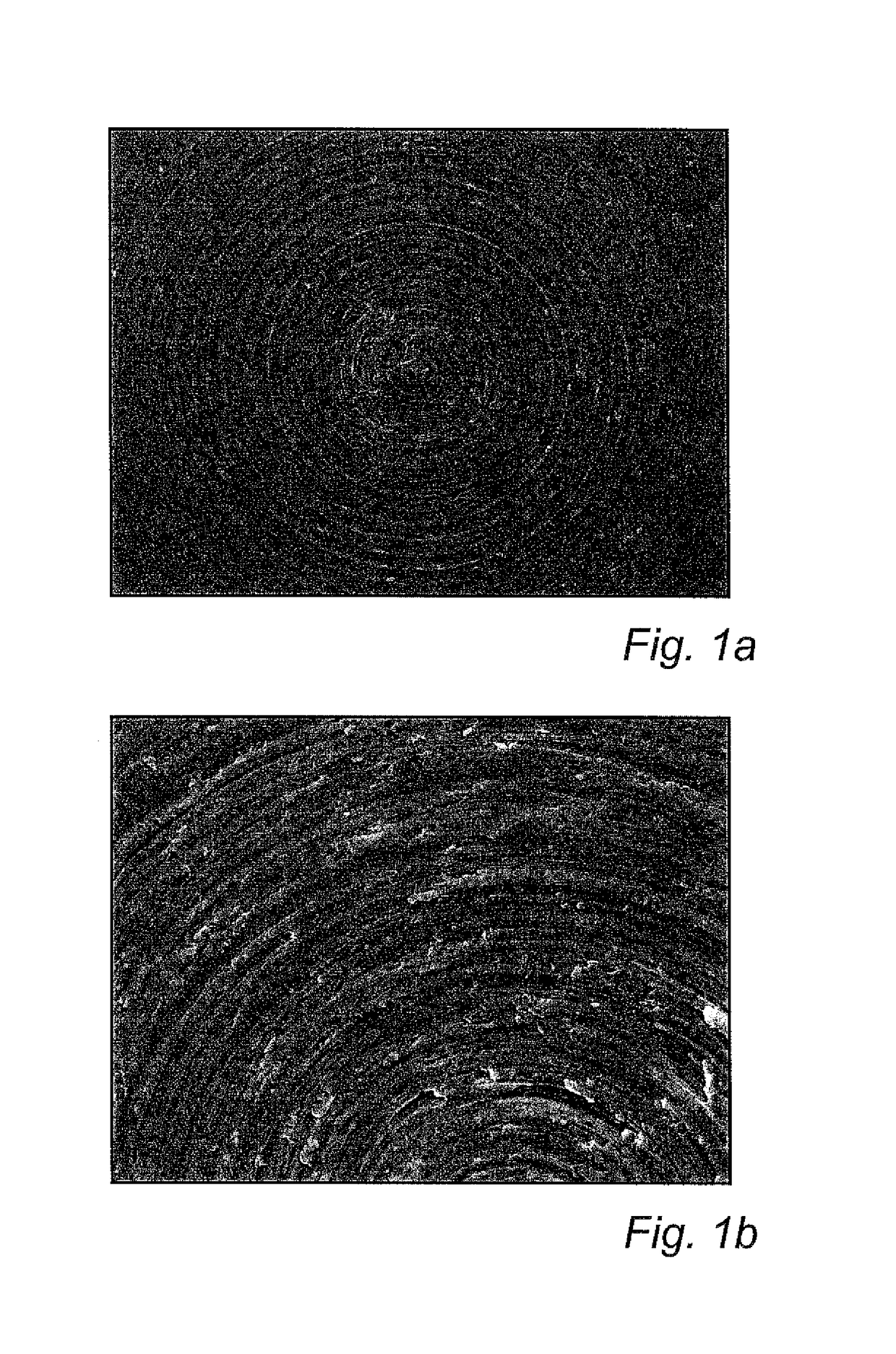
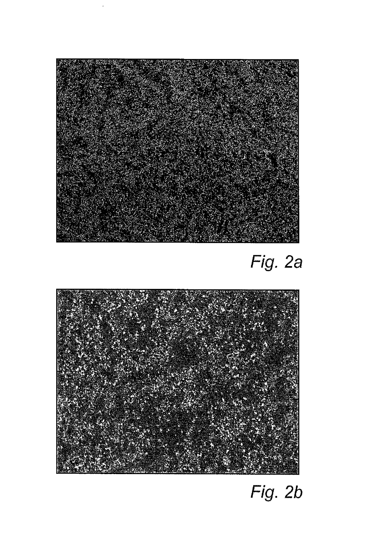
[0112]FIGS. 1a and 1b show an uncoated specimen at 100× (FIG. 1a) and 500× (FIG. 1b) magnification, respectively. Machining tracks can be seen, but no particles. In contrast, FIGS. 2a and b show a coated specimen at the same magnifications. As can be seen in FIGS. 2a-b, the coated specimens were partly, but not completely, covered by dispersed nanoparticles.
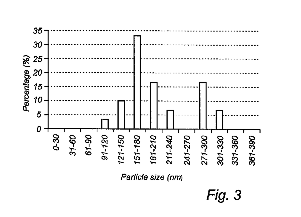
[0113]The diameter of the Bi2O3 particles present on th...
example 3
bial Effect
[0117]a) Inhibition of Bacterial Growth Using Film Contact Method
[0118]A film contact method as described in M. Yasuyuki, K. Kunihiro, S. Kurissery, N. Kanavillil, Y. Sato, Y. Kikuchi. Biofouling 26 (2010) 851-858, was used for evaluating the antimicrobial effect. Streak plates of Pseudomonas aeruginosa (PA01) were made and 1 colony was inoculated to 5 ml tryptic soy broth (TSB) in culture tubes and grown under shaking conditions for 18 hours. Cell density was measured in a spectrophotometer at OD 600 nm and counted using a cell counting chamber. The cell culture was adjusted with sterile TSB to 1-5×10⁶ cells / ml. Specimens of commercially pure (cp) titanium coins (diameter of 6.25 mm) and cp titanium coins spin coated with Bi2O3 nanoparticles according to Example 1 above were aseptically prepared and placed each in a respective well of a 12 well plate. Thin transparent plastic film was punched, and sterilized using 70% ethanol and UV irradiation on each side. A 15 μl drop...
PUM
| Property | Measurement | Unit |
|---|---|---|
| particle size | aaaaa | aaaaa |
| thickness | aaaaa | aaaaa |
| particle size | aaaaa | aaaaa |
Abstract
Description
Claims
Application Information
 - R&D
- Intellectual Property
- Life Sciences
- Materials
- Tech Scout
- Unparalleled Data Quality
- Higher Quality Content
- 60% Fewer Hallucinations
Browse by: Latest US Patents, China's latest patents, Technical Efficacy Thesaurus, Application Domain, Technology Topic, Popular Technical Reports.
© 2025 PatSnap. All rights reserved.Legal|Privacy policy|Modern Slavery Act Transparency Statement|Sitemap|About US| Contact US: help@patsnap.com


