

Preparation method of function surface strength laman scattering probe
A surface-enhanced Raman, functional technology, used in Raman scattering, biochemical equipment and methods, and microbial determination/inspection. It can solve problems such as narrow excitation spectrum, low fluorescence intensity, and background fluorescence flooding, avoiding the Measurement error, the effect of improving reliability
- Summary
- Abstract
- Description
- Claims
- Application Information
AI Technical Summary
Problems solved by technology
Method used
Image

Examples
Embodiment 1
[0043] Embodiment 1, preparation of functional SERS probe
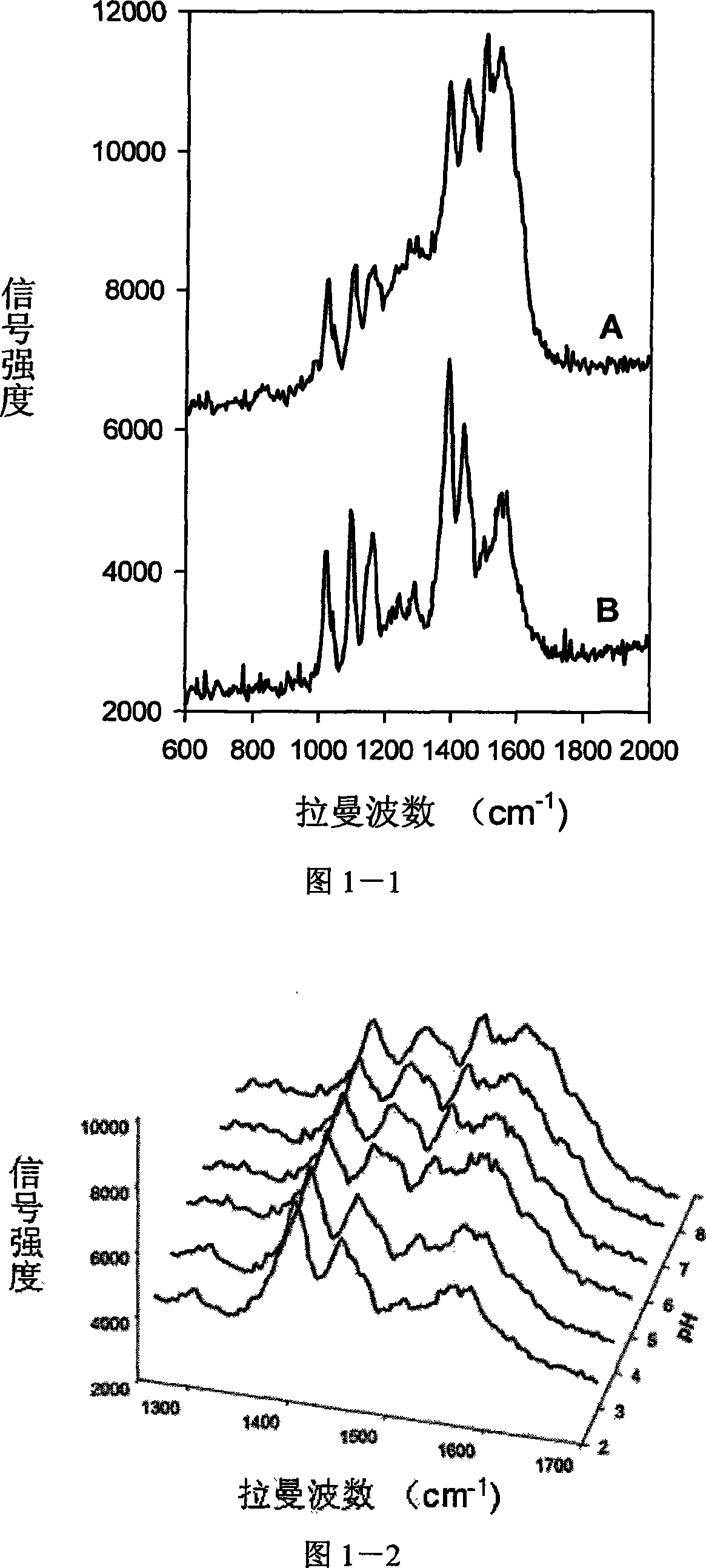
[0044] In the first step, first prepare silver colloid solution according to the method described by Lee and Meisel: under ice bath condition, make 50mL concentration 5×10 -3 Add 150mL of silver nitrate solution of M to a concentration of 2×10 -3 M in sodium borate solution and mix well, and stir at room temperature for 1 hour. Obtain a yellow-green transparent solution, that is, a silver colloid solution, for use.
[0045] In the second step, the concentration is 10 -5 The methanol solution of 2-aminophenol sulfur of M and the silver colloid solution prepared in the first step were uniformly mixed in a volume ratio of 3:1000, and reacted for 20 minutes. 2-Aminophenol-sulfur molecules are easily intercalated on the surface of silver nanoparticles in silver colloid through -HS bonds. Silver nanoparticles labeled with 2-aminophenol sulfur molecules were separated from solution by centrifugation. The rotational spee...
Embodiment 2
[0046] Example 2, Functional SERS Probe Used for pH Sensing in Pancreatic Cancer Cells
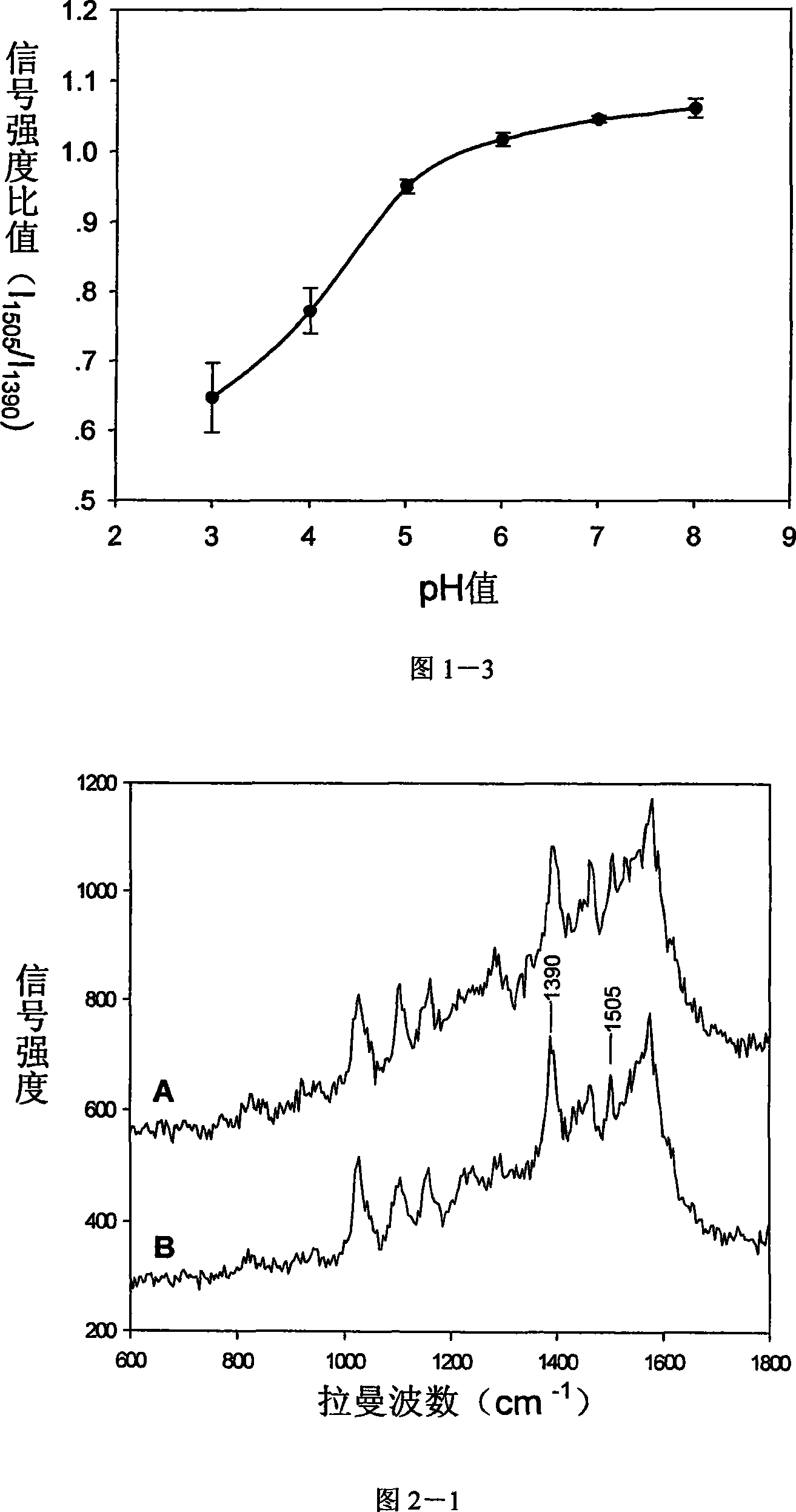
[0047] In the first step, the response curve of the SERS probe to pH is measured. The functional SERS probe solution was added to the buffer solution of pH3.0~pH8.0, the pH value interval was 1.0. After 10 minutes, the mixed solution was dropped onto the silicon chip and fixed on the carrier of the Raman confocal microscope. on the object stage to measure its SERS spectrum. Repeat the measurement 10 times corresponding to each pH value, and calculate the 1505cm in each SERS spectrum -1 at 1390cm -1 The ratio of signal strength at the place, that is, I 1505 / I 1390 , taking the average of 10 experimental results. Taking pH as the abscissa, I 1505 / I 1390 Draw the response curve of the SERS probe to pH as the ordinate, as shown in Figure 1-3. The probe has a high acid detection limit, and the linear response range is 3.0-6.0.
[0048] In the second step, pancreatic cancer cells were...
Embodiment 3
[0050] Example 3, Functional SERS Probe Used for Multicomponent Detection in Pancreatic Cancer Cells
[0051] In the first step, pancreatic cancer cells were cultured in culture medium (37°C, 5% CO 2 ). After 24 hours, the SERS probe solution was added to the culture medium in a volume ratio (3:1), shaken gently, and placed in the incubator again. At this time, the SERS probe enters the interior of the cell by being phagocytized by the cell. One hour later, the medium was discarded, and the cells were gently washed 3 times with phosphate buffered saline (PBS, 7.4) to remove the SERS probes remaining in the medium that were not phagocytized by the cells. stand-by.
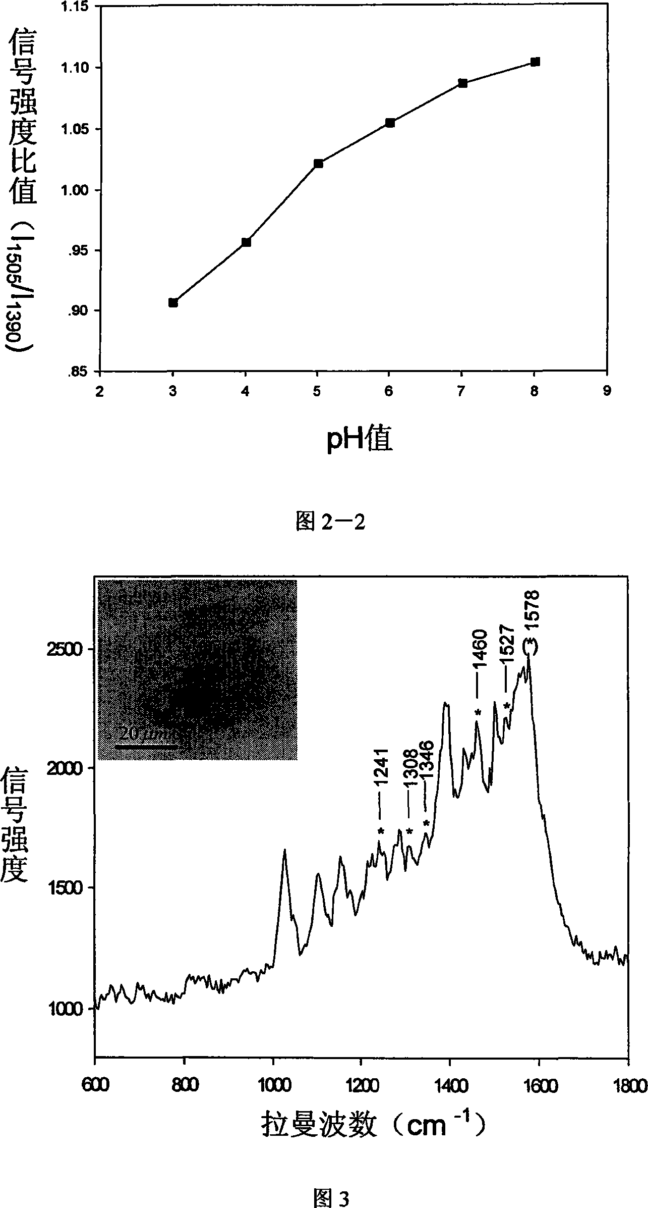
[0052] In the second step, the cells treated in the first step were placed in phosphate buffered saline (PBS, 7.4), and fixed on the stage of a confocal Raman microscope. Select a single-cell region to detect the SERS spectrum in the cell, as shown in Figure 3. In addition to the original signal of the probe, a...
PUM
| Property | Measurement | Unit |
|---|---|---|
| size | aaaaa | aaaaa |
Abstract
Description
Claims
Application Information