Whole layer biological cornea as well as construction method and use thereof

A kind of cornea and biological technology, applied in the field of medical materials, can solve the problem of cornea not being ideal

Active Publication Date: 2009-05-20
SHANGHAI NINTH PEOPLES HOSPITAL AFFILIATED TO SHANGHAI JIAO TONG UNIV SCHOOL OF MEDICINE
View PDF1 Cites 31 Cited by
  • Summary
  • Abstract
  • Description
  • Claims
  • Application Information

AI Technical Summary

Problems solved by technology

The commonly used carriers for corneal construction are not optimal due to parameters such as tra

Method used


Image

  • Whole layer biological cornea as well as construction method and use thereof
  • Whole layer biological cornea as well as construction method and use thereof
  • Whole layer biological cornea as well as construction method and use thereof

Examples


Embodiment 1

[0028] Example 1 Preparation and Biocompatibility of Porcine Corneal Acellular Matrix

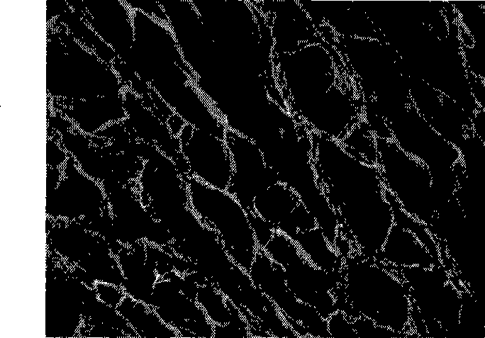
[0029] Take fresh porcine cornea, mechanically remove the epithelial layer, tear off the elastic layer, soak in 1% TritonX-100, shake at 4°C for 72 hours to decellularize, then repeatedly shake and wash with PBS buffer, and the tissue is taken to remove the front lamellar layer And the back plate layer, keep the middle third of the thick matrix layer, freeze at -80°C overnight, and then transfer to vacuum drying for 24 hours. The acellular matrix was rehydrated in PBS buffer and serum-free DMEM medium before application, and stored at 4°C for future use. The prepared porcine corneal acellular matrix maintains the original basic shape and toughness of the cornea, and is edematous and translucent. Under the scanning electron microscope, the collagen fibers in the longitudinal section of the matrix layer are neatly arranged, wavy, dense, and the fibers can be seen in different sizes pores ...

Embodiment 2

[0032] Example 2 Isolation and Culture of Rabbit Corneal Epithelial, Stromal and Endothelial Cells

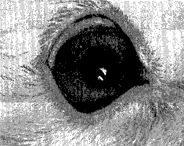
[0033] Rabbit corneas were taken aseptically, and the corneal epithelium and stroma were peeled off under a microscope, and the endothelial layer was torn off along Descemet's membrane. Tissue block planting method to cultivate primary corneal epithelial cells: after removing the superficial epithelial cells, cut the corneal limbus into 1mm×2mm tissue blocks, stick the epithelium side down on the bottom of the culture dish, dry in the incubator for half an hour, add a small amount of 10% The DMEM / F12 (1:1) medium of fetal bovine serum is better just submerging the tissue block, and the culture medium is added after overnight. The primary stromal cells were obtained by combined digestion and tissue block method: cut the stromal layer tissue block into pieces, put them in a centrifuge tube, add 3% type II collagenase for thermal digestion, and shake at 37°C for about 40 minute...

Embodiment 3

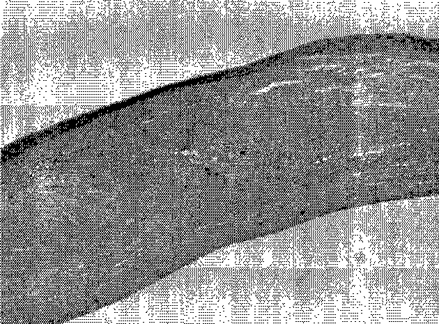
[0035] Example 3 Cultivation of three-dimensional corneal stroma in a bioreactor

[0036] First, the corneal stromal cells were transfected with green fluorescent protein (GFP), and the cells were fluorescently labeled. When the corneal stromal cells adhere to the wall and grow nearly 70% to 80% confluent, discard the culture medium, add fresh culture medium and GFP recombinant adenovirus solution at a ratio of 2:1 to the culture dish, and place at 37°C, 5% CO 2 Overnight in a saturated humidity incubator, discard the mixture the next day, wash twice with PBS buffer to remove residual virus particles, then add fresh serum-containing medium again, continue to culture for 24 hours, and observe the high expression of green fluorescence under a fluorescent microscope After that, it can be used to plant the corneal acellular matrix carrier.

[0037] GFP-labeled corneal stromal cells were digested and centrifuged to make a concentration of 2×10 6 / ml of cell suspension, injecte...


PUM


Abstract

The invention aims at providing a novel full-thickness biological cornea used for transplantation. The full-thickness biological cornea takes an animal cornea acellular matrix as a carrier, and the acellular matrix comprises animal cornea matrix cells, epithelial cells and endothelial cells which are cultured and augmented in vitro. The cornea is characterized in that the xenogenic corneal acellular matrix prepared by a biochemical method can be used as a good carrier for in vitro constructing the biological cornea in the aspects of shape, structure and biological compatibility, the matrix cells, epithelial cells and endothelial cells of the cornea are planted respectively, and dynamically cultured in the simulated in vivo environment of a biological reactor, so that the full-thickness biological cornea with nearly normal tissue structure and characteristics can be constructed in vitro, and the biological cornea can be used to simulate the physiological cornea for fundamental research on physiology, pathology and pharmacology; moreover, the biological cornea can also be directly used as the donator for corneal transplantation.

Description

technical field [0001] The invention relates to the field of medical materials in medicine and bioengineering, in particular to a full-thickness biological cornea that is cultured and constructed in a bioreactor using heterogeneous corneal acellular matrix as a carrier. This biological cornea can be used in physiology, pharmacology and pathology For basic research, it can also be used as a donor for corneal transplantation and refractive surgery. Background technique [0002] Corneal disease is a refractory blinding eye disease with high incidence and difficult treatment. Corneal injury, ulcer, scar and edema turbidity caused by various reasons are one of the main causes of blindness in the world today. At present, lamellar or penetrating keratoplasty is the main method for treating corneal opacity, but it is limited by the lack of corneal donor sources, aging corneal donors, postoperative complications, and postoperative immune rejection. The clinical application of polyme...

Claims


Application Information

Patent Timeline
IPC IPC(8): A61F2/14A61L27/38C12N5/06A61L31/00C12N5/071
Inventor 范先群傅瑶
Owner SHANGHAI NINTH PEOPLES HOSPITAL AFFILIATED TO SHANGHAI JIAO TONG UNIV SCHOOL OF MEDICINE
Why Eureka
Try Eureka