
Liquid phase chip for joint detection of multiple tumor markers and preparation method thereof

A technology for joint detection of tumor markers, which is applied in the field of multiple tumor marker joint detection liquid phase chips and its preparation, can solve the problems of unfavorable joint detection liquid phase chips for commercialization and separate storage, and achieve stable results and easy operation , Long shelf life effect

Active Publication Date: 2011-04-20
HENAN YUKANG BIOTECH
View PDF3 Cites 0 Cited by
  • Summary
  • Abstract
  • Description
  • Claims
  • Application Information

AI Technical Summary

Problems solved by technology

[0012] However, the various components of the existing multi-tumor marker combined liquid chip can only be stored separately, and can only be mixed together during detection
For example, Chinese patent document CN1766615A discloses a liquid phase chip for parallel detection of colorectal cancer protein markers, and CN101216491A discloses a liquid phase chip for rapid detection of Mycobacterium tuberculosis. In this type of joint detection chip, various coupling microspheres and The detection antibody can only be stored separately, and can only be mixed together for joint detection when it is in use. This drawback brings a lot of troubles to storage, transportation, operation and use, and is not conducive to the commercialization of joint detection liquid phase chips.

Method used


Image

  • Liquid phase chip for joint detection of multiple tumor markers and preparation method thereof

Examples


Embodiment 1

[0049] Example 1 A liquid chip for joint detection of multiple tumor markers, mainly including:

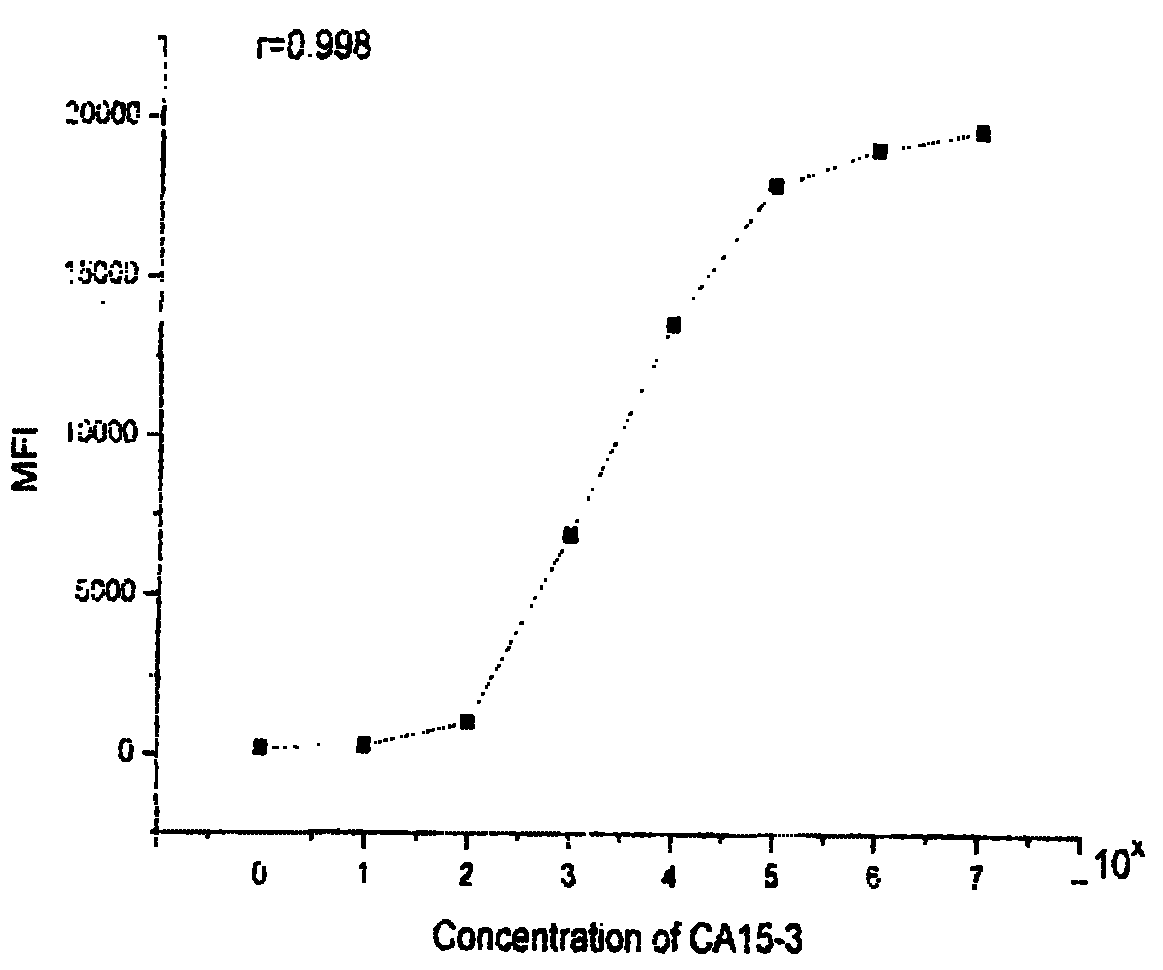
[0050] ① Microspheres coupled with capture antibody include microspheres coupled with AFP capture antibody, microspheres coupled with CEA capture antibody, microspheres coupled with CA125 capture antibody, microspheres coupled with CA153 capture antibody, Microspheres coupled with CA19-9 capture antibody, microspheres coupled with CA242 capture antibody, microspheres coupled with CA72-4 capture antibody, microspheres coupled with PSA capture antibody, microspheres coupled with HGH capture antibody Microspheres, microspheres coupled with beta-HCG capture antibodies, and microspheres coupled with different antibodies correspond to different color codes;

[0051] ②Biotin-labeled detection antibodies are divided into biotin-labeled AFP, CEA, CA125, CA153, CA19-9, CA242, CA72-4, PSA, HGH, beta-HCG detection antibodies;

[0052] ③ streptavidin phycoerythrin; and

[0053] ④ Baijijiao s...

Embodiment 2

[0076] Embodiment 2 A liquid chip for combined detection of multiple tumor markers, mainly including:

[0077] ① Microspheres coupled with antibodies are the same as in Example 1;

[0078] ②The biotin-labeled detection antibody is the same as in Example 1;

[0079] ③ streptavidin phycoerythrin;

[0080] The above components are evenly dispersed or dissolved in the white glue solution with a solute content of 5-8wt‰.

[0081] The preparation method is basically the same as that of Example 1, except that in the preparation step, the required various coupling microspheres are mixed in a certain proportion, and biotin-labeled antibodies corresponding to the type and quantity are added, as well as the corresponding quantity streptavidin phycoerythrin, and then adjust the concentration of microspheres with appropriate concentration of white and hydrosol, after dispersing and mixing, the concentration of each coupling microsphere is 1×10 3 ~2×10 3 Each / ml, white and solute conten...

Embodiment 3

[0083] Example 3 A liquid phase chip for combined detection of multiple tumor markers, which is basically the same as Example 1, except that microspheres coupled with antibodies, biotin-labeled detection antibodies, and streptavidin phycoerythrin dissolve / Dispersed in the refined seaweed gel aqueous solution with a solute content of 2-4wt‰. The preparation method of the seaweed glue used is: take the seaweed glue above CP grade, add water to soak for 6 to 10 hours, make it fully dissolved, filter and remove impurities with an EK filter plate, adjust the concentration to a solute content of about 5wt%, and sterilize It is ready for later use, and during the preparation process, the introduction of divalent ions is avoided. Its preparation method is basically the same as Example 1.


PUM


Abstract

The invention relates to a liquid phase chip for joint detection of multiple tumor markers and a preparation method thereof. The liquid phase chip comprises micro-balloons of a coupled antibody (at least two of the followings: AFP, CEA, CA125, CA153, CA19-9, CA242, CA72-4, PSA, HGH, Beta-HCG), corresponding biotin-labeled detection antibodies, streptavidin phycoerythrin and vegetable hydrosol or polysaccharide hydrosol which has a solute content of 1-10 wt per thousand and does not contain protein. The preparation of the liquid phase chip comprises refining and purification of hydrosol, coupling between captured antibodies and micro-balloons, preparation of biotin-labeled antibodies, dispersing the coupled micro-balloons into the vegetable hydrosol or the polysaccharide hydrosol, and the like. The liquid phase chip has the advantages of stable performance, good micro-balloon dispersivity, long preservation time, fast and convenient use and operation, small amount of samples in use, high detection sensitivity, wide linear scope and low detection cost, can detect ten tumor markers at most at one time, and requires a cost which is a quarter of the total fee of conventional methods.

Description

technical field [0001] The invention relates to the field of biochips, in particular to a liquid phase chip for combined detection of multiple tumor markers and a preparation method thereof. Background technique [0002] Tumor markers refer to a large class of substances produced by tumor tissue that exist in the tumor tissue itself or are secreted into blood and other body fluids, or are produced by host cells due to tumor cell stimulation and have significantly abnormal levels. Tumor markers play an important role in the clinical diagnosis and treatment of tumors. The detection of the presence or content of tumor markers in blood or body fluids can not only be used for general screening, early diagnosis, auxiliary diagnosis, benign and malignant differentiation, and clinical staging of tumors. At the same time, it is also of great value in monitoring curative effect, judging prognosis, and predicting tumor recurrence and metastasis. The most commonly used clinical tumor m...

Claims


Application Information

Patent Timeline
Patent Type & Authority Patents(China)
IPC IPC(8): G01N33/569G01N33/546G01N33/531
Inventor 王韫烨王学铭
Owner HENAN YUKANG BIOTECH
Learn More