

Method for detecting protein content in 2-keto-L-gulonic acid
A technology of protein content and gulon acid, which is applied in the field of chemical analysis, can solve the problems of inability to detect protein content, and achieve the effects of low detection equipment requirements, good reproducibility, and good time stability
- Summary
- Abstract
- Description
- Claims
- Application Information
AI Technical Summary
Problems solved by technology
Method used
Image

Examples
Embodiment 1
[0054] Example 1: Detection of protein content in 2-keto-L-sodium guronate fermentation broth;
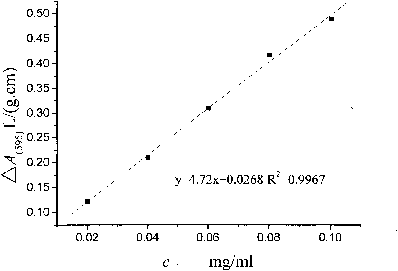
[0055] Take the 2-keto-L-gulonic acid sodium fermentation liquid in production and centrifuge at 2000r / min for 20min, pipette 2ml of the supernatant into a test tube, then add 2ml of 40% KOH solution, boil in boiling water for 30min, wash it with deionized water into a 100ml volumetric flask, then add 4.5ml of 20% nitric acid solution and 10ml of a buffer solution with a pH of 7, and dilute to volume with deionized water.
[0056] Pipette one part of 1 mL of deionized water and two parts of the test solution into three test tubes, add 5 mL of Coomassie Brilliant Blue reagent to the first two test tubes, add 5 mL of deionized water to the third test tube, and detect the deionized water at 595nm and the absorbance of test tube 2 are 0.037 and 0.058L / (g.cm), adjust the absorbance of test tube 1 to 0, and detect the absorbance of test tube 3 within 30 to 60 minutes to be 0.162L / (g.cm),...
Embodiment 2
[0057] Example 2: Detection of protein content in 2-keto-L-sodium guronate ultrafiltrate
[0058] Take 20ml of post ultrafiltrate to a 50ml volumetric flask, then add 10ml of buffer solution with a pH of 7, and dilute to volume with deionized water.
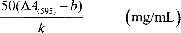
[0059] Pipette one part of 1 mL of deionized water and two parts of the test solution into three test tubes, add 5 mL of Coomassie Brilliant Blue reagent to the first two test tubes, add 5 mL of deionized water to the third test tube, and detect the deionized water at 595nm and the absorbance of test tube 2 are 0.037 and 0.046L / (g.cm), adjust the absorbance of test tube 1 to 0, and detect the absorbance of test tube 3 within 30 to 60 minutes to be 0.205L / (g.cm). The protein content in the liquid is 0.089mg / ml.
Embodiment 3
[0060] Example 3: Detection of protein content in dry product of 2-keto-L-gulonic acid
[0061] Weigh 5g of the dry product of 2-keto-L-gulonic acid in production, dissolve it in deionized water and transfer it to a 100ml volumetric flask, then add 6.5ml of 20% KOH solution and 10ml of buffer solution with a pH of 7, and dilute to volume with deionized water .
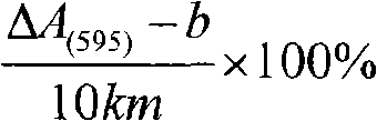
[0062] Pipette one part of 1 mL of deionized water and two parts of the test solution into three test tubes, add 5 mL of Coomassie Brilliant Blue reagent to the first two test tubes, add 5 mL of deionized water to the third test tube, and detect the deionized water at 595nm The absorbance of test tube 2 and test tube 2 are respectively 0.037 and 0.039 L / (g.cm), adjust the absorbance of test tube 1 to 0, and detect the absorbance of test tube 3 within 30 to 60 minutes to be 0.115 L / (g.cm), which can be calculated by formula 1 The protein content in the dry product of 2-keto-L-gulonic acid is 0.036%.
PUM
| Property | Measurement | Unit |
|---|---|---|
| Absorbance | aaaaa | aaaaa |
| Absorbance | aaaaa | aaaaa |
| Absorbance | aaaaa | aaaaa |
Abstract
Description
Claims
Application Information